Continuous regulation rather than an occasional emergency switch
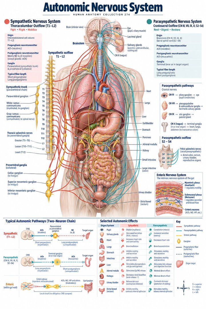
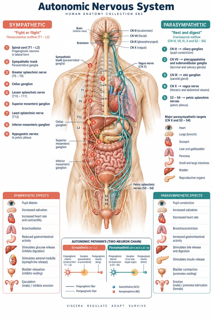
The autonomic nervous system helps regulate internal functions such as cardiovascular activity, gastrointestinal function, gland secretion, pupil size, and bladder behavior. Its activity continues during ordinary daily life, including sleep, eating, standing, and movement. “Fight or flight” and “rest and digest” are useful introductory phrases, but neither describes the full system. Autonomic outputs can change differently across organs, and baseline neural activity is important even when a person is resting. A medical learner should therefore think in terms of pathways, target tissues, receptors, and feedback rather than two whole-body switches that simply alternate between on and off.
This perspective matters for healthcare teams because one observation cannot reveal the complete autonomic state. Heart rate, sweating, pupil appearance, gastrointestinal activity, and blood pressure reflect overlapping influences. They are not interchangeable measurements of one hidden dial. Anatomical education provides a framework for relating each observation to a pathway and effector, while clinical interpretation requires context. The best foundation begins with a question: which information is coming toward the central nervous system, which signal is leaving it, and what structure ultimately changes its behavior? That route-based explanation remains useful as the details become more advanced.
Afferent information and efferent pathways must be distinguished
Visceral sensory pathways convey information about internal conditions toward central integrating regions. Autonomic efferent pathways carry commands toward smooth muscle, cardiac tissue, glands, and related targets. These are different directions of communication. An internal-organ reflex can include sensory input, central processing, and autonomic output without every participating neuron being an autonomic motor neuron. The brainstem, hypothalamic regions, spinal circuits, and peripheral ganglia occupy different positions within the overall organization. A sensory nerve ending measuring stretch is not the same structure as a motor terminal releasing a transmitter onto an effector.
For a simple teaching diagram, draw the sensory route and motor route as separate arrows. Place the central integrating region between them, then identify the target organ. This avoids a misleading diagram in which the organ appears to send and receive information along one unlabeled wire. The distinction also supports more precise workplace language. If a clinician discusses a reflex pathway, the learner should ask which part is being described: receptor, afferent route, integrating center, efferent neuron, or target response. Different findings can arise from different parts of the same functional loop, so a single anatomical label should not stand for the entire loop.
The usual two-neuron motor chain and its ganglion
The typical autonomic motor route has a preganglionic neuron with its cell body in the central nervous system and a postganglionic neuron with its cell body in a peripheral ganglion. Their synapse creates a relay before the signal reaches the target. This differs from the usual somatic motor route to skeletal muscle, where a lower motor neuron reaches the muscle directly. Ganglia are collections of neuronal cell bodies outside the central nervous system; nuclei are neuronal cell-body groups within it. These anatomical terms should not be exchanged merely because both involve clusters of neurons.
The two-neuron organization is useful numerically because it describes a pathway model, not the total number of neurons serving an organ. A diagram with two circles should therefore be labeled as a representative efferent chain. An organ may receive many fibers, divergent connections, and additional local circuits. Learning the model first simplifies the route, but the explanation should retain the distinction between a representative chain and the complete network. When reviewing a micrograph of nervous tissue, the image may demonstrate cell or tissue structure without showing an entire functional autonomic pathway. Captioning the image honestly preserves that distinction.
Sympathetic origins and routes through peripheral ganglia
Sympathetic preganglionic cell bodies occupy the thoracolumbar spinal cord, commonly described as T1–L2. Fibers leave through anterior roots and access the sympathetic trunks through white rami communicantes. They can synapse at the entry level, travel within the trunk to other levels, or pass toward prevertebral ganglia through appropriate splanchnic routes. The sympathetic trunk therefore helps distribute outputs beyond the spinal levels where the preganglionic neurons originate. A target in the head does not imply that its sympathetic preganglionic cell body begins in the brain. NCBI describes sympathetic anatomical routes.
This origin-versus-destination distinction is one of the most important ideas in autonomic anatomy. A map may place ganglia near the neck and show fibers entering a cranial region, but the diagram still needs to identify the thoracolumbar origin of the preganglionic component. Different colors for preganglionic and postganglionic fibers make the relay visible. The same logic applies to abdominal routes: a fiber passing through a trunk without synapsing there is different from one that has already become postganglionic. The anatomical route should be traced before transmitter rules are added, because the type of neuron depends on which side of the ganglion it occupies.
Parasympathetic origins and ganglia near targets
The conventional parasympathetic arrangement has cranial outflow associated with cranial nerves III, VII, IX, and X, plus sacral outflow from S2–S4 through pelvic splanchnic nerves. Ganglia are generally near or within target organs, producing relatively long preganglionic and short postganglionic components in many pathways. The vagus contributes extensive thoracic and abdominal innervation, but it does not account for every parasympathetic route. Pelvic organs and parts of the distal intestinal region involve sacral pathways. NCBI describes parasympathetic origins and peripheral routes.
The four named cranial nerves are a teaching count of outflow routes, not four interchangeable nerves performing identical jobs. Their destinations and ganglia differ. Likewise, the three sacral segment labels describe a source region, not a count of only three fibers serving the pelvis. When explaining the arrangement to a colleague, state both the origin and principal destination rather than saying that all involuntary control travels in the vagus. A simplified body map can make the cranial and sacral components clear, provided it identifies itself as a regional overview rather than a complete nerve atlas.
Transmitters, receptors, and important exceptions
Both major divisions use acetylcholine at their preganglionic synapses, acting on nicotinic receptors in autonomic ganglia. Parasympathetic postganglionic terminals generally use acetylcholine acting through muscarinic receptors at targets. Most sympathetic postganglionic terminals use norepinephrine at adrenergic receptors. Eccrine sweat glands are a notable sympathetic cholinergic exception. Neuroscience explains visceral neurotransmission. The adrenal medulla is another specialized arrangement: preganglionic sympathetic input acts on chromaffin cells, which release catecholamines into blood rather than sending a conventional postganglionic axon to a local target. Endocrinology describes the adrenal medulla. These examples make a one-line transmitter rule incomplete.
Receptor identity matters because the same chemical messenger can produce different effects in different tissues. A transmitter name alone does not tell whether a muscle contracts, relaxes, or changes the timing of its activity. For medical students, it is helpful to separate three columns: neuron category, transmitter, and target receptor. Add the organ response only after these relationships are clear. This prevents the common error of describing acetylcholine as intrinsically calming and norepinephrine as universally stimulatory. The response belongs to the complete receptor-and-tissue arrangement, not to an emotional label attached to the chemical.
Cardiovascular reflexes connect pressure sensing with coordinated output
The baroreceptor reflex links changes in arterial stretch with neural processing and adjustments in cardiovascular activity. Carotid sinus and aortic arch sensory information reaches brainstem integrating regions through relevant cranial-nerve routes. Changes in autonomic output can alter heart activity and vascular tone. The mechanism contributes to short-term regulation during changes such as moving from lying to standing. A reflex diagram should distinguish the stretch-sensitive input from the efferent effects on heart and vessels. NCBI reviews carotid sinus anatomy and the baroreceptor pathway.
Standing provides a useful conceptual example because gravity changes the distribution of blood and the circulatory system responds. The illustration should not promise one exact heart-rate or pressure change for every person. The visible cardiovascular measurement is an outcome of several interacting conditions, including posture, volume status, medications, and the individual's physiology. For healthcare learners, the anatomical value is to see a coordinated loop rather than assume that a rapid pulse identifies a single autonomic cause. A real observation belongs in the appropriate assessment context, while the pathway explains why central integration and several effectors may be involved simultaneously.
Pupil size uses two iris muscles and two neural arrangements
The sphincter pupillae has a circular arrangement and participates in pupil constriction under parasympathetic influence. The dilator pupillae has a radial arrangement and participates in dilation under sympathetic influence. Pupil size is therefore the result of muscular and neural relationships within the iris, not a direct measurement of the size of a hole in the skull. The light reflex also includes an afferent visual route and central connections before the parasympathetic output reaches the sphincter. The Clinical Methods chapter describes pupil muscles and their innervation.
This example separates an effector from a full reflex. A picture showing a constricted pupil demonstrates an appearance, but it does not show the complete route from light entering the eye to central processing and motor output. It also cannot establish why that pupil has a particular size. A teaching graphic can use circular and radial muscle arrows to make the mechanics clear, while keeping the neural arrows separate. Such an approach supports the accurate use of terms in professional communication without turning an educational illustration into a substitute for a pupil examination.
Enteric circuits organize local gastrointestinal activity
The enteric nervous system contains local neural circuits in the gastrointestinal wall. The myenteric plexus lies between major smooth-muscle layers, while the submucosal plexus occupies the submucosal region. These interconnected circuits participate in motility, secretion, and local coordination. Sympathetic and parasympathetic influences interact with them, so gastrointestinal control is not merely a long motor cable from the brain to every intestinal muscle cell. The enteric arrangement is sometimes described as a division of the autonomic system and sometimes presented separately because of its extensive local organization. NCBI reviews gastrointestinal nervous control.
A bowel-wall diagram is especially informative because it places the networks at their tissue depths. Draw muscle layers, submucosa, and mucosa first, then add the neural plexuses. This shows how an organ wall contains a local coordinating network in addition to effector tissues. A general autonomic body diagram cannot convey that detail. For students, comparing the two representations explains why several levels of organization are necessary. For healthcare staff, the practical insight is that intestinal behavior emerges from local and extrinsic inputs together, so “parasympathetic activity” alone is not a complete description of every change in motility or secretion.
Bladder storage and emptying require coordinated systems
Bladder control illustrates the interaction between autonomic regulation and somatic motor control. The detrusor is smooth muscle within the bladder wall. Parasympathetic activation supports its contraction during emptying, while sympathetic influences contribute to storage-related functions. The external urethral sphincter's striated muscle belongs to somatic control and participates in the coordinated outlet arrangement. Central pathways help coordinate the timing of bladder and outlet activity; these components should not be drawn as unrelated switches. The Neuroscience chapter explains the interaction of voluntary and visceral motor pathways.
The anatomical lesson is coordination, not a simplistic rule that contracting every muscle empties the bladder. A storage reservoir and its outlet must change their behavior in a compatible way. This same reasoning helps distinguish the muscle in the bladder wall from muscles surrounding the outlet. Urinary observations in a clinical setting can have many causes and should be documented within the relevant assessment framework. An educational diagram can establish which tissues and pathways are involved without giving individualized interpretation, catheter instructions, or treatment advice. The route and effector labels remain useful across different professional roles.
Tone, organ specificity, and the limits of opposing-arrow diagrams
Autonomic tone describes continuing baseline activity that can be increased or decreased. In many organs, the two major divisions influence different aspects of function rather than producing a perfectly symmetric tug-of-war. Some targets receive predominantly one type of autonomic influence. Different receptor distributions further complicate any assumption that sympathetic means activation of everything and parasympathetic means inhibition of everything. A paired-arrow chart can introduce a relationship, but it should be expanded with target tissue and receptor context before it is used to explain a whole organ's behavior.
For example, a gland secreting fluid, a vascular smooth-muscle layer changing diameter, and a cardiac pacemaker changing timing are different effectors. “More activity” has a different physical meaning in each case. A useful explanation names the actual response: secretion, contraction, relaxation, or altered rate. This precision prevents vague statements about the body being simply sped up or slowed down. It also explains why autonomic anatomy links so many specialties: the common signaling framework operates through highly specific organ designs. Understanding the shared pathway and the distinct effector together is more informative than learning a slogan for each division.
A clinically useful explanation also retains the time course. The pupil response to a light change, cardiovascular adaptation to posture, and gastrointestinal coordination during a meal are not interchangeable events simply because all involve visceral regulation. A diagram should identify the stimulus being modeled and the response being illustrated. It should not imply that observing one response proves that every organ changed in the same direction or at the same moment. This discipline helps students describe mechanisms accurately and helps healthcare teams distinguish a recorded observation from a broad inference about autonomic function. It is especially valuable when several physiological measurements are discussed together.
Latin vocabulary and numerically honest pathway diagrams
Systema nervosum autonomicum names the autonomic system; pars sympathica and pars parasympathica identify its main extrinsic divisions. Ganglion, truncus sympathicus, nervus vagus, nervi splanchnici pelvici, plexus myentericus, and plexus submucosus identify important relays or routes. The two-neuron chain, four cranial parasympathetic routes, and S2–S4 source region are anatomical teaching facts with different counting units. They should never share one unlabeled numerical chart. A final pathway explanation should state direction, origin, relay, transmitter, receptor, and effector. That framework connects anatomy with meaningful physiological regulation while preserving organ-specific complexity.
Anatomical Latin: quick reference
| Latin term | English meaning |
|---|---|
| Pars sympathica | Sympathetic division |
| Pars parasympathica | Parasympathetic division |
| Ganglion | Group of peripheral neuron cell bodies |
| Nervus vagus | Vagus nerve |
Sources and image credits
Sources reviewed on 9 October 2026. Counts, estimates, study statistics and hypothetical calculations retain their context in the text.
- NCBI: autonomic anatomy
- NCBI: autonomic physiology
- NCBI: parasympathetic nervous system
- Neuroscience: visceral neurotransmission
- Endocrinology: adrenal gland
- NCBI: carotid sinus and baroreflex
- Clinical Methods: pupils
- NCBI: gastrointestinal nervous control
- Neuroscience: autonomic bladder regulation
- NCBI: myenteric plexus
- Review: enteric nervous system
Explanatory prose and SVG graphics are original. Photograph authors, source records and licenses are credited beside each image. Catalog illustrations provide educational context; anatomical facts are supported by the linked sources.