A connected route with specialized segments
Blood vessels form a continuous transport network, but their walls and functions vary along the route. Large arteries receive pulsatile flow from the heart, smaller arteries and arterioles distribute and regulate delivery, capillaries create exchange interfaces, and venules and veins return blood. This sequence connects organ-scale circulation with microscopic tissue support. Medical learners need to understand why vessel size, wall composition, pressure environment, and local tissue needs belong together. A diagram that labels every red tube an artery and every blue tube a vein captures a drawing convention, not the defining biology of the vessels.
For healthcare staff, vessel terminology supports location descriptions and communication about circulation without requiring every observer to interpret vascular disease independently. The most useful starting questions are directional: where does the blood come from, where is it going, and which segment is being discussed? Then ask structural questions: what forms the wall, what controls its diameter, and what crosses the exchange barrier? These questions produce a more complete explanation than a list of vessel names. They also help distinguish a visible superficial vessel from the deeper network and avoid assuming that one local appearance represents the whole circulation.
Artery and vein are defined by flow direction
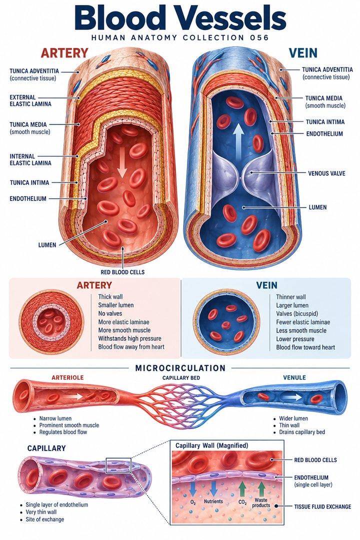
An artery carries blood away from the heart; a vein carries blood toward it. Oxygen content does not define the category. Pulmonary arteries carry relatively oxygen-poor blood from the right ventricle toward the lungs, while pulmonary veins return oxygenated blood to the left atrium. Systemic arteries and veins generally show the opposite oxygen pattern. The pulmonary and systemic circuits are connected in sequence through the heart, although each has its own pressure and tissue environment. NCI SEER explains the directional distinction and the two circuits.
This definition resolves a common problem in teaching diagrams. Red and blue are convenient colors for relative oxygenation, but they should not replace directional arrows or vessel labels. A pulmonary artery remains an artery even when drawn blue. Similarly, a pulmonary vein remains a vein when drawn red. The same lesson applies to photographs: blood within living vessels is not bright blue in the way a schematic line may be. When discussing an anatomical route, name its origin, destination, and direction before adding the oxygenation context. The route remains interpretable even if the illustration uses a different color palette.
Intima, media, and adventitia are different tissue relationships
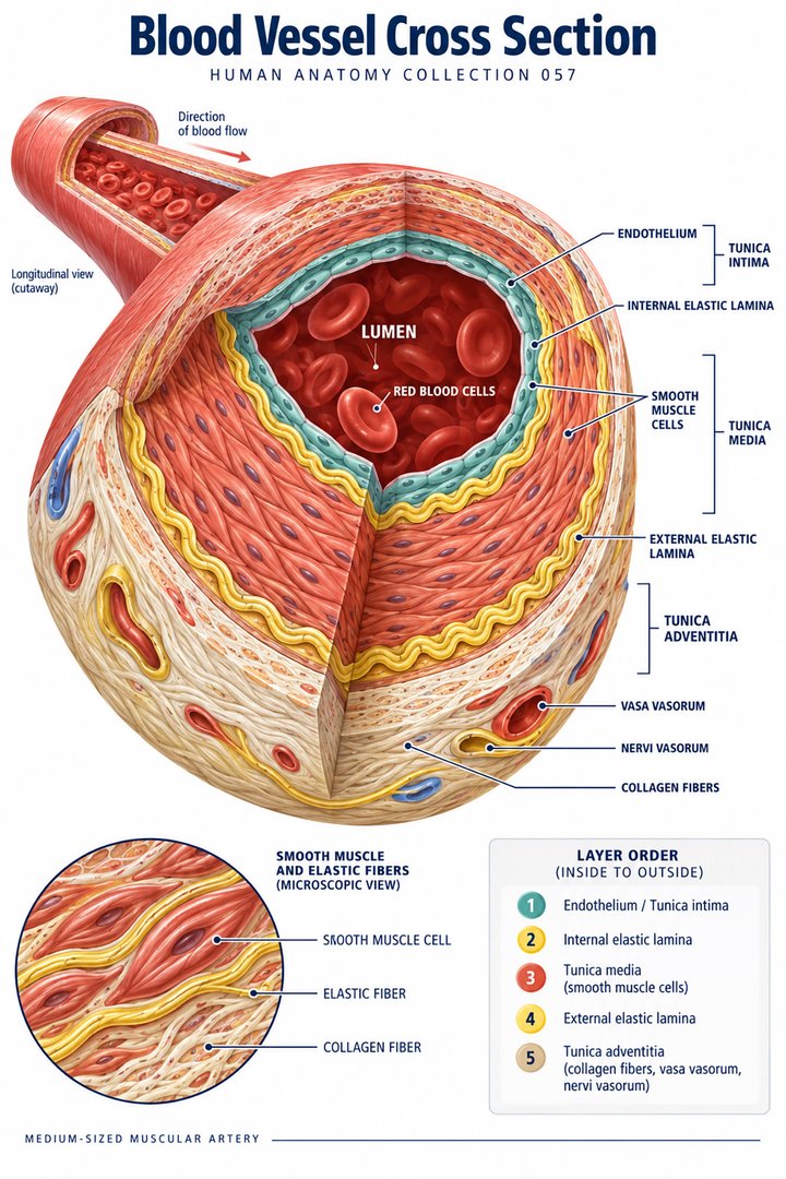
The wall of a typical substantial artery or vein can be described through three tunics. Tunica intima includes the endothelial interface and supporting elements. Tunica media contains smooth muscle and, in varying proportions, elastic material. Tunica adventitia or externa provides connective-tissue support and continuity with surrounding tissues. The relative prominence of each component changes by vessel class. Capillaries do not have the same complete three-tunic arrangement. NCBI describes the anatomical organization of vessel walls.
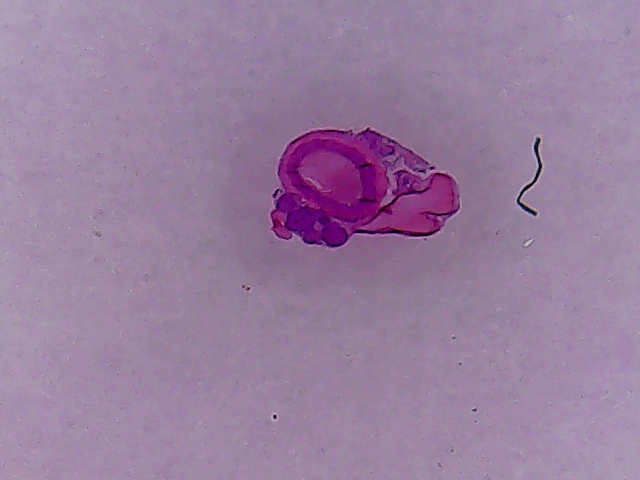
The tunics should not be drawn as equally thick universal rings. In a muscular artery, the media is prominent; a vein has a different relative wall arrangement and often a wider lumen in relation to its wall. A histological section may show a partly collapsed vein, so lumen shape alone is not sufficient for identification. Compare wall composition, surrounding tissue, and the relative thickness of muscular layers. Specimen preparation can alter the appearance of the lumen, whereas the layered organization provides more durable clues. In clinical learning, this distinction supports a careful separation of observed shape from inferred vessel function.
Elastic arteries buffer pulsatile output
Large elastic arteries, including the aorta, have abundant elastic components in their walls. Expansion during ventricular ejection and recoil afterward help distribute pulsatile input over the cardiac cycle. They do not generate blood flow independently of the heart, but their mechanical behavior influences how that flow is transmitted downstream. A rigid-pipe image therefore misses an important feature of the living arterial system. The wall's ability to respond to changing pressure belongs to its function, not simply to its appearance in a cross-sectional drawing. NCBI reviews vascular histology and elastic versus muscular wall patterns.
An educational animation can show wall expansion and recoil with the caption “schematic behavior,” rather than imply an exact change in diameter for every person. The teaching point is the temporal relationship between ejection, wall deformation, and downstream movement. This is distinct from average flow, and distinct again from pressure measured at a particular site. For a medical student, keeping these quantities separate helps prevent equations and diagrams from becoming contradictory. For healthcare staff, the same concept explains why a pulse is a propagating pressure-related event rather than a discrete packet of blood traveling unchanged to the fingertips.
Muscular arteries and arterioles distribute flow
Muscular arteries contain substantial smooth muscle and distribute blood to regions and organs. Arterioles provide an important site of adjustable resistance before capillary networks. Smooth-muscle contraction narrows the lumen, while relaxation can increase it; the resulting effect depends on the broader network and driving pressure. Local metabolic influences, endothelial signals, neural input, and circulating factors interact in regulating vascular tone. An arteriole is therefore more than a small miniature of the aorta. Its wall organization and location give it a different role in controlling tissue delivery.
Consider two tissues with different activity at the same moment: working skeletal muscle and a relatively quiet neighboring region. Their local delivery requirements need not change identically. The vascular network can distribute flow differently rather than forcing every organ to receive the same proportionate increase. A useful diagram shows one upstream distribution route branching into separate tissue beds with adjustable arteriolar segments. This makes regional control visible. It also avoids an overly simple explanation in which a change in one vessel diameter must imply the same change in blood pressure, flow, and oxygen delivery everywhere else in the body.
Pressure gradients and resistance: a transparent numerical example
Flow through a pathway depends on a driving pressure difference and the resistance of that pathway. In the idealized Hagen–Poiseuille model, resistance varies inversely with the fourth power of radius when other assumptions are fixed. Reducing a model tube's radius from one relative unit to one-half gives sixteen times the resistance. Increasing it from one unit to two gives one-sixteenth the resistance. These are mathematical teaching calculations, not measured clinical changes in a person's artery. NCBI explains the radius relationship in peripheral vascular resistance.
The assumptions matter: living blood flow is pulsatile, blood is not always a simple Newtonian fluid, vessels are deformable, and networks contain branching pathways. The equation helps reveal why radius is influential, but it should not be presented as a patient prediction without the relevant conditions. A chart can label normalized radius and normalized resistance explicitly, showing the calculation beside the bars. Such a figure is more defensible than claiming one exact vessel length or one universal whole-body resistance value. Quantitative anatomy becomes most useful when its units, assumptions, and intended level of explanation remain visible.
Capillaries create exchange interfaces, not thick muscular tubes
Capillaries have thin endothelial walls with associated supporting structures suited to exchange. Continuous, fenestrated, and discontinuous or sinusoidal patterns describe important differences in endothelial organization. Continuous endothelium occurs in many tissues, fenestrations support particular exchange settings, and discontinuous sinusoidal arrangements occur in organs such as the liver, spleen, and bone marrow. Permeability is not identical across all capillary beds. The structural category must be related to the organ and the substances being exchanged. The Endothelium chapter describes this structural diversity.
The three-pattern count is another categorical fact rather than a set of three equally common vessel populations. A graphic should compare the interfaces, not suggest that every organ contains the same mixture. Different exchange needs require different barriers. A capillary serving brain tissue belongs to a different structural and cellular environment from a hepatic sinusoid. Medical learners should therefore pair a capillary label with the local tissue context. In a real micrograph, the visible lumen, surrounding cells, and stain provide only part of the information; the source's organ identification remains important for accurate interpretation.
Diffusion, filtration, and lymphatic return are distinct processes
Exchange across vascular interfaces includes movement of solutes and water through several mechanisms. Diffusion follows concentration-related gradients, while fluid filtration depends on pressure relationships and the properties of the barrier. The endothelial glycocalyx and subglycocalyx environment are important in modern explanations of fluid exchange. The older universal story of filtration at every capillary's arterial end and sustained reabsorption at its venous end is an oversimplification. Lymphatic return is central to tissue fluid balance in many steady-state settings. The revised Starling-principle article explains why the traditional picture needs refinement.
An exchange diagram should consequently show several routes without forcing them into one identical pair of arrows. Oxygen movement toward tissue, carbon-dioxide movement toward blood, fluid filtration, and lymphatic drainage are related but not interchangeable. The amount and direction of movement depend on tissue conditions and barrier properties. A teaching image can distinguish solute arrows from fluid arrows using labels as well as colors. For clinical learners, this helps connect vascular anatomy with interstitial tissue and lymphatics. It also prevents a visually tidy capillary picture from implying that every form of tissue swelling has the same anatomical mechanism.
Venules and veins collect blood and provide capacity
Venules receive blood from microvascular beds and merge into larger venous routes. Veins operate at lower pressure than systemic arteries in ordinary physiology and can accommodate substantial changes in contained volume. Their role in capacitance means that venous tone and blood distribution influence the return of blood to the heart. A large lumen and comparatively less muscular wall do not make veins passive or biologically unimportant. Their function belongs to the whole circulatory loop, where return must be coordinated with cardiac output and arterial delivery. The Control of Cardiac Output chapter explains venous return relationships.
The network context is crucial. A vein seen beneath skin is part of a larger drainage route, with superficial and deep systems interacting regionally. Its local fullness can change with position and surrounding conditions, but a photograph alone cannot determine every pressure relationship or establish a diagnosis. For healthcare staff, accurate descriptions identify the region, visible finding, and relevant circumstances without silently substituting a complete physiological conclusion. For students, drawing the venous route back to the heart makes clear why “return” is directional, while “low pressure” describes a usual physiological environment rather than the definition of a vein.
Valves and the skeletal muscle pump support directed return
Many limb veins contain valves that help limit backward movement. Contraction of surrounding skeletal muscle can compress veins and promote movement toward the heart in coordination with these valves. The skeletal muscle pump is therefore a relationship among muscle, vessel, pressure, and valve orientation. Not all veins contain valves, and the mechanism should not be drawn as universal for every venous bed. The skeletal-muscle circulation chapter describes the muscular pump and regional valve differences.
A two-panel schematic can show the vein before and during surrounding muscle contraction. Its arrows should reflect valve orientation rather than simply point upward on the page. “Toward the heart” is the functional direction; upward is only the direction chosen in that particular drawing. This matters when teaching with a person lying down, a limb elevated, or an illustration rotated horizontally. The same conceptual clarity helps healthcare teams discuss anatomical direction without confusing it with room coordinates. The diagram explains a normal supporting mechanism but does not prescribe a movement program or determine an individual's venous function.
Endothelium is an active interface
Endothelial cells line the luminal vascular surface and participate in signaling, barrier control, interactions with blood components, and regulation of vascular tone. They are not merely a frictionless coating. Their phenotype and surrounding structures vary between arterial, venous, and microvascular environments. This helps explain why vessel classification includes cellular identity and tissue context in addition to gross diameter. A large-vessel wall and a capillary interface share an endothelial component but organize it within different surrounding architectures. The arterial-versus-venous endothelial review discusses vascular cellular distinctions.
The clinical-learning implication is that vascular behavior emerges from several layers and interfaces. Smooth muscle can change lumen caliber, elastic material contributes mechanical behavior, connective tissue provides support, and endothelial signals influence local responses. A simple cross section should label these components distinctly rather than assign every function to the media. The vessel's surrounding tissue also matters. A superficial skin vessel, a vessel embedded within muscle, and a hepatic microvascular route occupy different biological environments. Relating wall structure to location makes the anatomy more useful than memorizing identical rings for every named vessel.
Naming routes and reading vascular images
Vessel names often identify regions, neighboring structures, or destinations. Arteria refers to an artery, vena to a vein, arteriola to an arteriole, and venula to a venule. Vas capillare identifies a capillary; tunica intima, tunica media, and tunica adventitia name wall layers. Lumen refers to the internal space. A vessel's name alone does not define every branch or anatomical variant. Learning a major route should include its origin, course, branches, and destination at the detail appropriate to the educational level.
Imaging and specimen photographs add their own conventions. A transverse vessel profile differs from a longitudinal view, and an injected or preserved specimen may show colors introduced during preparation. These should be identified in the caption rather than interpreted as the natural color of flowing blood. A final vascular explanation should connect direction, wall structure, regulation, exchange, and return. Adding a normalized resistance calculation can make one functional relationship quantitative without inventing patient statistics. The resulting map supports later study of circulation, organ perfusion, microscopy, and the vascular relationships described throughout medical practice.
Three broad categories and five commonly named sequential segments are compatible descriptions: artery, arteriole, capillary, venule, and vein refine the arterial–exchange–venous route. Neither list is a count of the body's individual vessels. A numerical graphic should state its classification level, because a learner could otherwise mistake five segment types for five major vascular routes. Similarly, one capillary profile in a tissue photograph is only one visible section through a much larger network. Connecting the classification with an actual route makes the numbers meaningful and prevents attractive but anatomically ambiguous infographics from replacing the underlying explanation.
Anatomical Latin: quick reference
| Latin term | English meaning |
|---|---|
| Arteria | Artery |
| Vena | Vein |
| Vas capillare | Capillary vessel |
| Tunica intima | Innermost vascular tunic |
Sources and image credits
Sources reviewed on 9 October 2026. Counts, estimates, study statistics and hypothetical calculations retain their context in the text.
- NCI SEER: blood vessel classification
- NCBI: blood vessel anatomy
- NCBI: vascular histology
- NCBI: peripheral resistance
- The Endothelium: cellular functions
- Physiological article: revised Starling principle
- Control of Cardiac Output: venous return
- Skeletal Muscle Circulation: vascular tone
- Review: arterial and venous endothelial cells
Explanatory prose and SVG graphics are original. Photograph authors, source records and licenses are credited beside each image. Catalog illustrations provide educational context; anatomical facts are supported by the linked sources.