Joints connect skeletal anatomy with real movement
A joint is an anatomical relationship between skeletal elements, not simply a place where movement is visible. Some joints permit broad movement, some allow small deformation, and some function mainly as firm connections. The adult skeleton combines these arrangements to support the body while preserving the motion needed for walking, reaching, breathing, speaking, and handling objects. For medical students and healthcare staff, understanding joint types provides a practical language for distinguishing the structure of an articulation from its permitted movement and from the functional activity performed by the surrounding limb or region.
Three questions establish the foundation. What material connects the skeletal elements? Is there a synovial cavity? Which movement does the complete arrangement permit? The answers need not collapse into one label. A joint can be described structurally as cartilaginous and functionally as slightly movable; another can be structurally synovial and have a predominantly hinge-like geometry. Surrounding ligaments, muscle actions, and neighboring joints influence the actual movement. Naming a shape is therefore the beginning of an explanation, rather than a complete account of how a living joint functions under load.
Structural and functional classifications answer different questions
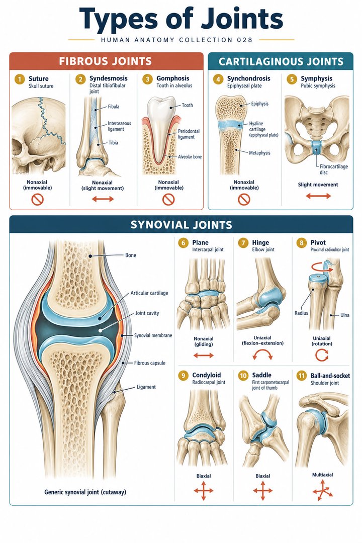
The three principal structural categories are fibrous, cartilaginous, and synovial. Fibrous articulations connect elements through fibrous connective tissue. Cartilaginous articulations use cartilage between the elements. Synovial articulations have a cavity enclosed by a capsule. Functional terminology instead describes mobility: synarthrosis indicates an essentially immobile connection, amphiarthrosis a slightly movable one, and diarthrosis a freely movable articulation. These terms overlap with structural categories but should not be treated as perfectly interchangeable lists. NCI SEER introduces articulation and functional mobility terms.
Consider a report or classroom question that asks for a joint's “type.” A strong answer first identifies which classification is being requested. If the example is the pubic symphysis, “cartilaginous” describes the connecting tissue, while “symphysis” identifies a more specific structural arrangement. If the example is an elbow articulation, “synovial” does not yet tell the listener whether the discussion concerns hinge-like elbow movement or forearm rotation. This precision is useful in multidisciplinary communication because a short label can otherwise conceal several different anatomical relationships. Clarifying the intended classification preserves the information that the label is supposed to convey.
Fibrous joints: sutures, syndesmoses, and gomphoses
Sutures connect cranial bones through fibrous tissue. Syndesmoses connect bones with ligaments or an interosseous membrane; the distal tibiofibular relationship is a familiar example. A gomphosis anchors a tooth in its socket through the periodontal ligament. These arrangements differ in shape and function even though all belong to the fibrous category. “Fibrous” does not mean that every example has identical movement or that living connective tissue is inactive. It identifies the tissue relationship that binds the elements. NCBI describes fibrous and cartilaginous joint subtypes.
The forearm and lower leg illustrate why a syndesmosis is worth distinguishing from a suture. An interosseous connection participates in the coordinated mechanical relationship of paired long bones rather than simply joining the edges of cranial plates. When analyzing a movement, learners should locate both the mobile articulations and the fibrous connections that guide or constrain the overall system. A person's hand can change orientation through forearm rotation while the radius and ulna remain related by several structures. Describing only the most visible joint misses the supporting network that makes controlled movement possible.
Cartilaginous joints: hyaline connections and fibrocartilage bridges
A synchondrosis is a cartilaginous connection based on hyaline cartilage. A growing long bone's epiphyseal plate is a developmental example; once that plate ossifies and the regions fuse, the adult relationship is different. A symphysis incorporates fibrocartilage, as in the pubic symphysis and connections between vertebral bodies. These joints can allow limited movement or deformation while maintaining continuity. The important distinction is the connecting material and arrangement, not the assumption that every cartilaginous connection behaves as the same flexible pad.
The vertebral column offers a particularly useful comparison. The relationship between neighboring bodies includes the intervertebral disc, while posterior articular processes participate in synovial facet joints. Two structural categories can therefore operate in one regional motion segment. A drawing of the disc alone is not a complete diagram of all spinal joints. The pubic symphysis similarly belongs within the wider pelvic ring, where its role should be considered with neighboring bones and articulations. Anatomical classification helps organize these relationships, but the functional explanation requires the complete regional arrangement rather than one isolated tissue specimen.
The common synovial plan: capsule, cavity, and cartilage
A synovial articulation has an enclosed cavity with a surrounding articular capsule. The capsule comprises an outer fibrous component and an inner synovial lining. The synovial membrane lines nonarticular internal surfaces rather than covering the articular cartilage itself. Cartilage provides a specialized contact surface; synovial fluid contributes to lubrication and the local environment. Ligaments reinforce the arrangement, and some joints contain additional discs, menisci, or labra. These structures differ between joints and should not be automatically added to every generic diagram. The National Library of Medicine defines the two capsule components.
The cavity is a controlled interface, not an empty gap that has no biological role. Joint movement changes the relative position of contact surfaces while the capsule and surrounding tissues maintain continuity. A cross-sectional drawing may enlarge the space to make the cavity visible; that visual exaggeration should be explained. Similarly, the surface cartilage should be distinguished from a fibrocartilage meniscus placed between surfaces. Different cartilage arrangements contribute different aspects of the joint's function. For clinical learners, this layered description supports accurate interpretation of terms such as capsular, intra-articular, periarticular, and synovial without treating them as synonyms.
Plane joints: small gliding movements within a larger system
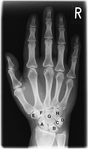
Plane synovial joints have relatively flat opposing surfaces and permit limited gliding. Examples occur between selected carpal bones and at other small articulations. “Plane” describes the basic surface relationship; it does not promise unlimited sliding or the absence of rotation in the complete regional system. Capsular attachments and ligament orientation constrain the available movement. Several small gliding contributions can collectively change the shape or position of a region. The resulting functional movement may be much more noticeable than the displacement at one individual articulation.
This principle is useful when examining hand motion. The wrist and carpal region cannot be explained by a single joint surface, even if one articulation dominates a simplified diagram. The bones change their relationships through coordinated movements, transmitting forces while preserving the continuity of the hand. A caregiver observing the hand during a task sees the functional result, not the isolated trajectory of every carpal bone. Anatomical teaching should therefore move between the local joint and the larger motion pattern. That approach prevents the mistaken expectation that a plane joint must be mechanically unimportant because its individual excursion is small.
Hinge and pivot: bending differs from rotation
A hinge-like articulation mainly permits flexion and extension around one principal axis. A pivot arrangement primarily permits rotation around a longitudinal axis. In the elbow region, humeroulnar movement contributes to bending and straightening, whereas the proximal radioulnar joint participates in pronation and supination with the distal radioulnar relationship. These functions coexist within the upper limb and should not be reduced to a single label for everything near the elbow. NCBI reviews elbow joint components, and the distal radioulnar reference explains the complementary distal relationship.
Hold a hand palm-up and then rotate it palm-down without substantially changing elbow flexion. This demonstrates a functional distinction between bending the elbow and rotating the forearm. The demonstration is educational, not a measurement of an individual's normal range. The radius changes its relationship to the ulna during the rotation, while the hand follows the distal radial arrangement. A movement name should identify what is moving relative to what. Calling every turning action “wrist rotation” can obscure the contribution of the forearm's proximal and distal joints and make the underlying anatomy less clear.
Condyloid joints: two principal movement directions
A condyloid, or ellipsoid, arrangement has an oval convex surface fitting a corresponding concavity. It permits movement in two principal directions, commonly represented as flexion–extension and abduction–adduction. Combining these directions produces circumduction, in which the distal segment describes a cone-like path. Circumduction is not identical to rotation around the segment's long axis. Metacarpophalangeal articulations of the fingers provide useful examples, although individual digits and joint geometry differ. NCBI describes the metacarpophalangeal joints and their regional differences.
The practical distinction can be seen when spreading and closing the fingers compared with bending them toward the palm. Those actions require different movement directions at related articulations. During a real grasp, the movements combine with muscle activity, tendon routing, and other finger joints. A clean two-axis diagram is therefore a model of surface geometry rather than a full simulation of a hand task. For students, the model helps establish terminology. For healthcare staff, recognizing the difference between digit movement, whole-hand position, and forearm rotation supports more precise observation and communication.
Saddle joints and the distinctive mobility of the thumb
A saddle joint has reciprocally concave and convex surfaces. The first carpometacarpal articulation, between the trapezium and first metacarpal, is the familiar example. Its geometry supports movement in more than one principal direction and contributes to opposition, allowing the thumb pad to face other digit pads. Opposition is a coordinated composite movement rather than a perfectly isolated rotation at one surface. The thumb's orientation also means that terminology cannot always be transferred mechanically from the other fingers without considering its different position. A detailed anatomical review describes thumb carpometacarpal geometry and biomechanics.
Compare holding a cup with touching the thumb pad to the little-finger pad. The second task reveals why the thumb cannot be represented adequately as one more parallel finger. Its joint geometry, muscles, and orientation support a different relationship to the palm. When drawing a saddle joint, show the reciprocal curvature in two planes rather than only one concave surface. Otherwise, the feature that distinguishes it from a simple ball-and-socket picture may disappear. Functional anatomy becomes clearer when the image and explanation together relate the surface design to the grasping tasks it enables.
Ball-and-socket joints: hip and shoulder share a class, not an identical design
Ball-and-socket articulations permit movement around multiple axes. The hip joins the femoral head with the acetabulum; the glenohumeral joint joins the humeral head with the glenoid cavity. Their shared category does not mean equal depth, congruence, loading, or soft-tissue support. The hip's deeper socket and surrounding structures support weight transfer, while the shoulder's arrangement supports extensive upper-limb positioning. Both rely on more than bony shape alone. NCBI reviews hip anatomy, while its glenohumeral reference describes static and dynamic stabilizers.
The comparison is clinically useful because a classification may conceal different priorities. A person reaching overhead and a person standing on one lower limb place different demands on joints that share the same broad shape category. Around the shoulder, rotator cuff muscles contribute to dynamic control of the humeral head; around the hip, capsule, ligaments, musculature, and the pelvic relationship contribute to support and movement. Labra modify the socket interface in both regions. These details explain why anatomy should progress from a shape name to the actual regional design instead of assuming that one textbook drawing applies unchanged to both joints.
The knee demonstrates why real joints exceed simple shape models
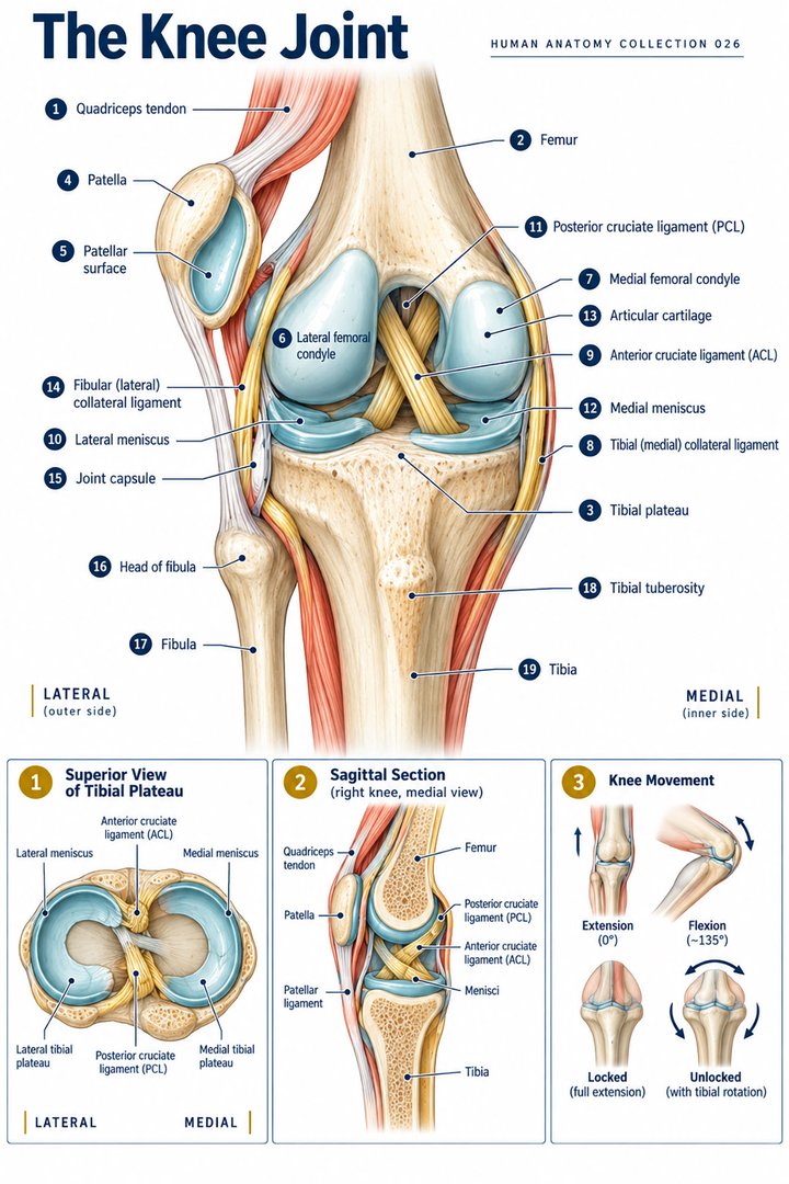
The knee is commonly described as a modified hinge because flexion and extension predominate, but its motion includes coupled rolling, gliding, and rotational relationships. Two menisci, medial and lateral, contribute to the interface between femoral and tibial surfaces. Collateral and cruciate ligaments constrain different aspects of movement. The patellofemoral relationship adds another articulation to the region. A complete knee lesson therefore requires more than a hinge symbol or a single drawing of two bone ends. IQWiG explains knee components, and NCBI details anterior cruciate ligament anatomy.
The two-meniscus count is an anatomical teaching fact, not a statement about injury frequency or a diagnosis based on symptoms. Similarly, identifying a ligament's role in limiting a movement does not imply that the ligament alone controls the entire joint. Muscle activity, contact geometry, and several soft tissues operate together. For healthcare students, the knee is an excellent example of the limits of classification: “synovial” and “modified hinge” are useful, but each captures only one level of description. Understanding the regional relationships makes subsequent study of movement, imaging, and clinical examination more coherent.
Latin vocabulary, movement language, and numerical context
Articulatio names an articulation. Articulatio fibrosa, articulatio cartilaginea, and articulatio synovialis identify structural classes. Capsula articularis names the capsule, cavitas articularis the cavity, and cartilago articularis the contact cartilage. Ligamentum, meniscus, and labrum identify distinct supporting structures. Flexio, extensio, abductio, adductio, pronatio, and supinatio describe movements, while articulatio sellaris identifies a saddle arrangement. Use the Latin term alongside its functional explanation so that terminology improves clarity rather than adding another disconnected memorization burden.
The familiar six synovial shape categories—plane, hinge, pivot, condyloid, saddle, and ball-and-socket—are six models, not six anatomical joints. A chart should show representative examples and movement directions instead of pretending that the models are rigidly isolated in every living region. Likewise, the three structural categories answer a different question from the three functional mobility terms. A strong final explanation can classify one joint, describe its connective tissues and cavity, identify its principal movements, and relate those movements to the surrounding limb. That sequence connects anatomical labels with meaningful function while preserving the complexity of the living body.
When reviewing movement documentation, identify the starting position and the segment being described before interpreting an angle or direction. Flexion measured from one reference position cannot be compared uncritically with an unlabeled diagram that starts elsewhere. Active movement also reflects the person's ability to generate and control motion, whereas passive movement describes a different observation. Neither can be inferred reliably from a static skeletal picture. The anatomical contribution is to define the joint surfaces, surrounding restraints, and moving segments precisely. This produces a clearer basis for professional discussion while leaving measurements and their clinical interpretation within the appropriate examination framework.
Anatomical Latin: quick reference
| Latin term | English meaning |
|---|---|
| Articulatio | Joint / articulation |
| Capsula articularis | Joint capsule |
| Cartilago articularis | Articular cartilage |
| Ligamentum | Ligament |
Sources and image credits
Sources reviewed on 9 October 2026. Counts, estimates, study statistics and hypothetical calculations retain their context in the text.
- NCI SEER: articulations
- NCBI: joints
- NLM MeSH: joint capsule
- NCBI: elbow joint
- NCBI: distal radioulnar joint
- NCBI: metacarpophalangeal joints
- Anatomical review: thumb CMC joint
- NCBI: hip joint
- NCBI: glenohumeral joint
- IQWiG: knee anatomy
- NCBI: ACL anatomy
Explanatory prose and SVG graphics are original. Photograph authors, source records and licenses are credited beside each image. Catalog illustrations provide educational context; anatomical facts are supported by the linked sources.