Skin is an active boundary with several linked jobs
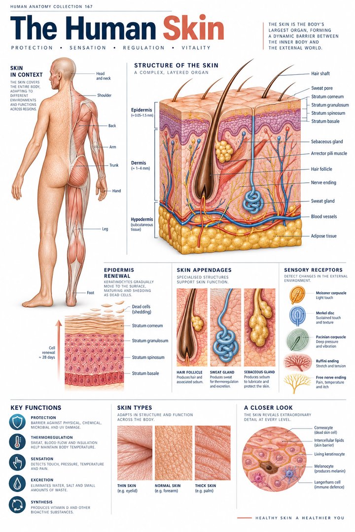
Skin separates the internal tissues from the external environment while maintaining exchange of information about temperature, touch, pressure, and injury. Its visible surface is only one component of a layered arrangement. The outer epithelial barrier, underlying connective tissue, associated nerves and vessels, and deeper subcutaneous tissues contribute different functions. Medical students benefit from following these layers from superficial to deep rather than treating “skin” as a single uniform sheet. For healthcare staff, the same anatomical map supports precise discussion of tissue depth, body location, sensory findings, and the different structures encountered beneath the surface.
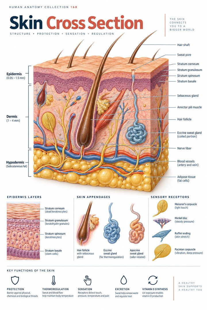
Two naming conventions are common. In a strict anatomical description, skin proper consists of epidermis and dermis, with hypodermis or subcutaneous tissue beneath. Many clinical and educational diagrams display all three as the skin's layers. Neither convention justifies silently changing what is being counted. State whether the subcutaneous compartment is included, then identify the tissue relationships clearly. The most useful question is what occupies each depth and how the compartments interact. A three-band illustration can orient a beginner, but a complete explanation must also show appendages, vessels, nerve routes, and the uneven interface between epidermis and dermis.
Epidermis: a renewing stratified epithelium
The epidermis is a keratinized stratified squamous epithelium. Keratinocytes dominate its cellular structure, differentiating as they move from deeper regions toward the surface. The epidermis has no blood vessels within its normal epithelial layers; its living cells depend on exchange with the vascular connective tissue underneath. This distinction separates cellular vitality from vascularity: an avascular tissue can contain living metabolically active cells. The mature outer barrier includes flattened corneocytes and extracellular lipid organization rather than a stack of identical living cells. NCBI describes skin histology and the epidermal barrier.
The renewing arrangement explains why the epidermis should be drawn as a progression, not merely five differently colored stripes. Cells in a basal region do not have the same shape or differentiation state as cells at the outer surface. The barrier depends on coordinated structure at both cellular and extracellular levels. A common classroom misunderstanding is to associate “dead surface cells” with biological uselessness. In fact, the organized outer material is essential to the protective interface. Another misunderstanding is to assume that all superficial damage automatically reaches a blood vessel. The depth relationship, rather than surface appearance alone, determines which tissue compartments are involved.
Four strata in ordinary epidermis and five in thick skin
From deep to superficial, the main epidermal strata are basale, spinosum, granulosum, and corneum. Thick skin on palms and soles also has a recognizable stratum lucidum between granulosum and corneum. Thus the familiar five-layer sequence applies specifically to thick epidermis, not to every skin sample. “Thick skin” is a histological classification, especially concerning epidermal structure; it should not be confused with the thickness of the entire skin and underlying fat at any body site. Regional comparisons need to specify which layer is being measured. NCBI distinguishes thick and thin skin anatomy.
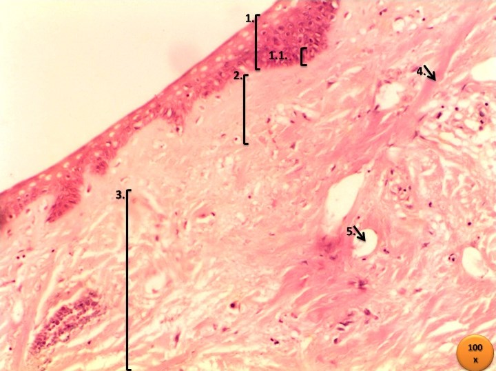
The four-versus-five count is a useful numerical fact because it describes a defined anatomical difference. A chart should name the strata and show where lucidum is present rather than turning the count into a ranking of strength or health. A palm and an eyelid meet very different functional demands, so their epidermal arrangements and associated structures differ. Both are valid examples of human skin. This is especially relevant when selecting a real micrograph: its body location influences which layers and appendages are expected. If the source does not state the region, the caption should avoid assigning a precise body site simply to fit the lesson.
Basal attachments, cell junctions, and differentiation
The stratum basale lies next to the epidermal basement membrane region. Basal keratinocytes contribute to renewal and attach to their underlying interface through specialized junctions. As cells differentiate in the stratum spinosum, prominent intercellular junctional relationships support cohesion. Granular-layer cells contain materials involved in the development of the surface barrier. The stratum corneum provides the mature outer interface. These stages are functionally connected: barrier maintenance requires production, adhesion, differentiation, and controlled loss of surface material. NCBI reviews epidermal cell layers and junctional organization.
This sequence explains why a simple peeling analogy is incomplete. Skin renewal is not just the removal of one old sheet and its replacement by another. Different cells and junctions are involved at successive depths, and the tissue must maintain continuity while renewing. For a learner examining histology, nuclei, cell shape, and staining differences provide clues to this progression. These clues should be interpreted together instead of identifying a layer from color alone. A bright stain at the surface may highlight keratinized material, but the exact appearance depends on the stain, specimen processing, orientation, and how the photograph was captured.
Dermis: papillary and reticular connective-tissue regions
The dermis is connective tissue supporting the epidermis and hosting vascular, neural, and appendage structures. Its superficial papillary region contains loose connective tissue arranged along the epidermal interface. The deeper reticular region has a stronger dense irregular connective-tissue organization. Collagen and elastic components contribute different mechanical properties, while fibroblasts maintain extracellular material. The two-region description is an anatomical framework, not a claim that each region has a perfectly straight border or equal thickness. NCBI explains dermal structure and function.
The epidermis–dermis relationship can be visualized as a tissue interface with interlocking contours rather than two flat laminated cards. This geometry helps distinguish the papillary region in a section, although the visible pattern depends on the plane of cutting. A tangential section can make a structure appear discontinuous even when it forms a continuous arrangement in three dimensions. Dermal support also explains why surface flexibility cannot be attributed to one epidermal layer alone. Mechanical behavior involves connective fibers, tissue hydration, regional organization, and the relationship with deeper structures. The complete layered design is more informative than the thickness of one colored stripe in a diagram.
Hypodermis: adipose tissue, connective partitions, and deeper routes
The subcutaneous compartment contains adipose tissue and connective-tissue partitions, with nerves and vascular routes passing through it. Its arrangement contributes insulation, energy storage, cushioning, and the relationship between the skin and underlying tissues. Thickness varies by body region and individual characteristics. It should not be represented as the same uniform yellow band beneath every skin site. Adipose tissue is a living tissue with biological functions; calling it simply packing material misses its metabolic role and its importance in local tissue organization.
The anatomical relevance of this compartment becomes clear when comparing the back of a hand with the abdomen or plantar region. The amount and organization of deeper tissue differ, changing the relationship between surface skin and structures underneath. A diagram used to teach injection compartments should distinguish epidermal, dermal, subcutaneous, and deeper muscular locations, but a general educational article should not supply a procedure-specific technique. The transferable anatomical lesson is depth: a needle route, a wound, and a tissue section can pass through different compartments even when they begin at the same visible skin surface.
Hair follicles and sebaceous units cross the layered map
Hair follicles are epidermally derived structures extending into deeper tissues. Their relationship with sebaceous glands and arrector pili muscle forms part of the pilosebaceous unit. A follicle therefore cannot be understood as a simple hole drilled through otherwise unrelated layers. The follicular epithelium, surrounding connective tissues, glands, muscle attachment, and hair shaft form an organized regional structure. The depth of its components and its appearance in a section vary with location and orientation. NCBI describes skin appendages and the pilosebaceous unit.
One real micrograph may show a circular follicle profile, while another shows a long oblique structure. These can represent different cutting planes through comparable anatomy. Learners should avoid concluding that the shapes are different organs solely because the profiles differ. The follicle's presence also helps distinguish many ordinary skin sites from the hairless thick skin of palms and soles. In a general schematic, the follicle should be labeled as an example for a hair-bearing site rather than inserted into a palm diagram without explanation. This small caption choice prevents the illustration from teaching a false regional combination.
Sweat glands and ducts: surface secretion follows an anatomical route
Eccrine sweat glands have a coiled secretory component and a duct leading toward the skin surface. Apocrine glands have a different regional distribution and commonly open into hair-follicle-associated routes. Their names identify different gland arrangements rather than two interchangeable symbols for a sweat droplet. Eccrine secretion is important in evaporative heat loss, while apocrine anatomy should be considered within its follicular and regional context. NCBI describes apocrine gland organization.
In histology, several small glandular circles can be cross sections through one coiled structure rather than many independent glands. This is an important quantitative caution: counting profiles in a single section does not automatically count whole anatomical units. A duct may also appear disconnected from the secretory coil because the intervening route lies outside the section plane. Pairing a micrograph with a simple three-dimensional coil diagram helps explain this. For healthcare learners, following secretion from gland to surface establishes a route that can later be connected with autonomic control and thermoregulation without confusing the gland with the epidermal barrier itself.
Vessels, sensory receptors, and temperature regulation
The dermis contains blood vessels, lymphatic vessels, and sensory neural structures. These routes support exchange, tissue drainage, information transfer, and heat distribution. The epidermis depends on the underlying vascular tissue while maintaining its own avascular epithelial arrangement. Sensory endings and receptors occupy different depths and respond to different features of stimulation. A generic drawing should not imply that every labeled receptor is present in equal numbers at every site. The functions of sensation and thermal exchange connect skin anatomy to the nervous and cardiovascular systems rather than creating an isolated skin-only process.
Imagine touching a cool object with a fingertip. The sensory experience requires peripheral receptors and neural signaling, while the local tissue also participates in heat exchange. The image of the skin alone cannot display the complete perceptual pathway to the brain. Likewise, increased or reduced blood flow changes heat transport but does not by itself describe every cause of a visible color change. Healthcare documentation should separate observations from inferred mechanisms. Record the location and observed characteristic precisely, while recognizing that the deeper physiological explanation may require examination, context, and additional information.
Pigmentation and immune surveillance involve specialized cells
Melanocytes produce pigment that is transferred to keratinocytes through melanosomes. Epidermal immune-associated cells, including Langerhans cells, contribute to surveillance at a boundary exposed to the environment. Merkel-cell-associated arrangements participate in tactile sensation. These examples show that the epidermis contains several specialized cell populations even though keratinocytes dominate its structure. Skin tone should not be explained by assuming that one population simply has many more melanocytes than another. Pigment production, handling, and distribution matter. The skin histology reference discusses pigmentation.
This cellular diversity changes how the barrier should be presented. A continuous outer layer can still include specialized cells coordinating information with other systems. Pigment, immunity, and touch are distinct functions, so one icon should not stand for all epidermal cells. In clinical education, photographs should show a range of skin appearances where appropriate, with lighting and color reproduction acknowledged. A diagram's pink or beige fill is a graphic convention, not a biological definition of normal skin. Accurate communication separates the structural layer from the color chosen to illustrate it and avoids equating one visual appearance with a universal baseline.
Repair links tissue depth with multiple overlapping processes
Skin repair involves hemostasis, inflammation, proliferation, and remodeling. These four phases overlap; they do not operate as four perfectly separated calendar blocks. Different cell populations, matrix changes, vascular responses, and epithelial movement contribute to restoring continuity. A superficial epithelial disruption and a deeper injury crossing into connective tissue do not involve identical anatomical compartments. Describing depth is therefore part of understanding the repair problem, although it is not sufficient to predict an individual outcome. NCBI reviews the four overlapping phases.
For an educational timeline, use overlapping bands rather than rigid day-by-day boxes. The diagram can show the sequence of dominant activities without inventing a universal healing schedule. A wound's appearance at one moment cannot determine its complete cellular state, and healing varies with many local and systemic factors. This is a useful anatomical distinction for practice assistants, nurses, and eldercare teams: surface closure, tissue strength, and the condition of surrounding tissues are different aspects of the process. Observation and documentation should follow the clinical framework in use, while the anatomy lesson explains why depth and tissue type matter.
Aging, anatomical vocabulary, and a clear layered summary
Age-related changes can make skin less elastic and more fragile, while vascular fragility contributes to easier bruising in older adults. Such changes vary and should not be treated as identical in every older person. The National Institute on Aging describes these general relationships without implying that every skin finding is simply caused by age. Read NIA's overview of skin and aging. In eldercare, the anatomical lesson is to understand why tissue condition and supporting compartments deserve attention, while avoiding diagnosis from a diagram or age category alone.
Cutis names skin; epidermis identifies the epithelial layer, dermis the supporting connective tissue, and tela subcutanea the underlying subcutaneous compartment. Stratum basale, stratum spinosum, stratum granulosum, stratum lucidum, and stratum corneum identify epidermal strata. Glandula sudorifera, glandula sebacea, and folliculus pili identify appendages. A complete summary should explain the barrier from deep to superficial, state where blood vessels are found, distinguish the four-versus-five epidermal count, and connect appendages with their routes. The result is a layered functional model that supports histology study, accurate workplace terminology, and thoughtful use of real skin images.
Anatomical Latin: quick reference
| Latin term | English meaning |
|---|---|
| Epidermis | Outer epithelial layer |
| Dermis | Connective tissue layer of skin |
| Tela subcutanea | Subcutaneous tissue |
| Stratum corneum | Outermost epidermal stratum |
Sources and image credits
Sources reviewed on 9 October 2026. Counts, estimates, study statistics and hypothetical calculations retain their context in the text.
- NCBI: skin histology
- NCBI: skin anatomy
- NCBI: epidermal anatomy
- NCBI: dermis
- NCBI: skin appendages
- NCBI: apocrine glands
- NCBI: wound healing
- NIA: skin care and aging
Explanatory prose and SVG graphics are original. Photograph authors, source records and licenses are credited beside each image. Catalog illustrations provide educational context; anatomical facts are supported by the linked sources.