Reading a complex framework from more than one view
The skull is a protective enclosure, a framework for the face, and an arrangement of passages connecting the head with the rest of the body. Its educational difficulty comes partly from this combination. A single bone may contribute to several spaces, and a single space may be constructed from several bones. Learning the skull therefore requires more than finding a forehead, cheekbone, and jaw. Students must relate outer surfaces to internal structures, understand which openings transmit nerves or vessels, and distinguish the age-dependent organization of the cranial vault from the appearance of a typical adult specimen.
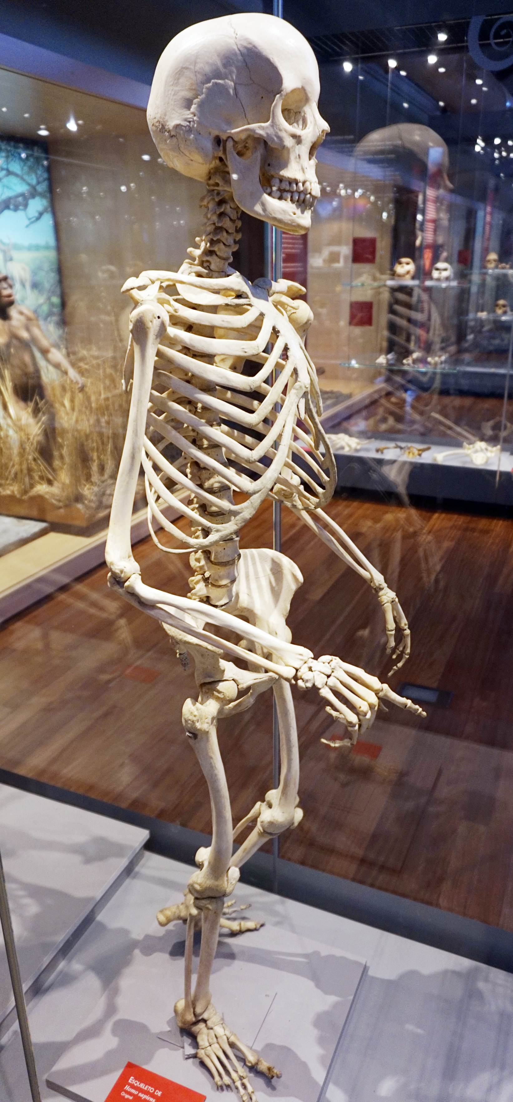
Start with the entire skull before separating its parts. An anterior view establishes the orbits and nasal opening; a lateral view reveals the relationship between the cranial vault and facial skeleton; an inferior view displays the base; and an internal view shows the cranial floor. These views complement rather than duplicate one another. When healthcare staff use skull terminology in conversation, the same principle applies: the name of a region becomes useful only when its position and neighboring structures are clear. A carefully oriented photograph and a simplified diagram can work together to make that relationship understandable.
Twenty-two bones: a convention with stated boundaries
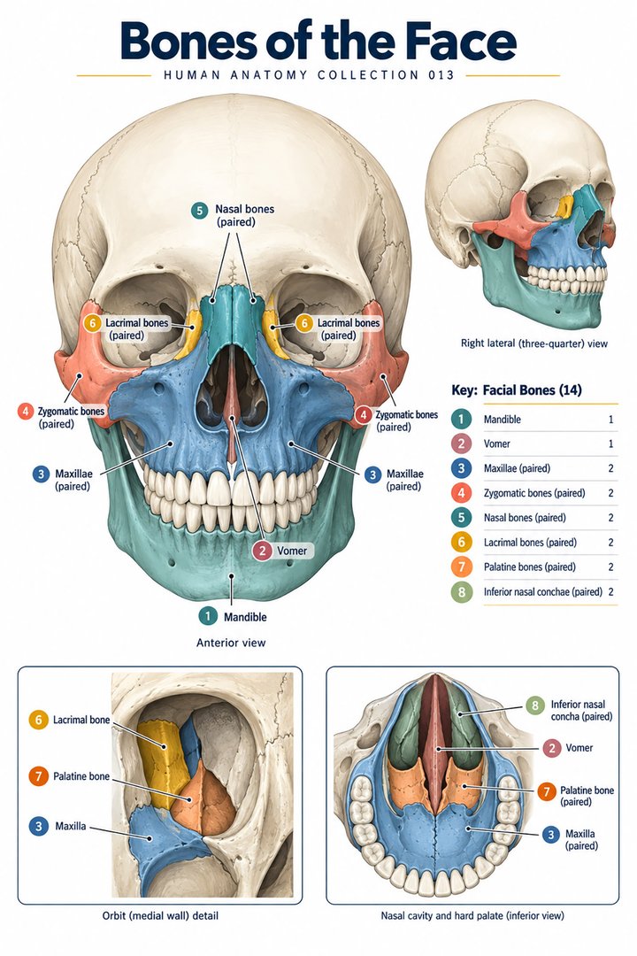
A common adult teaching count is twenty-two skull bones: eight cranial bones and fourteen facial bones. The cranial group includes paired parietal and temporal bones, plus the frontal, occipital, sphenoid, and ethmoid bones. The facial group includes paired maxillae, zygomatic, nasal, lacrimal, palatine, and inferior nasal conchal bones, plus the mandible and vomer. This count excludes the six auditory ossicles. Some skeleton lists include those ossicles within a broader skull grouping, producing twenty-eight. Teeth and the hyoid are not additional components of the twenty-two-bone convention. NCI SEER shows the component lists and its broader counting convention.
Numbers should clarify the classification, not become a contest between sources that count different things. If a learner obtains twenty-eight from one resource and twenty-two from another, the first question is whether auditory ossicles have been included. Additional sutural bones and other anatomical variants can also complicate literal counting of a particular specimen. A useful exercise is to separate the cranial and facial groups using two colors, then state whether paired structures have been counted as one name or two bones. This turns a memorization task into an explanation of what the number actually represents.
The cranial vault and the skull base
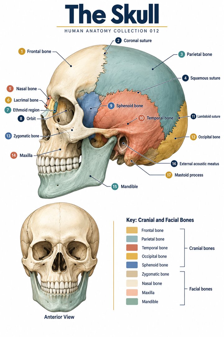
The calvaria is the dome-like cranial vault, while the skull base provides the floor and contains numerous passageways. The entire cranium is not equally thin, equally flat, or equally visible from outside. Frontal and parietal areas dominate a superior view; the sphenoid and ethmoid become more understandable through internal or inferior views. The temporal bone combines broad external surfaces with complex regions related to hearing and major vascular passages. The occipital bone contains the foramen magnum and articulates with the first cervical vertebra through its condyles. NCBI reviews skull regions and principal landmarks.
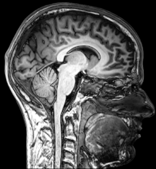
For an orientation exercise, imagine placing an adult skull upright and then rotating it to inspect the underside. The face remains anterior even when it points away from the viewer, and the occipital region remains posterior. This sounds obvious, but changing specimen orientation frequently leads learners to reverse directions. Before naming an unfamiliar opening, identify the anterior and posterior limits and the relevant bone. A label detached from orientation is difficult to retain and easier to misapply. The same caution matters when viewing imaging: the displayed side of a screen and the anatomical side of the person are separate concepts.
Sutures connect bones without creating ordinary mobile joints
The coronal suture lies between frontal and parietal bones; the sagittal suture lies between the two parietal bones; and the lambdoid suture marks the parietal–occipital relationship. The squamous suture contributes to the parietal–temporal boundary. These are fibrous articulations rather than the freely mobile synovial joints seen in limbs. A suture is a joint relationship and should not be confused with a fracture simply because it forms an irregular line. Its appearance varies with age, specimen preservation, and the specific location being examined. NCBI describes the coronal suture and related landmarks.
Instead of learning the suture names in isolation, follow each line across a specimen and identify the bones on both sides. Then compare the same boundary in another view. This practice makes the joint's location more memorable than a floating arrow on a diagram. It also illustrates why a suture may be partly hidden in a photograph: the visible surface only captures one portion of a three-dimensional junction. When discussing an image with colleagues, identify whether the line is a known anatomical boundary, an imaging finding, or something that requires formal interpretation. Anatomical learning supports accurate questions; it does not independently establish the meaning of an unfamiliar clinical image.
The developing skull is not a miniature adult skull
In infancy, sutures and fontanelles permit growth of the cranial framework in relation to the developing brain. Fontanelles are membranous regions at junctions between developing cranial bones; they are not holes exposing brain tissue. The anterior fontanelle lies near the frontal and parietal junctions, while the posterior fontanelle relates to the parietal and occipital junctions. Other paired fontanelles occur laterally. The organization and proportions of the infant skull differ from those of the adult, and developmental anatomy should be learned on an age-appropriate illustration. NCBI explains fontanelle anatomy.
The main educational point is change over time. A rigid adult model cannot demonstrate every feature that matters in a newborn. Similarly, an infant illustration should not be used as the default map of adult sutures, proportions, or spaces. For medical learners, comparing age-specific images helps explain why the face and cranial vault do not retain the same relative proportions through growth. For staff communicating with families, plain anatomical language is useful: a fontanelle is a normal developmental region with protective tissues. Any assessment of its appearance belongs within the person's clinical context and appropriate professional evaluation.
The facial skeleton supports several neighboring spaces
The maxillae form the upper jaw and contribute to the hard palate, nasal cavity, and orbital floors. Zygomatic bones create the cheek prominence and contribute to the lateral orbital framework and zygomatic arches. Nasal bones form part of the bridge of the nose, while the vomer contributes to the bony nasal septum. Inferior nasal conchae are separate facial bones, whereas the superior and middle conchae are parts of the ethmoid. This distinction is a useful example of why similar-looking structures do not always belong to the same bone category.
To build a mental map, trace the boundary between the mouth, nose, and orbit. The exercise shows that a bone is not assigned to only one cavity because its name appears in a particular list. Draw the upper jaw first, then add the orbital floor and nasal boundary in different colors. The shared framework becomes easier to understand than it is in separate organ illustrations. This relational approach also supports reading descriptions of location. A statement about a maxillary region is more precise when the learner knows whether the discussion concerns the alveolar process, sinus, palate, or contribution to the orbit.
The orbit: seven bones surrounding one important compartment
Each bony orbit is assembled from seven bones: frontal, sphenoid, zygomatic, maxilla, palatine, lacrimal, and ethmoid. The eye occupies only part of this space; muscles, nerves, vessels, fat, and associated structures also belong to the orbital contents. The roof, floor, medial wall, lateral wall, and posterior apex have different neighbors. The optic canal and orbital fissures illustrate how the orbit communicates with other regions. A bony orbit is therefore an anatomical compartment, not simply an empty cup holding an isolated eyeball. NCBI identifies orbital bones and boundaries.
A useful student exercise begins by removing the assumption that every orbital wall has the same thickness or shape. Compare the medial and lateral walls on a model, and identify which bone contributes to each surface. Then look at an external photograph and note what remains hidden: the posterior apex, the depth of the walls, and the internal routes. Such a comparison explains why a neat frontal illustration cannot show every clinically important relationship. It also encourages disciplined image captions. If a photograph displays a prepared skull, say so rather than describing it as a view of the living orbital tissues.
Three cranial fossae organize the internal floor
The anterior, middle, and posterior cranial fossae are major regions of the internal skull base. They are useful geographical divisions for relating the cranial floor to overlying neural structures and passageways. Their borders are not arbitrary horizontal stripes laid across a skull photograph. The anterior region relates prominently to frontal and ethmoidal structures; the middle region includes sphenoidal and temporal contributions; and the posterior region includes the occipital area around the foramen magnum. Understanding the overall floor first reduces the burden of memorizing openings without any regional context.
For practice, sketch the three fossae with a small number of major landmarks and leave detailed foramina for a second pass. Then compare an intact skull with a specimen in which the vault has been removed. The latter reveals the floor but changes the visual context: structures that were externally concealed are now exposed. This should be explained when teaching from real photographs. A colored schematic may intentionally flatten the depth of the fossae to make boundaries clearer. Its caption should state that it is an orientation diagram. Three-dimensional relationships become more durable when the learner recognizes how each visual representation simplifies the actual specimen.
Openings are routes, and route names need contents
Foramina, canals, and fissures are distinct forms of passageway. The foramen magnum connects cranial and vertebral regions; the optic canal provides an orbital route for the optic nerve and ophthalmic artery; and the carotid canal carries the internal carotid artery with associated sympathetic fibers. At the surface, supraorbital and infraorbital openings or notches relate to neurovascular structures supplying facial regions. Learning a route is more useful than memorizing the shape of a hole. The question should be where it begins, where it leads, and which structures are associated with it. A primary anatomical study maps orbital openings and their relationships.
A practical learning method uses a three-column note: landmark, connecting regions, and principal contents. Write only the major relationship initially, then refine the detail as your course requires. This avoids a common error in which a learner can name a foramen but cannot say why it matters. The method also helps identify uncertainty. If an unfamiliar specimen has a small opening near a familiar landmark, its identity should be checked against orientation and neighboring structures instead of guessed from size alone. Natural variability and preparation artifacts can influence appearances, so precise identification depends on several clues working together.
Paranasal sinuses and the meaning of a hollow bone
The paranasal sinus groups are frontal, maxillary, ethmoidal, and sphenoidal. They occupy different bones and communicate with the nasal cavity through particular routes. The ethmoidal group consists of multiple air cells rather than a simple single chamber. The common description of four paired groups is a classification, not a prediction of eight identical cavities of equal size in every person. Development and individual variation influence their shape. A frontal skull view does not display the full extent of these internal air spaces, and their positions are better understood with sectional illustrations. NCBI reviews paranasal sinus organization.
Sinus anatomy is also a useful lesson in interpreting names. A maxillary sinus belongs within the maxilla, but the maxilla has many functions beyond enclosing that sinus. Likewise, sphenoidal air spaces are part of a central skull bone whose other landmarks may be learned in a different lesson. To connect these ideas, add a transparent cavity outline to a bone diagram and identify neighboring spaces. Label the outline as schematic rather than implying a universal shape. This improves anatomical teaching without moving into diagnostic interpretation of a scan or recommending an approach to symptoms.
The mandible and its paired joint relationships
The mandible forms the lower jaw and articulates with the temporal bones at the two temporomandibular joints. The body, rami, angle, condylar processes, and coronoid processes provide a practical set of orientation landmarks. The condylar process participates in the joint; the coronoid process provides an important muscle attachment. Jaw movement combines rotation and translation, with an articular disc contributing to the joint arrangement. Calling this simply a door hinge loses the sliding component and the relationship between the two sides. The National Academies' masticatory-system chapter explains coordinated jaw movement.
Try describing opening the mouth without reducing the explanation to one muscle or one joint surface. Both sides of the mandible belong to the same bone, so their movement must be considered together. Teeth, muscles, and surrounding soft tissues also contribute to the masticatory system. A dry skull demonstrates bony geometry well but cannot show an intact disc or living muscle coordination. This limitation should be visible in the lesson rather than left for the learner to infer. For healthcare staff, recognizing the difference between a bony landmark and a functional joint system supports clearer communication about location and anatomy.
Latin terms and an integrated study routine
Cranium refers to the skull; calvaria to its vault. Os frontale, os parietale, os temporale, os occipitale, os sphenoidale, and os ethmoidale identify cranial bones. Maxilla, mandibula, os zygomaticum, os nasale, os lacrimale, os palatinum, vomer, and concha nasalis inferior identify facial components. Sutura coronalis, sutura sagittalis, and sutura lambdoidea name principal sutures. Fossa cranii anterior, media, and posterior identify cranial floor regions. Foramen magnum and canalis opticus preserve a route's identity across languages. These terms should be linked to structures already recognized in more than one view.
For a final review, explain the skull using five tasks: state the twenty-two-bone convention, follow a major suture, identify the boundaries of one orbit, relate a cranial fossa to the skull base, and trace a named passageway between regions. Then describe what a selected photograph actually reveals and what it conceals. This approach joins bone lists with spatial reasoning and image literacy. It is especially useful when moving from classroom specimens to imaging or workplace terminology. The skull becomes a connected map of supporting structures and routes rather than a long list of isolated names.
Anatomical Latin: quick reference
| Latin term | English meaning |
|---|---|
| Cranium | Skull |
| Mandibula | Mandible |
| Maxilla | Maxilla / upper jaw bone |
| Foramen magnum | Large opening in the occipital bone |
Sources and image credits
Sources reviewed on 9 October 2026. Counts, estimates, study statistics and hypothetical calculations retain their context in the text.
- NCI SEER: cranial and facial bone counts
- NCBI: skull anatomy
- NCBI: coronal suture
- NCBI: fontanelles
- NCBI: orbital bones
- Primary anatomical study: orbital rule of seven
- NCBI: paranasal sinuses
- National Academies: masticatory anatomy
Explanatory prose and SVG graphics are original. Photograph authors, source records and licenses are credited beside each image. Catalog illustrations provide educational context; anatomical facts are supported by the linked sources.