Tissue is an organized relationship, not a pile of identical cells
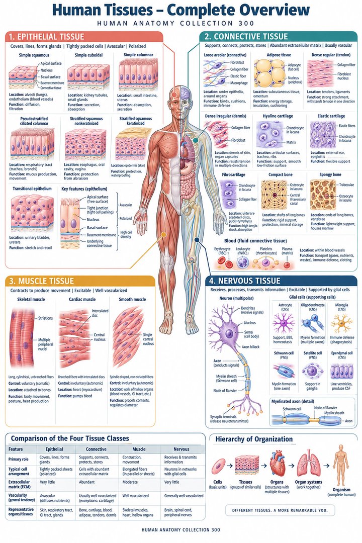
A tissue is an organized biological arrangement in which cells and extracellular components work together. Four principal categories provide the usual foundation: epithelial, connective, muscle, and nervous tissue. These categories describe broad structural and functional patterns, while each contains specialized forms. An organ generally combines several categories, so the stomach is not simply epithelial tissue and the heart is not simply muscle. Histology helps reveal how the components are arranged, and gross anatomy places that arrangement within the whole organ and body. NCI SEER introduces the four tissue categories.
For healthcare learners, the useful question is how a tissue contributes to an organ's task. A lining creates an interface; a matrix supports or connects; a muscle generates force; and neural arrangements process or transmit information. These functions often occur next to one another. A section through an organ wall therefore should not be interpreted as a contest to find one winning tissue label. First identify the major compartments, then examine the cells and extracellular organization within each. This approach connects microscopic details with clinically meaningful structures instead of leaving tissue names separate from the anatomy encountered in practice.
Epithelial tissue creates surfaces and selective interfaces
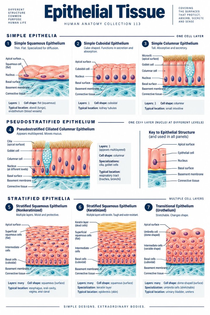
Epithelial tissues cover surfaces, line hollow structures, and form glandular components. Their cells are closely arranged with relatively little extracellular matrix between them. An apical surface faces a lumen or external environment, while the basal interface relates to a basement membrane and underlying tissue. Protection, absorption, secretion, filtration, and exchange are different roles that require different epithelial designs. A thin exchange surface should not be expected to resemble a thick protective surface. NCI SEER describes epithelial organization and functions.
A useful anatomical comparison places an intestinal lining beside the epidermis. Both are epithelial arrangements, but their tasks and structures differ. One must support selective movement between a lumen and the body, while the other creates a protective external boundary. The shared category does not erase the functional specialization. Healthcare students should also distinguish a surface epithelium from the whole mucosa, which includes additional supporting components. A label such as mucosal injury refers to a regional tissue arrangement, and its exact depth cannot always be inferred by replacing “mucosa” with “epithelium” in conversation.
Epithelial classification combines cell shape with layer arrangement
Squamous, cuboidal, and columnar describe familiar cell-shape patterns. Simple epithelium has one cell layer; stratified epithelium has multiple layers. Pseudostratified arrangements can appear layered because nuclei lie at different levels, even though the cells relate to one underlying basement-membrane interface. Transitional epithelium, or urothelium, is adapted to changes in the urinary tract's distension. Cell shape and layering are separate descriptors, so a complete label combines them where appropriate. NCBI reviews epithelial classification and polarity.
To identify an epithelium in a micrograph, locate its free surface and basal relationship before counting apparent nuclear rows. A slanted cut can make a single layer look more complicated, and not every nucleus lies within the particular plane photographed. Cell outlines, tissue orientation, and the organ context should be interpreted together. Classification becomes more accurate when it answers a structural question rather than relying on one visual clue. This also explains why a schematic may show cleaner cell boundaries than the real tissue section. The drawing is an aid to recognition, while the micrograph preserves the complexity of the specimen.
Connective tissue is defined partly by its extracellular organization
Connective tissues include cells dispersed within extracellular matrix that varies greatly in composition and arrangement. Fibers and ground substance contribute to mechanical support, continuity, exchange, and local tissue environments. Fibroblasts, macrophages, mast cells, and other cell populations perform different roles. Loose connective tissue, dense connective tissue, adipose tissue, cartilage, bone, and blood illustrate the category's diversity. The name should not imply that all connective tissues have the same stiffness, vascularity, or regenerative behavior. NCI SEER describes connective-tissue components and examples.
The extracellular matrix is not simply blank material between meaningful cells. Its architecture is part of the tissue's function. Compare a flexible loose connective-tissue region with a strongly aligned tendon and a mineralized bone. Their cells may occupy relatively little of the visible area, while their supporting material dominates the mechanical behavior. This is why an image that crops out most matrix can give a misleading impression of the tissue. In medical learning, matrix organization should be identified alongside the cell type. Clinical terms involving fibrosis, connective support, or tissue planes become more understandable when this extracellular contribution is retained.
Tendons, fascicles, and organized transmission of force
Dense connective arrangements can organize fibers to support directional loading. Tendons link muscle-generated force with skeletal attachments through a hierarchical collagen-based architecture. Connective tissues also surround muscle cells, fascicles, and whole muscles, contributing continuity between microscopic and gross levels. Endomysium, perimysium, and epimysium identify different levels of this organization. These supporting tissues are not themselves the contractile muscle cells, although they participate in transmitting the effects of contraction. NCBI explains connective-tissue organization around muscles and tendons.
This relationship clarifies why an entire muscle should not be described as nothing but a bundle of contractile fibers. It also contains vascular routes, nerves, and supporting connective structures. A section through a fascicle can demonstrate the local arrangement without showing the full tendon route. To connect the scales, follow force conceptually from a contracting cell through the surrounding organization toward the skeletal attachment. The explanation remains anatomical rather than prescriptive: it describes how the system is connected, without inferring a diagnosis or proposing an exercise from the appearance of a tissue image alone.
Cartilage and bone show different matrix designs
Cartilage contains chondrocytes within a specialized matrix and is normally avascular. Hyaline, elastic, and fibrocartilage have different matrix arrangements suited to different roles. Articular cartilage belongs to the hyaline category, while fibrocartilage participates in structures such as intervertebral discs and menisci. Cartilage's lack of vessels within its matrix does not mean its cells lack nutritional requirements. Exchange with surrounding environments remains important. Columbia University's histology teaching resource describes the three cartilage types.
Bone combines cells with a mineralized matrix and a vascular tissue organization. It supports loading while undergoing ongoing biological maintenance. The contrast with cartilage makes clear that “supportive tissue” covers several strategies. A tissue can be firm through its matrix composition without all firm tissues sharing the same blood supply or organization. In a joint lesson, distinguish the cartilage contact surface from the underlying bone and from fibrocartilage structures between surfaces. Their proximity does not make them identical. Accurate tissue labeling therefore helps explain the different contributions seen within one skeletal region.
Blood and adipose tissue broaden the connective category
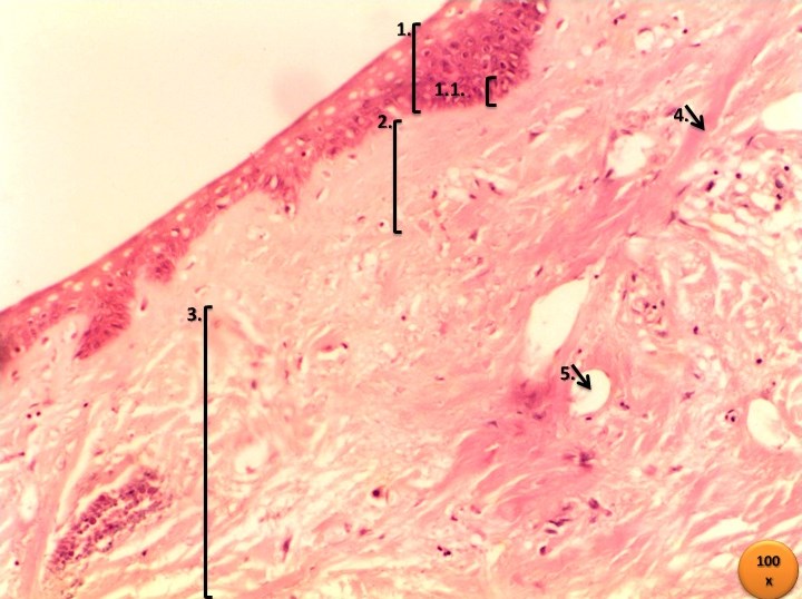
Blood is commonly classified as a connective tissue with cells and formed elements suspended in a fluid extracellular environment, plasma. Its transport and defensive roles differ dramatically from those of a tendon, yet both belong within the broad connective framework. Adipose tissue organizes specialized cells that store and mobilize energy and contribute to endocrine and local tissue functions. Its cushioning and insulating roles depend partly on where it is situated. Neither blood nor fat should be treated as an exception that is biologically inactive simply because it looks different from a fibrous supporting tissue.
These examples help learners recognize the difference between category and appearance. A smear of blood, a section of adipose tissue, and a tendon image may have almost nothing visually in common at first glance. Their classification becomes understandable through cells, matrix, organization, and role rather than through a universal connective-tissue color. In clinical communication, this prevents vague references to tissue from erasing important differences. A discussion of a blood component, an adipose compartment, or a fibrous tissue plane should preserve the specific structure involved rather than imply that all have the same physical properties.
Muscle tissue converts cellular organization into force
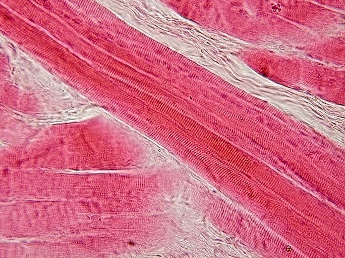
The three principal muscle types are skeletal, cardiac, and smooth. Skeletal muscle fibers are striated and commonly multinucleated; cardiac cells are striated, branching, and connected through intercalated-disc relationships; smooth-muscle cells lack the same visible striation pattern and are often spindle-shaped. Their different arrangements fit different functional settings. Skeletal muscle contributes to body movement and posture, cardiac tissue forms the contractile myocardium, and smooth muscle participates in many hollow-organ and vascular walls. NCI SEER compares the three muscle tissues.
The phrase muscle fiber can be confusing because in skeletal muscle it commonly refers to one long muscle cell, whereas a visible strand in a gross specimen may include many cells and supporting structures. A learning diagram should identify the scale it represents. Striations also require appropriate specimen orientation and imaging conditions to be evident. The absence of clearly visible stripes in one poorly oriented photograph is not enough to classify the tissue as smooth muscle. Cell shape, nuclear arrangement, branching, surrounding tissue, and source information contribute to a more reliable interpretation.
Nervous tissue combines signaling cells with active support
Neurons have specialized processes and membrane properties supporting information transfer. Their cell bodies, dendrites, and axons occupy different relationships within a network. Glial cells contribute essential support, insulation, immune-associated functions, and interactions with the local environment. Nervous tissue therefore should not be represented as neurons alone. The distribution of cell bodies and processes differs between regions of the central and peripheral nervous systems. NCI SEER introduces neurons and neuroglia.
A micrograph may show a neuronal cell body but not the complete extent of its axon, or may show a nerve profile with many fibers and surrounding connective tissue. These are different views of nervous organization. The appearance of one neuron is not a diagram of a whole sensory or motor pathway. Medical learners should separate cellular anatomy from circuit anatomy and from gross regional anatomy. That separation becomes especially useful when studying peripheral nerves: the organ contains neural elements, vessels, and connective sheaths, even though nervous tissue provides its characteristic signaling role.
An organ wall demonstrates the partnership of all four categories
A hollow organ wall can combine an epithelial lining, connective-tissue support, smooth muscle, and neural structures. These tissues contribute barrier and secretion, structural continuity, movement, and coordination respectively. Vascular and lymphatic routes cross the arrangement, supplying its living components and supporting exchange. The overall function emerges from the arrangement of tissues rather than from one category acting independently. An intestinal wall illustrates this particularly well because the lining, muscle layers, and local neural plexuses occupy recognizable depths.
For a practical explanation, follow material in a lumen and identify the barriers it crosses before it becomes part of the internal environment. Then follow an instruction to the muscle and ask which tissue generates force and which structures coordinate the response. Finally, identify the connective support that keeps the layers related. This route-based reasoning connects tissue categories with the organ's task. It also prevents a misleading assumption that an organ system is built from just one tissue type. A digestive organ contains muscle and nerves; a skeletal muscle contains connective support and vessels; and a nerve contains supporting structures as well as neural elements.
Tissue interfaces, vascularity, and repair are context dependent
Tissue boundaries are biologically important because different compartments have different cells, extracellular environments, and exchange routes. Epithelial surfaces relate to basement membranes; connective matrices relate to embedded cells and vascular supply; muscle fibers relate to supporting sheaths; and neural elements relate to specialized local environments. Tissue depth therefore changes what structures are encountered. Repair cannot be predicted simply from the name of the organ. The type and extent of disruption, local environment, and systemic conditions all matter.
For healthcare staff, this is a reason to document specific location and tissue involvement within the appropriate clinical framework. “Surface disruption,” “subcutaneous involvement,” and “muscle involvement” describe different anatomical depths. The terminology does not by itself determine management, but it preserves information that a generic word such as damage can conceal. For students, comparing vascular connective tissue with avascular cartilage also prevents oversimplified claims that every connective tissue has an abundant blood supply. Biological categories support organization, while actual tissue properties require attention to the specialized subtype and location.
Reading real micrographs without counting imaginary structures
Histological images are sections, not transparent complete organs. Cutting plane, stain, processing, magnification, and field selection influence what appears. One coiled gland can generate several circular profiles; one vessel can look elongated in an oblique cut; and an empty-looking region may be a processing artifact rather than a living anatomical cavity. A numerical count of profiles should not automatically become a count of entire cells, glands, or vessels. The scale bar is more reliable for size interpretation than a magnification claim detached from display dimensions.
An informative caption identifies the tissue or organ, the stain if known, the source, and the relevant viewing limitation. Do not assign a human species label if the source merely says tissue without specifying species. Similarly, a photograph of a model should be described as a model, even when it accurately represents an organ. These distinctions matter because medical education depends on comparing idealized diagrams with actual biological material. The diagram emphasizes organization; the micrograph demonstrates a specimen. Used together, they build stronger understanding than either can provide when its limitations are left unexplained.
Latin terminology and useful categorical numbers
Textus epithelialis, textus connectivus, textus muscularis, and textus nervosus identify the four major tissue categories. Epithelium, membrana basalis, fibroblastus, chondrocytus, osteocytus, myocytus, and neuron identify important structures or cells. The four-category count and the three-muscle-type count refer to different levels of classification, so they should not be added together as if they count equivalent things. A final explanation should identify cells, matrix, arrangement, location, and function, then show how the tissue cooperates with other components in an organ. This joins histology vocabulary to useful anatomical reasoning.
Anatomical Latin: quick reference
| Latin term | English meaning |
|---|---|
| Textus epithelialis | Epithelial tissue |
| Textus connectivus | Connective tissue |
| Textus muscularis | Muscle tissue |
| Textus nervosus | Nervous tissue |
Sources and image credits
Sources reviewed on 9 October 2026. Counts, estimates, study statistics and hypothetical calculations retain their context in the text.
- NCI SEER: four tissue types
- NCI SEER: epithelium
- NCBI: epithelial cell histology
- NCI SEER: connective tissue
- NCBI: connective tissue anatomy
- Columbia University: cartilage histology
- NCI SEER: muscle tissue
- NCI SEER: nervous tissue
Explanatory prose and SVG graphics are original. Photograph authors, source records and licenses are credited beside each image. Catalog illustrations provide educational context; anatomical facts are supported by the linked sources.