Systems organize functions without dividing the body into isolated boxes
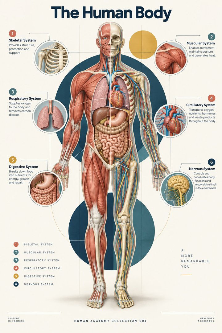
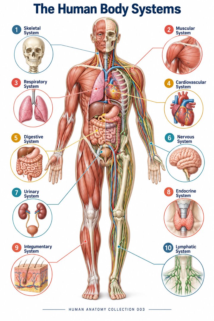
A body system is a useful way to group organs around related functions, but the living body does not operate as eleven independent machines. Circulation supports nearly every tissue; nervous and hormonal signals connect distant regions; digestive absorption changes the materials available to cells; and renal regulation influences the environment in which other organs work. Medical students benefit from learning the groups and then deliberately tracing their connections. Healthcare staff use the same approach when relating anatomical language to observations, procedures, and communication across professional roles.
This guide uses eleven familiar groups: integumentary, skeletal, muscular, nervous, endocrine, cardiovascular, lymphatic and immune, respiratory, digestive, urinary, and reproductive. The grouping is a teaching convention, not a universal biological census. Some resources combine or separate categories differently; NCI SEER's introductory list presents ten major systems and does not list integumentary as its own entry. The difference should be stated rather than concealed. NCI SEER explains organs and systems. An organ can also participate in more than one functional network, so counting the groups does not assign each organ to exactly one box.
Integumentary system: the body boundary is a functional interface
Skin, hair, nails, and associated glands form the integumentary grouping. Epidermal organization supports a protective boundary, dermal connective tissue provides support, and vessels and nerves relate the interface to the rest of the body. Skin contributes to sensation, thermal exchange, and the regulation of water loss at the surface. Subcutaneous tissues add deeper structural and metabolic relationships. The visible skin surface cannot demonstrate all these functions alone. NCBI describes the skin's layered anatomical organization.
The system illustrates how anatomy and environment meet. A change in external temperature creates thermal demands; a touch provides sensory input; and the barrier limits uncontrolled exchange with the surroundings. These tasks require collaboration with neural pathways and circulation. In eldercare and hospital work, skin observations also become part of broader assessment rather than belonging exclusively to a skin-only specialty. The anatomical contribution is to identify location, tissue depth, and the structures involved. It does not infer an individual's hydration, perfusion, or diagnosis solely from a photograph or the appearance of one small area.
Skeletal system: living support, protection, and internal resources
Bones and related skeletal structures provide support, protect selected organs, and create lever relationships with muscles. Bone tissue is alive, vascular, and subject to ongoing maintenance and remodeling. Mineralized matrix contributes mechanical properties, while marrow provides important blood-cell-production relationships. The commonly taught adult count of 206 bones reflects a standard counting convention, including 80 axial and 126 appendicular bones; anatomical variation and fusion affect literal counts in particular people. NCI SEER explains skeletal functions, and its division framework identifies the major skeletal groups.
This system cannot be understood as an inert frame surrounding more important organs. Its blood supply links it with circulation, its mineral handling links it with endocrine regulation, and its marrow connects it with hematology and immunity. A skeleton photograph effectively shows shape and alignment but conceals marrow, living cells, and local blood vessels. In teaching, place the photograph beside a tissue or function schematic to make those hidden relationships explicit. The 206 figure is useful when its convention is clear; it should not be turned into a diagnostic expectation that every individual must match exactly.
Muscular system: force generation depends on coordinated supply and control
Skeletal muscles generate force for movement, contribute to posture, and produce heat during activity. Their relationship to tendons and bones converts cellular contraction into movement of body segments. The muscular grouping in a body-system overview usually emphasizes skeletal muscles, while smooth and cardiac muscle are also tissue types functioning within other organ systems. A tissue category and an organ-system category therefore describe different levels of organization. NCI SEER introduces muscular-system functions.
Standing from a chair demonstrates the connections. Muscles require coordinated neural activation, skeletal leverage, adequate circulation, and substrates for cellular activity. The observed motion is a whole-body task rather than the isolated action of one named muscle. A model displaying red muscle groups can show attachments and broad positions, but it cannot demonstrate living neural control or energy supply. Healthcare teams discussing mobility benefit from keeping those relationships visible. An anatomical explanation can describe the participating structures while leaving functional assessment, support needs, and individual rehabilitation decisions to the appropriate clinical context.
Nervous system: information pathways connect sensing with response
The brain and spinal cord form the central nervous system, while peripheral nerves and related ganglia connect central regions with the rest of the body. Sensory pathways carry information toward integrating networks; motor pathways influence muscles and other effectors. Autonomic organization helps regulate visceral functions, and local circuits contribute further coordination. Neurons and glia have different roles within the tissue architecture. NCI SEER describes the nervous system's coordinating functions.
The key systems-level lesson is direction and integration. Feeling a surface, recognizing a signal, choosing an action, and activating a muscle belong to connected processes, but not to one identical pathway. The nervous system also interacts with endocrine regulation and bodily feedback. A static brain image is therefore not a map of every function or a demonstration of an individual's neurological state. Medical learners should relate gross structures to pathways and then to the task being explained. Healthcare staff can use the same clarity when distinguishing a reported sensation, an observed movement, and a formal examination finding.
Endocrine system: distributed messages depend on target receptors
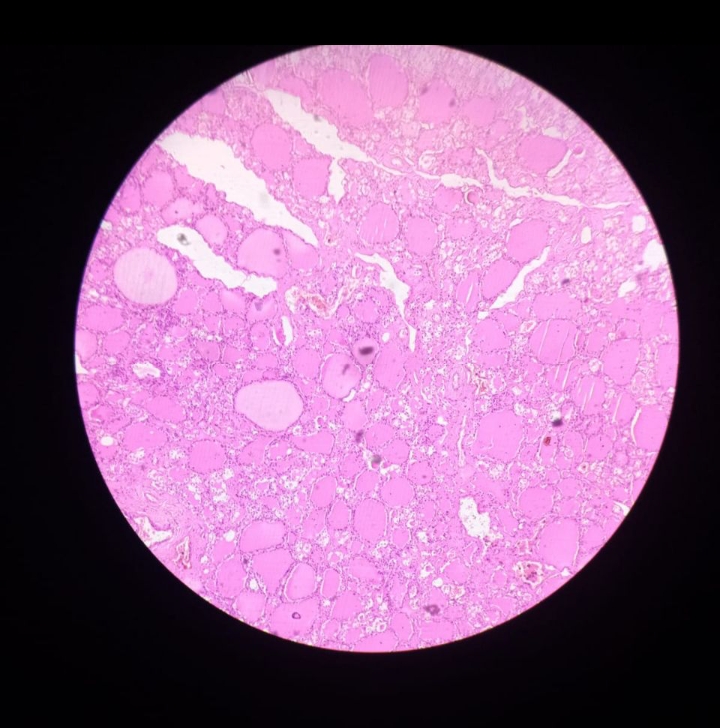
Endocrine tissues release hormones into the circulation, influencing cells with appropriate receptor arrangements. The pituitary, thyroid, parathyroids, adrenal glands, pancreatic endocrine tissue, and gonads provide familiar examples, while additional organs also produce endocrine signals. Hormones contribute to metabolism, growth, reproduction, and regulation of the internal environment. Being carried widely in blood does not mean that a hormone has the same effect on every cell. NCI SEER introduces endocrine regulation, and its hormone chapter explains target-receptor relationships.
The endocrine system shows why a glands-only body map is incomplete. The target organ, circulating route, receptor, and feedback relationship are also part of the explanation. The pancreas belongs in a digestive discussion because of exocrine secretion and in an endocrine discussion because of hormonal secretion. This is overlapping participation, not an anatomical contradiction. For medical students, separating the two routes clarifies ducts versus blood-borne signaling. For healthcare staff, recognizing shared organ roles helps explain why one organ may appear in more than one clinical conversation without requiring an assumption that the lists are inconsistent.
Cardiovascular system: transport is a continuous circuit
The heart, blood, and vessels provide a transport network linking tissue needs with exchange organs. Pulmonary circulation connects the heart with the lungs, while systemic circulation connects it with body tissues. Arteries carry blood away from the heart and veins toward it; oxygen content does not define the vessel category. Microvascular interfaces connect the large-scale route with local exchange. NCI SEER describes cardiovascular functions.
At the systems level, circulation is the shared route through which oxygen, nutrients, hormones, and many metabolic products move. The heart alone cannot perform every exchange function, and the lungs alone cannot distribute their oxygen throughout the body. A circulation diagram should therefore include the relevant exchange beds rather than stop at a decorative heart icon. Clinical measurements associated with circulation also answer different questions: a pulse, pressure reading, and observation of tissue condition are not identical measures. Anatomical education supports understanding those distinctions without replacing individual assessment or assigning a cause to one isolated finding.
Lymphatic and immune functions connect tissue fluid with defense
Lymphatic vessels return fluid from tissue environments toward the venous circulation. Lymph nodes, spleen, thymus, marrow, and distributed lymphoid tissues contribute to immune organization. Intestinal lymphatic routes also participate in transport of selected absorbed fats. The immune system includes more than a list of lymphatic organs; defensive functions also occur at barriers and within tissues throughout the body. Combining lymphatic and immune topics is a useful overview convention, provided their functions are not treated as identical. NCI defines the lymphatic system and its major roles.
This network helps explain why tissue fluid balance and immune response belong together spatially while remaining different functions. A lymph node is not a pump that sends oxygenated blood to tissue, and the spleen's relationship with blood differs from a node's relationship with lymphatic drainage. In teaching, use labeled arrows to distinguish fluid routes from the location of immune cell interactions. For healthcare staff, the overview supports precise identification of a region or structure. It does not establish why a particular node is enlarged or interpret symptoms from anatomy alone.
Respiratory system: ventilation and gas exchange are related, not identical
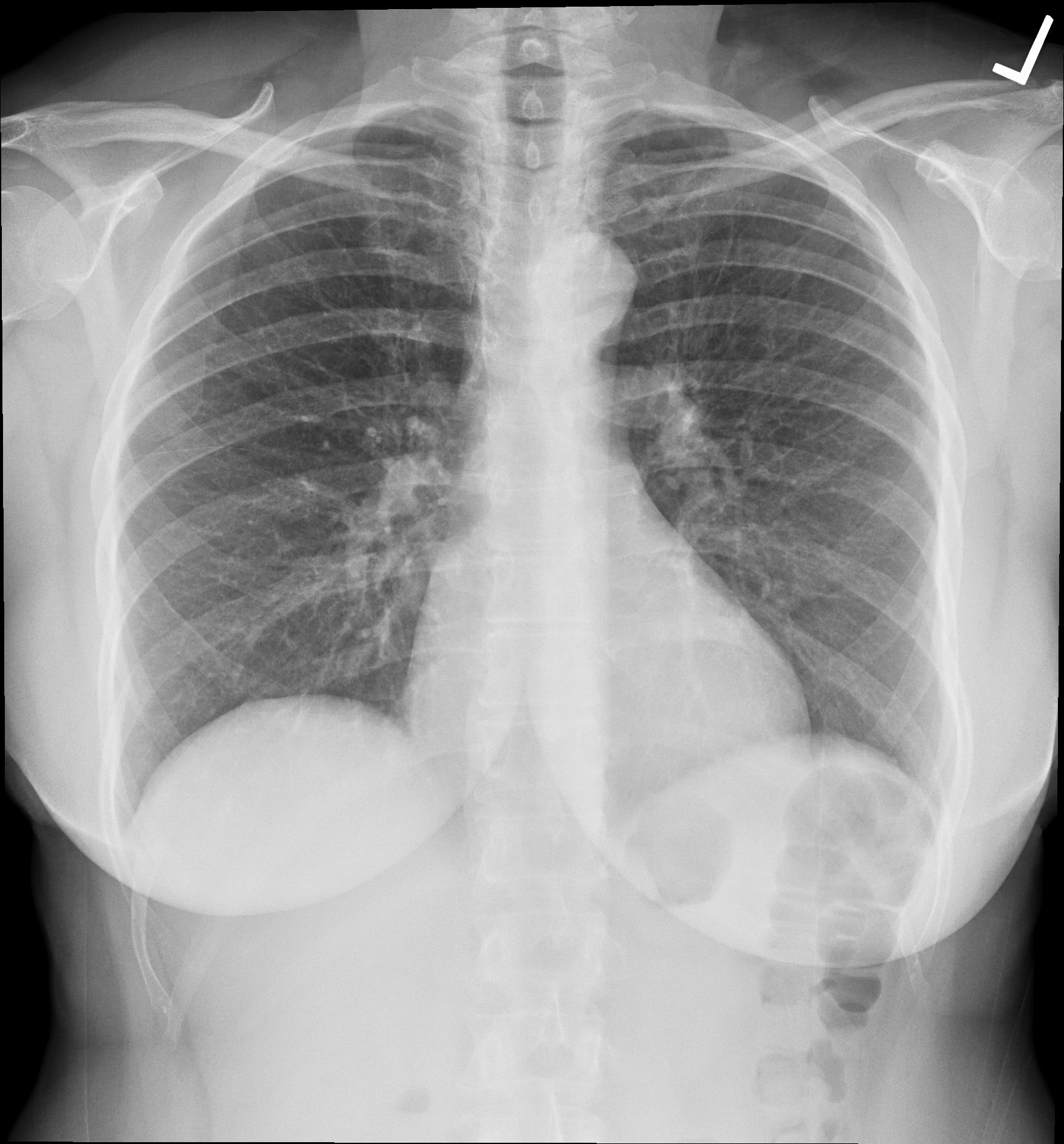
The respiratory grouping includes conducting airways and lung exchange structures. Ventilation moves air, while pulmonary exchange relates gases in alveolar environments to blood. Circulation then connects this exchange with the tissues. Carbon-dioxide handling links respiration with acid–base regulation as well as metabolic waste removal. Respiratory movement also depends on skeletal, muscular, and neural components outside the lung tissue itself. NCI SEER distinguishes ventilation and stages of respiration.
The integrated explanation prevents a common shortcut: assuming that moving air into the chest automatically guarantees delivery to every cell. Airway patency, exchange interfaces, vascular flow, blood transport, and tissue utilization are different relationships. A healthy-anatomy diagram can show how they connect without diagnosing which relationship is affected in an individual. The diaphragm provides a clear cross-system example because it is skeletal muscle contributing to respiratory mechanics under neural control. Naming the respiratory system should therefore not imply that its function belongs only to lungs viewed as isolated bags of air.
Digestive system: luminal processing and absorption enter different routes
The digestive tract processes ingested material through coordinated mechanical and chemical activities. The mouth, pharynx, esophagus, stomach, intestines, and terminal outlet form the major tract route. Accessory organs and glands provide secretions that support processing, including salivary glands, liver, gallbladder, and pancreas. Absorption transfers suitable products across specialized interfaces into internal transport routes. Material inside the gastrointestinal lumen has not automatically entered the internal tissues simply because it is inside the body's external outline. NCI SEER describes digestive functions and components.
Following a meal at the systems level involves several routes rather than one straight food tube. Some absorbed products enter portal blood, other selected lipids use intestinal lymphatic pathways, and hormonal or neural signals coordinate changing activities. The liver's relation to incoming portal blood illustrates the link between digestion and metabolism. This route-based explanation helps medical learners distinguish swallowing, digestion, absorption, and later cellular use. For healthcare staff, it supports clearer vocabulary when describing intake, gastrointestinal function, and the anatomical location under discussion without inferring nutritional status from one isolated observation.
Urinary system: regulation extends beyond making and storing urine
Kidneys regulate aspects of water, electrolyte, waste, and acid–base balance while producing urine. Ureters transport it, the bladder stores it, and the urethra provides the outlet route. Renal endocrine functions include erythropoietin signaling and participation in blood-pressure-related regulation through renin. The urinary system is consequently part of maintaining the internal environment, not merely a drain attached to the body. NCI SEER explains urinary homeostatic and endocrine functions.
The distinction between formation, transport, storage, and emptying is clinically useful because these stages occupy different anatomical sites. The bladder does not perform the same filtering and regulatory tasks as renal tissue. Emptying also involves neural and muscular coordination. A general systems diagram should therefore keep the kidney route connected with circulation and endocrine signaling while representing the lower tract separately. Healthcare learners can use this organization to preserve information in professional communication. It provides an anatomical foundation for discussing a clinical finding without assuming that every urinary observation points to the same organ or mechanism.
Reproductive system: gametes, support routes, and endocrine partnerships
Reproductive anatomy includes gonads, ducts, accessory glands, and structures supporting transport, sexual function, and, in the relevant anatomy, gestation and birth. Ovaries and testes contribute both gamete-related and hormonal functions. Reproductive roles therefore intersect with endocrine regulation and with broader tissue and vascular support. They should not be presented as the sole functions that determine an individual's health or identity. The anatomical description concerns structures and physiological relationships, while individual variation and life stage remain important. NCI SEER outlines major reproductive functions.
At the systems level, the gonadal hormone route differs from the duct route carrying gametes or associated secretions. A diagram that uses one unlabeled arrow for both obscures the distinction. Pregnancy additionally involves changing cardiovascular, renal, endocrine, and respiratory relationships, illustrating how a reproductive function can alter several systems at once. These are reasons to keep the system boundaries flexible in explanation. For medical students and healthcare teams, clear structural language and respectful communication are both important: identify the anatomy relevant to the discussion without assuming a person's reproductive goals, function, or life history.
Homeostasis is a coordinated relationship across the map
Homeostasis describes regulated maintenance of the internal environment through changing inputs and responses. It does not mean that every variable is motionless or that every organ works at one constant rate. Temperature regulation links skin, circulation, muscles, and neural control. Water balance links intake, gastrointestinal absorption, renal handling, and hormonal signals. Gas handling links respiratory exchange, cardiovascular transport, and tissue metabolism. These examples show why the systems should be learned as a network of dependencies rather than separate chapters that never meet.
For a useful review, trace one physiological task across three or more systems and label the material or information moving at each step. Distinguish blood flow from nerve signaling, secretion into a duct from hormone release into blood, and barrier exchange from movement through a lumen. The method reveals the connections that a colored full-body poster can only introduce. It also keeps numerical facts honest: eleven teaching groups, four major tissue categories, and a conventional adult bone count measure different things. None should be used as if it were a population statistic or a guarantee about every individual body.
Anatomical language supports collaboration across specialties
Systema nervosum, systema cardiovasculare, systema respiratorium, systema digestorium, systema urinarium, and systema lymphaticum identify familiar system groupings. Cutis, os, musculus, cor, pulmo, ren, hepar, and intestinum identify skin, bone, muscle, heart, lung, kidney, liver, and intestine respectively. The practical value of Latin-derived terminology is consistency across descriptions, not the replacement of plain explanations. A strong systems overview names the structures, identifies their major roles, and follows the routes connecting them. This gives healthcare learners a shared anatomical map while preserving the complexity of whole-body function.
Anatomical Latin: quick reference
| Latin term | English meaning |
|---|---|
| Systema nervosum | Nervous system |
| Systema cardiovasculare | Cardiovascular system |
| Systema respiratorium | Respiratory system |
| Systema digestorium | Digestive system |
Sources and image credits
Sources reviewed on 9 October 2026. Counts, estimates, study statistics and hypothetical calculations retain their context in the text.
- NCI SEER: organs and systems
- NCBI: skin anatomy
- NCI SEER: skeletal functions
- NCI SEER: skeletal divisions
- NCI SEER: muscular functions
- NCI SEER: nervous functions
- NCI SEER: endocrine functions
- NCI SEER: hormone targets
- NCI SEER: cardiovascular functions
- NCI: lymphatic definition
- NCI SEER: respiration
- NCI SEER: digestion
- NCI SEER: urinary functions
- NCI SEER: reproductive functions
Explanatory prose and SVG graphics are original. Photograph authors, source records and licenses are credited beside each image. Catalog illustrations provide educational context; anatomical facts are supported by the linked sources.