1. The CNS is an anatomical division with connected functions
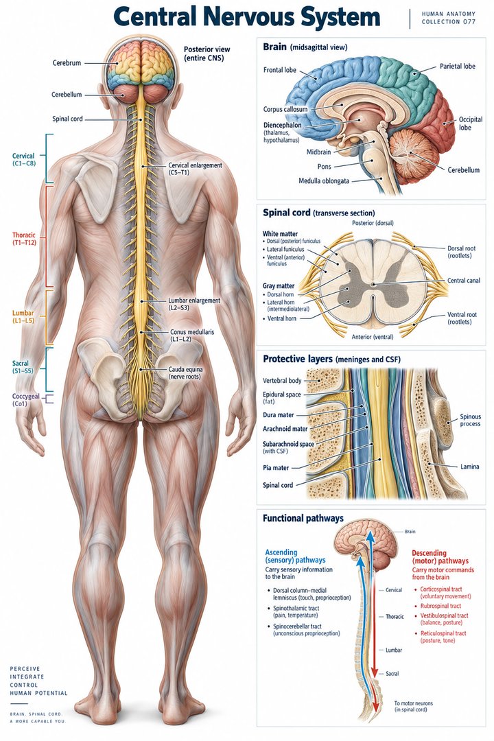
The central nervous system consists of the brain and spinal cord. Its functions depend on organized networks that receive, integrate, and influence information across the body. A drawing can establish the central division, but it should not imply that the CNS is disconnected from peripheral nerves or operates as one uniform tissue. The division is anatomical; the communication is continuous.
For healthcare learners, begin with location and then add organization. Identify the skull and vertebral canal, the major neural structures inside them, and the connections leaving the central division. This sequence makes later study of sensation, movement, cognition, and autonomic regulation easier. It also helps clinical teams distinguish a reported anatomical site from a functional observation that may involve several connected levels.
2. Major brain regions provide a starting framework
The cerebrum includes cortical and deeper structures, while the diencephalon, brainstem, and cerebellum add further organization. The brainstem includes midbrain, pons, and medulla oblongata. These broad regions should be distinguished before adding smaller nuclei or pathways. A lobe diagram of the cerebral surface shows only part of the CNS and does not replace a map of deeper or posterior structures. [OpenStax: central nervous system]
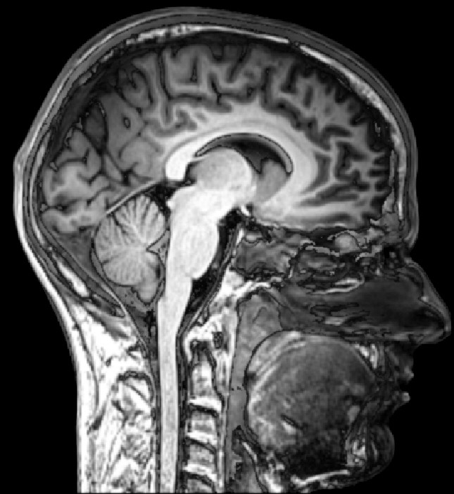
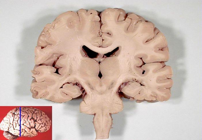
Compare a lateral surface view with a sagittal section. Ask which structures the first view conceals and which relationships the section exposes. A change of view can make a familiar structure appear very different without changing its identity. For students, explaining that transformation is often a stronger test of understanding than recognizing the same colored label repeatedly.
3. Gray matter and white matter describe tissue organization
Gray matter contains prominent neuronal cell bodies and associated elements, while white matter includes many myelinated axons. Both also contain glial cells and supporting structures. These names refer to general tissue organization rather than absolute purity. A white-matter region is not composed of axons alone, and a gray-matter region is not simply a collection of disconnected cell bodies. [OpenStax: neurons and glia]
When viewing a diagram, ask whether the colors represent these tissue categories or merely separate anatomical regions. Artificial color can conceal the reason for the traditional names. In a microscopic discussion, identify what the preparation can show before extending it to a pathway or function. The category helps organize the anatomy, but it does not tell the entire story of the cells and connections present.
4. The spinal cord has a regional and cross-sectional map
The spinal cord contains central gray matter and surrounding white-matter pathways. Its regional organization is described through cervical, thoracic, lumbar, sacral, and coccygeal segments. Spinal-cord segments and vertebral levels are related but are not identical labels, especially inferiorly. A drawing should state which structure its level markers describe. [OpenStax: spinal cord organization]
Use one longitudinal map for segments and one transverse view for tissue organization. A longitudinal line tells you where a level lies along the cord; a transverse section tells you what is arranged within that level. Combining the views supports spatial reasoning. It also prevents a learner from assuming that the familiar butterfly-like gray-matter profile is the same size or shape throughout the entire cord.
5. Spinal nerve-pair counts provide a quantitative anchor
The conventional spinal segment grouping associated with spinal nerves is eight cervical, twelve thoracic, five lumbar, five sacral, and one coccygeal pair, totaling thirty-one pairs. The presence of eight cervical nerve pairs is a useful reminder that nerve numbering should not be substituted directly for a count of cervical vertebrae. These are anatomical classification counts, not population prevalence figures. [OpenStax: spinal cord organization]
Add the regional counts before relying on the total. The arithmetic checks whether a group has been omitted, but it does not explain the connections within the cord. The graphic below compares groups using the same unit, nerve pairs. Keep that unit visible so that a bar value of twelve is not mistaken for twelve individual nerves or twelve spinal-cord structures of another kind.
6. Meninges and fluid compartments surround neural tissue
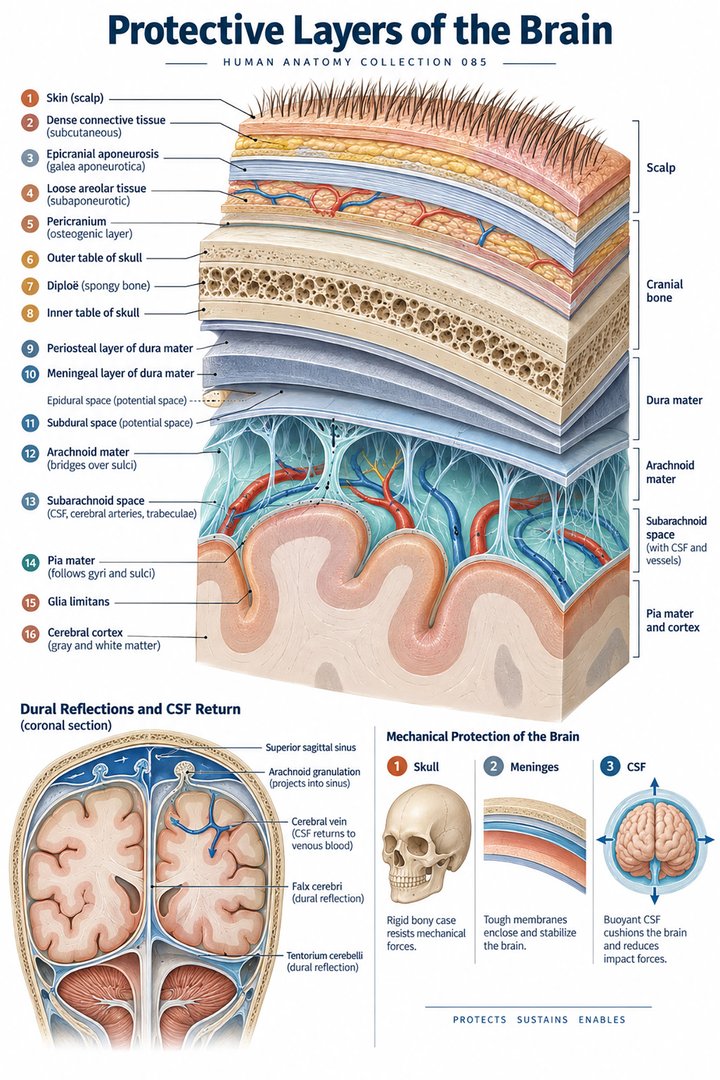
The meninges include dura mater, arachnoid mater, and pia mater. Cerebrospinal fluid occupies the ventricular system and subarachnoid relationships around the CNS. Choroid plexuses participate importantly in CSF production. These coverings and fluid spaces should be drawn separately from neural tissue rather than grouped into one generic protective shell. [OpenStax: CNS circulation and fluid compartments]
A layered diagram is especially useful here. Begin with neural tissue, add the pia, distinguish the subarachnoid space, and continue outward. State where the figure is schematic and which cranial or spinal context it represents. A layer name and a potential or actual space name are different kinds of terms, so the drawing should preserve the boundary that gives each its meaning.
7. Barriers and blood supply are different relationships
The blood–brain barrier involves specialized vascular interfaces that regulate exchange between circulation and neural tissue. It is not another meningeal sheet drawn outside the brain. Blood supply and CSF spaces provide different anatomical routes, even though both interact with the CNS environment. Keeping the vascular interface separate from the coverings prevents a common confusion in protective-layer illustrations. [OpenStax: CNS circulation and fluid compartments]
Place a small capillary-interface sketch beside a meningeal layer diagram. Ask where the barrier operates in each representation. The answer should name the vessel–tissue relationship rather than pointing only to the skull or outer membrane. For a clinical explanation, retain the distinction between the anatomical interface and the particular physiological or pathological finding established by an investigation.
8. Central and peripheral structures work together
Peripheral nerves connect the CNS with receptors, muscles, and other tissues. Sensory information can travel centrally, while motor and autonomic influences travel through connected routes toward peripheral targets. A reflex can be organized through spinal circuitry while remaining subject to wider influences. The term central does not mean that every action requires a conscious decision or a single cortical location. [OpenStax: nervous system organization]
For teaching, choose a simple sensory-to-response example and identify which structures belong to the central division and which belong to the peripheral route. Then explain which parts the simplified example leaves out. This makes the division useful without turning it into a wall. Healthcare communication benefits from the same habit when a report names a peripheral nerve but the discussion concerns a broader functional task.
9. Networks are more informative than isolated function labels
Cortical regions, deeper nuclei, cerebellar circuits, and brainstem structures contribute to connected processes. An association between a region and a function is a learning aid, not proof that the function exists only there. Complex tasks require several inputs and outputs. A general CNS poster should therefore introduce relationships rather than promise a one-to-one map from every observed difficulty to a single colored region. [OpenStax: central nervous system]
Medical students can revise by explaining a pathway with its endpoints and one intermediate relationship. In a case discussion, identify the established observation before adding the anatomical hypothesis or confirmed finding. This protects the difference between what a person did during assessment, what an image showed, and what interpretation the clinical team reached.
10. Latin terms help retain structural precision
Encephalon refers to the brain as a whole, while cerebrum, cerebellum, and medulla spinalis retain distinctions among major structures. Truncus encephali identifies the brainstem. Dura mater, arachnoidea mater, and pia mater are useful names for the meningeal layers. Learn a term with its extent and neighboring structures so that “brain” and “cerebrum” do not become automatic synonyms. [FIPAT: Terminologia Anatomica, second edition]
Build a table with whole structure, subdivision, and view. For each term, name the image that best demonstrates it and one feature concealed by that view. This makes terminology portable across posters and scans. When explaining established findings to a patient, translate the term into ordinary language while preserving whether it describes the whole CNS, a major region, or a covering.
| English | Selected Latin |
|---|---|
| Brain as a whole | Encephalon |
| Cerebellum | Cerebellum |
| Spinal cord | Medulla spinalis |
| Brainstem | Truncus encephali |
11. Real images teach orientation and evidence limits
The sagittal MRI below shows an approximately midline human head section with the face toward the image’s right. The coronal specimen photograph uses a different plane and preparation. Neither image is a complete view of every CNS pathway. Their value lies in comparing real anatomical relationships with the cleaner organization of a teaching illustration.
Practice assistants, hospital staff, physicians, and eldercare teams can use such comparisons to clarify terminology in established reports and discussions. An educational image does not determine a person’s neurological diagnosis. End a session by naming the plane, locating a major structure, and explaining one connection or covering. Those three tasks connect orientation, anatomy, and interpretation without claiming more than the evidence shows.
Sources and image credits
Sources checked on 8 October 2026. Numerical examples, anatomical classification counts, and research estimates are identified separately in the text.
- OpenStax: nervous system organization
- OpenStax: neurons and glia
- OpenStax: central nervous system
- FIPAT: Terminologia Anatomica, second edition
- OpenStax: CNS circulation and fluid compartments
- OpenStax: spinal cord organization
Explanatory prose and simplified SVG graphics are original. Photographs retain their stated Creative Commons licenses; creator and source links appear beside each image. Catalog artwork is educational and decorative; no clinical authorship or peer-review credential is claimed.