1. Liver function connects circulation with metabolism
The human liver participates in processing absorbed nutrients, synthesizing circulating substances, handling metabolic by-products, and producing bile. Its position and blood supply make these functions closely connected with the digestive and circulatory systems. A whole-organ picture provides orientation, but understanding function requires another scale: hepatocytes, sinusoids, and small channels within the tissue.
For healthcare learners, avoid reducing the liver to a universal toxin filter. A more useful framework identifies the substance, process, and destination. Glucose handling, nitrogen metabolism, protein synthesis, bilirubin processing, and bile secretion involve different pathways. The same organ contributes to all of them, but the contribution cannot be measured or explained with one undifferentiated arrow.
2. Two inflow routes meet within the liver
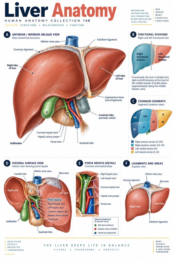
The liver receives blood through the portal venous system and hepatic arterial supply. In broad introductory descriptions, approximately seventy-five percent of inflow volume is portal and twenty-five percent arterial. These are blood-flow fractions, not the fractions of oxygen delivered. Blood from the supplies mixes in sinusoids and drains toward central and hepatic venous routes. [Review: hepatic microvascular anatomy and function]
Trace the incoming routes and then the outgoing hepatic veins separately. The portal vein is an inflow vessel in this organ relationship, despite the everyday tendency to treat every vein as a simple organ outlet. This example reinforces the need to follow connections rather than infer direction only from color or a vessel name detached from the actual circulation.
3. Blood and bile follow different microscopic routes
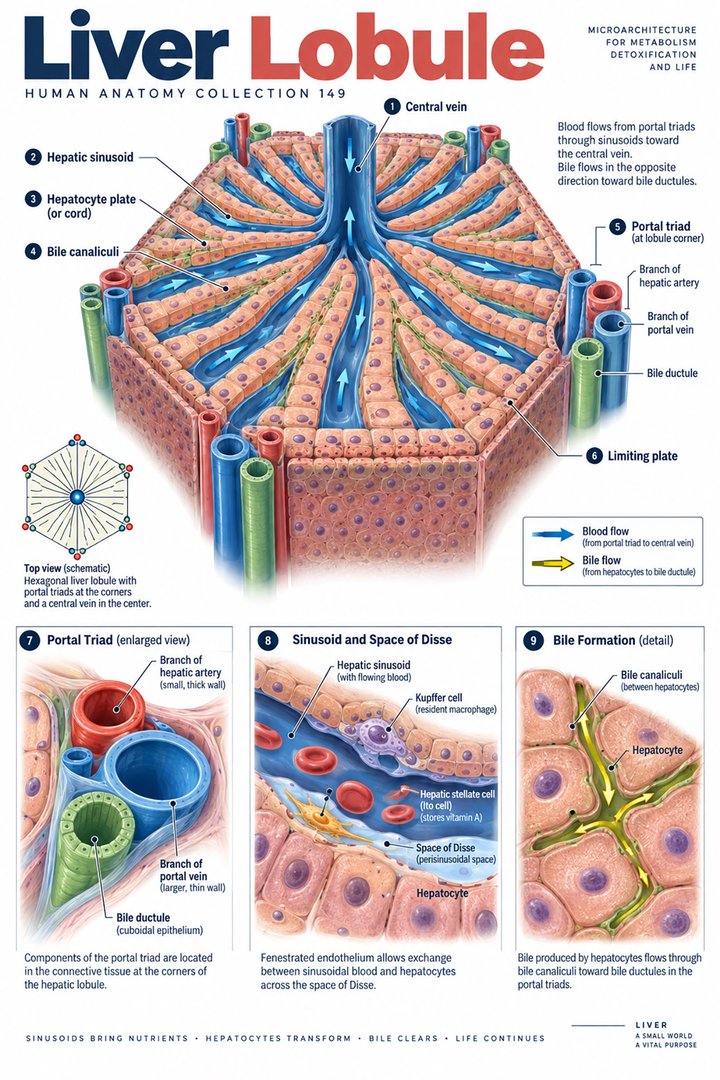
Hepatocytes produce bile, which enters canaliculi between neighboring hepatocytes and proceeds toward ductular and bile-duct routes. In a conventional lobule schematic, this direction is broadly opposite the sinusoidal blood route toward the central vein. The gallbladder stores and concentrates bile; it is not the organ that produces it. [Review: hepatocyte cell biology]
Use separate arrows for sinusoidal blood and bile. Explain the starting compartment of each before drawing their destinations. A crowded two-dimensional diagram can make the channels appear to intersect, so the caption should preserve their distinct lumens. This is one of the most useful exercises for linking a whole liver-and-gallbladder poster with a microscopic lobule image.
4. Glucose handling changes with metabolic circumstances
The liver can store glucose-related energy as glycogen and mobilize it when circumstances require. It also contributes to making glucose from other substrates. These processes connect meals, fasting intervals, hormonal signaling, and the needs of other tissues. The liver should not be imagined as a warehouse that only stores material without actively changing its handling over time. [InformedHealth: how the liver works]
For a teaching exercise, place two hypothetical circumstances beside the same organ diagram and describe which broad process is being emphasized. State that the exercise explains regulation rather than predicting a particular person’s blood glucose. A measurement at one time records a result within the wider system; the picture alone cannot determine the person’s metabolic state.
5. Nitrogen metabolism links liver and kidneys
Protein-related metabolism generates nitrogen-containing products. Hepatic processing converts ammonia into urea, which is subsequently excreted through renal handling. This is a useful example of connected organ roles: production of a circulating metabolic product and its eventual excretion are not necessarily performed in the same organ. [InformedHealth: how the liver works]
Draw a route from tissue metabolism through hepatic processing to renal excretion. Name the changing substance instead of drawing one generic “waste” label from beginning to end. This helps medical students connect biochemical learning with anatomy. For healthcare communication, retain the actual test or substance named in a report rather than replacing several different measurements with a broad statement about detoxification.
6. Protein synthesis supports circulating functions
The liver synthesizes albumin and many proteins involved in coagulation. These products connect the organ with fluid relationships, transport, and hemostatic processes. A synthesis role is distinct from an injury-associated laboratory marker. One should therefore avoid treating every result commonly grouped in a liver panel as a direct measurement of the same physiological function. [NCBI Bookshelf: liver physiology]
Create a table organized by question: tissue injury, synthesis-related information, or handling of a particular substance. Name the measurement before discussing its meaning, and retain the possibility of influences outside the liver. This is a useful professional habit because a familiar panel heading can conceal several different biological questions and does not itself provide a complete interpretation.
7. Bilirubin handling has several stages
Hepatic bilirubin processing includes uptake, conjugation, and biliary excretion. These stages should not be compressed into “the liver destroys bilirubin.” They describe a changing substance and a route through cellular and biliary compartments. The broader pathway also connects the liver with the handling of products arising from heme-related turnover. [NCBI Bookshelf: liver physiology]
For study, use separate boxes for the process and the anatomical route. Ask what changes chemically and where the material proceeds afterward. This method makes it easier to understand why different observations may concern different stages. In a clinical discussion, use the established findings and appropriate interpretation rather than assigning a cause from a general pathway drawing.
8. Biotransformation is specific rather than universal
Hepatic enzymes participate in modifying and conjugating many substances. The process can change how a substance behaves and how it is eliminated, but it should not be described as a guarantee that the liver removes every harmful material. Some processes can generate biologically active products. A function guide becomes more accurate when it names the transformation rather than relying only on the slogan “detox.” [NCBI Bookshelf: liver physiology]
This distinction is especially useful when explaining why a substance’s route and chemical form matter. For educational examples, identify an input, a processing step, and an output without selecting a drug dose or treatment. Medication decisions depend on the actual product, person, measurements, and clinical plan. The anatomical explanation provides context rather than a prescribing rule.
9. Functional tissue models use different organizing principles
The classic hepatic lobule emphasizes a central venous relationship and surrounding portal regions. Acinar models emphasize perfusion relationships and are commonly described with three zones: periportal zone one, intermediate zone two, and pericentral zone three. These are different ways to organize tissue relationships, not three separate organs or necessarily equal-sized compartments. [Review: hepatic microvascular anatomy and function]
Place the two model outlines over a simplified tissue map and explain what each highlights. The exercise shows why apparently different textbook figures can both be useful. It also prevents a colored zone from being treated as a complete physiological diagnosis. A model selects a relationship for teaching; it does not replace the complexity and variation of actual tissue.
10. Latin terms connect the whole organ with its routes
Hepar names the liver. Porta hepatis identifies the major hilar region, vena portae hepatis the portal vein, and venae hepaticae the hepatic veins. Lobulus hepaticus and hepatocytus connect larger anatomy with tissue and cell scales. Vesica biliaris identifies the gallbladder. These names become useful when each is paired with a direction or role. [FIPAT: Terminologia Anatomica, second edition]
For a vocabulary exercise, assign each term to organ, junction, vessel, tissue unit, or cell. Then explain one connection between categories. This retains the difference between a cell producing bile and a duct carrying it. When discussing a report with a patient, translate the term while preserving whether the finding concerns hepatic tissue, vascular supply, or the biliary route.
| English | Selected Latin |
|---|---|
| Liver | Hepar |
| Portal vein / hepatic veins | Vena portae hepatis / venae hepaticae |
| Hepatic lobule / hepatocyte | Lobulus hepaticus / hepatocytus |
| Gallbladder | Vesica biliaris |
11. Real histology supports a careful comparison
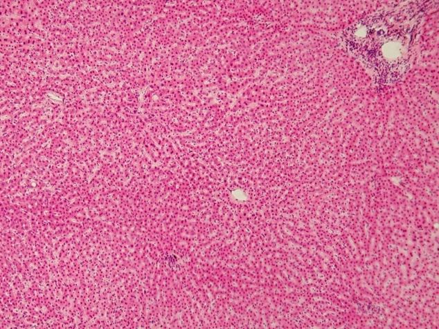
The real human liver micrograph below records stained tissue. Its visible organization can be compared with the simplified lobule poster, but it does not directly display every metabolic pathway. Colors reflect preparation, and a single field does not show the whole organ. The image is structural evidence within a defined method, not a complete assessment of hepatic function.
Medical students can use the micrograph to connect cell and vascular organization with the organ’s roles. Practice assistants, hospital staff, physicians, and eldercare teams can use the route vocabulary to clarify established information. End revision by explaining a blood route, a bile route, and one metabolic connection to another organ. Together, these relationships make liver function understandable beyond a list of slogans.
Sources and image credits
Sources checked on 8 October 2026. Numerical examples, anatomical classification counts, and research estimates are identified separately in the text.
- FIPAT: Terminologia Anatomica, second edition
- NCBI Bookshelf: liver physiology
- InformedHealth: how the liver works
- Review: hepatic microvascular anatomy and function
- Review: hepatocyte cell biology
Explanatory prose and simplified SVG graphics are original. Photographs retain their stated Creative Commons licenses; creator and source links appear beside each image. Catalog artwork is educational and decorative; no clinical authorship or peer-review credential is claimed.