1. Immunity is a coordinated set of processes
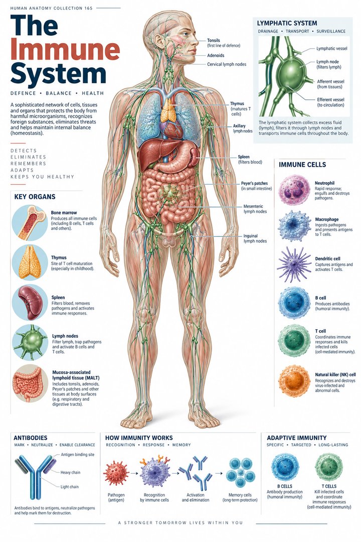
The human immune system combines barriers, cells, signaling molecules, and organized tissues. A whole-body poster can identify some organs, but many important events occur at microscopic interfaces or through communication between cells. Learning immunity therefore requires both an anatomical map and an account of coordinated responses. No single organ or white-cell type represents the entire system.
For healthcare learners, begin by separating a structure from a process. A lymph node is a tissue organization; antigen recognition is a process; an antibody is a molecular product. These entities can appear close together on a diagram without being interchangeable. A useful explanation follows how they interact while retaining the limits of the image or measurement being discussed.
2. Barriers create the first anatomical interface
Epithelial barriers, secretions, and local protective mechanisms contribute to innate defense. Skin and mucosal surfaces are active interfaces rather than inert packaging around internal organs. Innate responses also include phagocytes, natural killer cells, complement, and inflammatory signaling. These components interact, so a rigid picture of one barrier acting completely alone is incomplete. [OpenStax: innate immunity]
Choose a mucosal surface and describe the interface before adding immune cells. Ask what separates the body’s internal environment from the material encountered there. This establishes why the location of a response matters. For clinical communication, keep the general protective role separate from the observed condition of a particular person’s tissue or the result of a specific test.
3. Innate and adaptive responses interact
Innate and adaptive immunity describe related forms of defense, not two isolated departments that act only in a fixed sequence. Innate sensing and antigen presentation influence adaptive responses, while adaptive products can alter later interactions. The distinction remains useful when it is explained through recognition mechanisms and cellular roles rather than a simplistic “first system switches off when the second starts.” [InformedHealth: innate and adaptive immunity]
A timeline can introduce the idea of different response dynamics, but label it as simplified. Then add cross-connections to show that timing alone does not define a component. This helps students avoid classifying every rapidly acting cell as one category and every later event as another without understanding the mechanism. The goal is a connected model that can accommodate more detailed immunology later.
4. Phagocytes and antigen-presenting cells have distinct roles
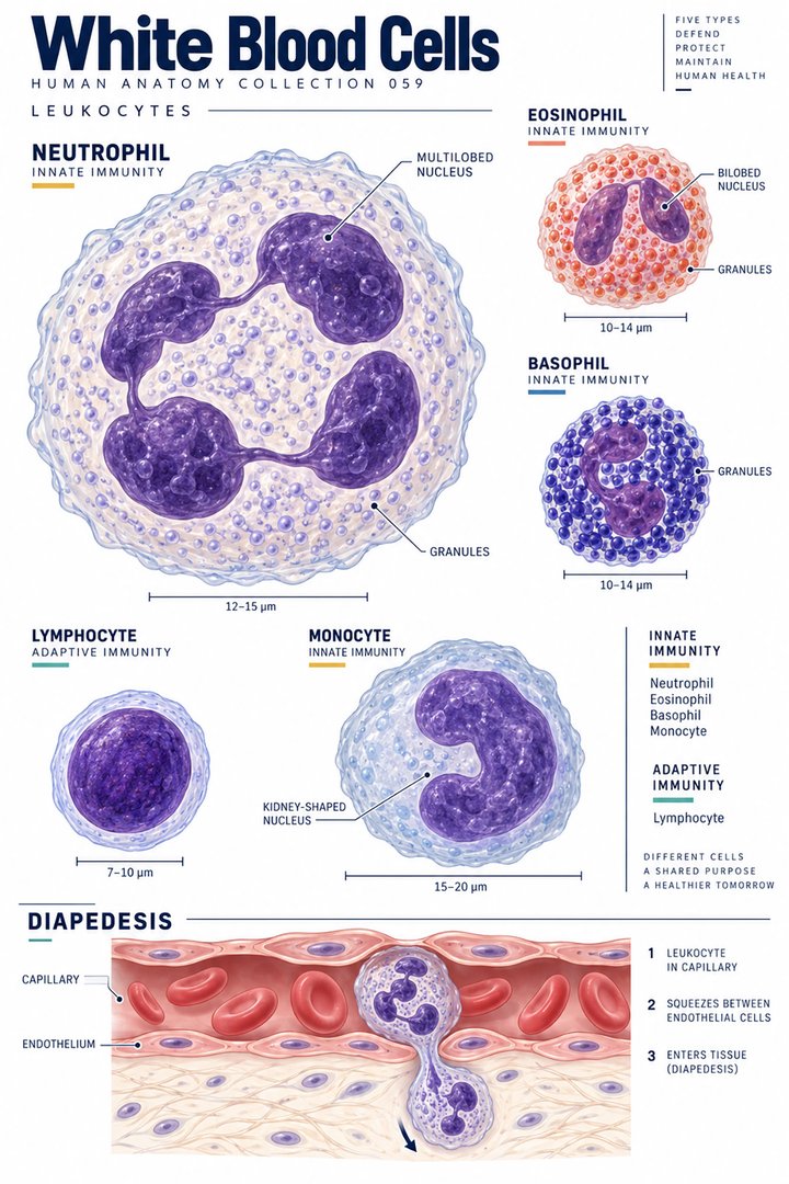
Phagocytosis involves taking material into a cell. Neutrophils and macrophages are prominent examples of cells involved in such processes. Dendritic cells contribute importantly to antigen presentation and communication with adaptive responses. The same general category of defensive cell can participate in several tasks, so a diagram should not reduce every cell to one isolated action. [OpenStax: innate immunity]
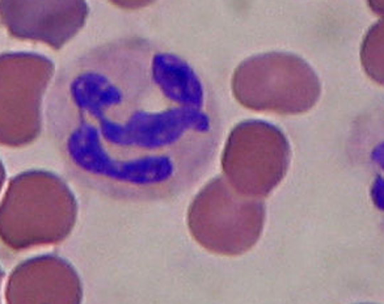
When viewing a blood smear, distinguish what can be recognized from what a functional experiment would establish. A characteristic cell profile can support a morphological identification, but it does not show every signaling event or the cell’s complete current activity. Explain the observed image first, then connect it with the general function described in the source.
5. T lymphocytes include several functional populations
T-cell populations include helper, cytotoxic, and regulatory functions. Their activities depend on recognition and communication within a larger response. Antigen presentation and relevant cellular interactions provide context; a generic lymphocyte picture does not visibly distinguish every T-cell subtype. Functional names are therefore not simply labels that can be assigned from an unlabeled surface micrograph. [OpenStax: T lymphocytes]
For study, pair a function with the interaction required to support it. Then identify which part is represented anatomically and which part requires molecular or functional evidence. This approach prevents a poster’s colored cell icons from being treated as literal microscopy. It also makes terminology clearer when students encounter laboratory methods that measure a population using markers rather than appearance alone.
6. B cells connect recognition with antibody production
B cells can differentiate into antibody-secreting plasma cells. Antibodies interact with particular targets and participate in several defensive relationships, rather than acting as universal substances that destroy anything unfamiliar. The relationship between B cell, plasma cell, and antibody should be drawn as a cellular differentiation and product pathway, not as three names for the same object. [OpenStax: B lymphocytes and antibodies]
An introductory schematic can show recognition, activation, and later products while acknowledging that important interactions are omitted. Ask learners to identify whether each box contains a cell type, a process, or a molecule. This simple check prevents a surprisingly common misunderstanding: interpreting an antibody icon as another immune cell or treating every lymphocyte as an antibody producer.
7. Natural killer cells challenge an easy classification shortcut
Natural killer cells are lymphocytes associated with innate immunity. This is an important exception to the shortcut that “lymphocyte” automatically means adaptive B or T cell. Cell morphology, lineage, recognition mechanism, and function describe different aspects of identity. A correct classification states which aspect is being considered. [OpenStax: innate immunity]
Make a revision card containing the term lymphocyte and several possible populations. Add the broad immune relationship beside each. The exercise tests whether a category name has been mistaken for a single function. In a professional discussion, retain the precise population identified by the laboratory or clinical assessment rather than extending a broad white-cell label into an unsupported functional conclusion.
8. Organized lymphoid tissues support immune interaction
Primary lymphoid organs include bone marrow and thymus, while nodes, spleen, and mucosal lymphoid tissues provide important secondary sites. Their anatomical relationships differ: nodes are associated with lymphatic routes, and the spleen interacts with blood. A whole-body immune map should preserve these distinctions even when it groups the tissues together for readability. [OpenStax: lymphatic and immune anatomy]
Use the map to locate a site, then add a small section showing that the tissue has internal organization. State when the detail is enlarged. This combines anatomical and cellular learning without implying that every response occurs inside a conspicuous node. The distributed nature of immune activity is a reason to avoid treating the few organs highlighted in a poster as a complete inventory.
9. Five antibody classes provide a quantitative teaching anchor
The five major antibody classes are IgG, IgA, IgM, IgE, and IgD. The count is a classification fact, not a claim that each contributes an equal percentage of circulating antibody. Differences in structure, distribution, and roles require their own context. The table lists classes without inventing prevalence values or comparing their names as if they were measurements. [OpenStax: B lymphocytes and antibodies]
A numerical graphic can compare selected counts, provided the units remain explicit. Here it distinguishes five antibody classes from the two broad response categories used in introductory teaching. Those numbers describe different classification systems and should not be read as proportions, importance scores, or evidence of a patient’s immune strength. A meaningful clinical measurement requires the actual test and its interpretation.
| Major antibody class | Learning note |
|---|---|
| IgG | One of five major classes |
| IgA | One of five major classes |
| IgM | One of five major classes |
| IgE | One of five major classes |
| IgD | One of five major classes |
10. Memory and tolerance complicate a simple battle metaphor
Adaptive memory can alter responses to later encounters. It does not imply guaranteed lifelong protection against every exposure. Immune regulation also matters: useful responses must be controlled in relation to the body’s own tissues and the surrounding environment. A battle metaphor can introduce defense, but it leaves out coordination, limitation, and the consequences of inappropriate activity. [InformedHealth: innate and adaptive immunity]
Use the terms memory and regulation in a case discussion without making a prediction from a general guide. Separate a known observation from the mechanism being taught. For example, a record of prior exposure or vaccination is not the same as a direct measurement of every future response. This distinction is useful across medical education and patient-facing communication.
11. Latin vocabulary and real images connect scales
Leucocytus and lymphocytus are Latinized cellular terms; medulla ossium rubra, thymus, and lien identify associated anatomical structures. Antigen, cytokine, antibody, and complement belong to modern scientific vocabulary rather than a single list of Latin organ names. Learn each with its category: cell, tissue, molecule, or process.
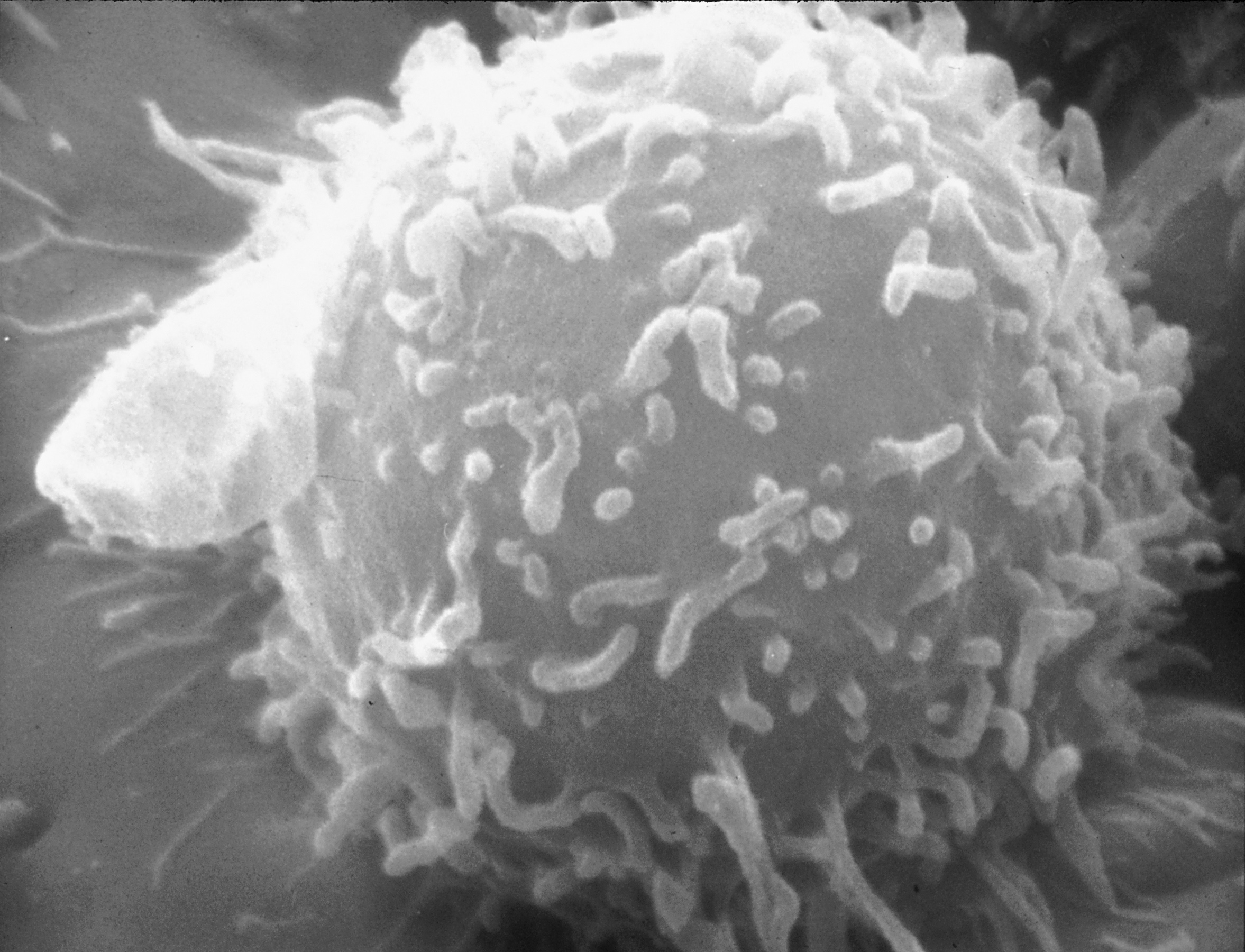
The real neutrophil blood-smear image and lymphocyte electron micrograph below use different preparations and imaging methods. Their visible colors and surfaces cannot be compared as if both were natural appearances under the same conditions. The lymphocyte image does not establish B-cell or T-cell identity. Preserve those limitations with the creator credits when using the images in professional teaching.
12. Make immune learning useful in clinical communication
Medical students can follow a response across anatomical and cellular levels. Practice assistants and hospital personnel can use the terminology to understand documented laboratory populations and the purpose of investigations. Physicians can explain why a test of one component does not describe every aspect of immunity.
For eldercare teams, connect educational discussion with the person’s established clinical information and care plan. A general immune guide does not select treatment or define an individual’s infection risk. End revision by distinguishing a cell from its product, innate from adaptive mechanisms, and a classification count from a clinical measurement. These distinctions provide a strong foundation for later immunology.
Sources and image credits
Sources checked on 8 October 2026. Numerical examples, anatomical classification counts, and research estimates are identified separately in the text.
- OpenStax: lymphatic and immune anatomy
- OpenStax: innate immunity
- OpenStax: T lymphocytes
- OpenStax: B lymphocytes and antibodies
- InformedHealth: innate and adaptive immunity
Explanatory prose and simplified SVG graphics are original. Photographs retain their stated Creative Commons licenses; creator and source links appear beside each image. Catalog artwork is educational and decorative; no clinical authorship or peer-review credential is claimed.