1. Ear anatomy serves hearing and balance
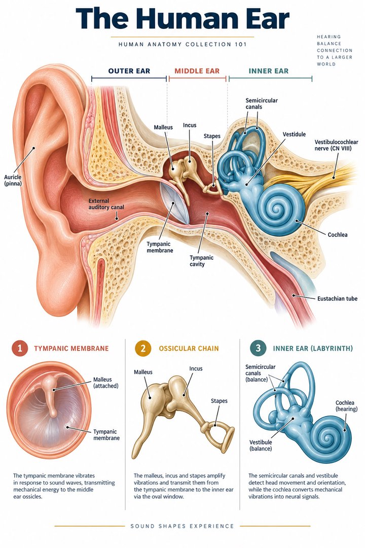
The human ear combines a sound-transmission system with sensory structures involved in hearing and equilibrium. The external outline is familiar, but most of the important internal organization lies within the temporal bone. A poster that exposes the middle and inner ear changes the view and scale to make those structures visible. The real external photograph at the end of this guide cannot reveal the same internal detail.
For healthcare learners, begin by separating the pathway of sound-related mechanical movement from the pathway of neural information. Then distinguish the auditory structures from the vestibular apparatus. These relationships make the anatomy useful in discussions of examinations and reports without assuming that every ear-related symptom involves the same structure or mechanism.
2. External, middle, and inner regions have different contents
Three broad anatomical regions are external, middle, and inner ear. The external canal leads toward the tympanic membrane, which forms a boundary with the middle-ear cavity. The middle ear is an air-containing space with an ossicular chain. The inner ear contains specialized auditory and vestibular arrangements with fluid compartments. The labels refer to organization, not three equally sized areas. [NCBI Bookshelf: middle-ear anatomy and physiology]
In a learning diagram, trace the external canal before naming the structures hidden beyond its endpoint. Mark the membrane as a boundary rather than placing it at the center of every auditory process. This clarifies what an external view or a standard examination can show. A clinical description of a visible membrane is not the same as direct observation of the cochlea or vestibular sensory organs.
3. The ossicular chain transmits mechanical movement
The three auditory ossicles are malleus, incus, and stapes. Their chain connects tympanic-membrane movement toward the oval-window interface, where the stapes footplate acts. These structures should be described through their connections rather than as three tiny bones floating independently in an empty chamber. Their movement contributes to transferring sound-related energy toward the inner ear. [NCBI Bookshelf: middle-ear anatomy and physiology]
Use a schematic to identify each junction in order. Then cover the labels and explain which structure precedes and follows the incus. This is a stronger test than recognizing three isolated bone shapes. Keep the oval window distinct from the tympanic membrane: both are important interfaces, but they lie at different points in the route and have different surrounding relationships.
4. The auditory tube connects the middle ear to another region
The auditory tube connects the middle-ear cavity with the nasopharynx and supports ventilation and pressure equalization. It is a separate relationship from the external auditory canal. A two-dimensional poster can place their outlines near one another, so labels and endpoints are essential. The tube does not provide the route by which external sound normally enters the canal and reaches the tympanic membrane. [NCBI Bookshelf: middle-ear anatomy and physiology]
Trace both channels with different line styles, naming their beginning and destination. This small comparison makes a useful teaching exercise for practice assistants and hospital teams. When a report refers to one route, retain its actual name and the established finding. A general anatomical connection does not determine the cause of pressure symptoms or select a treatment for an individual.
5. Cochlear organization converts mechanics into signaling
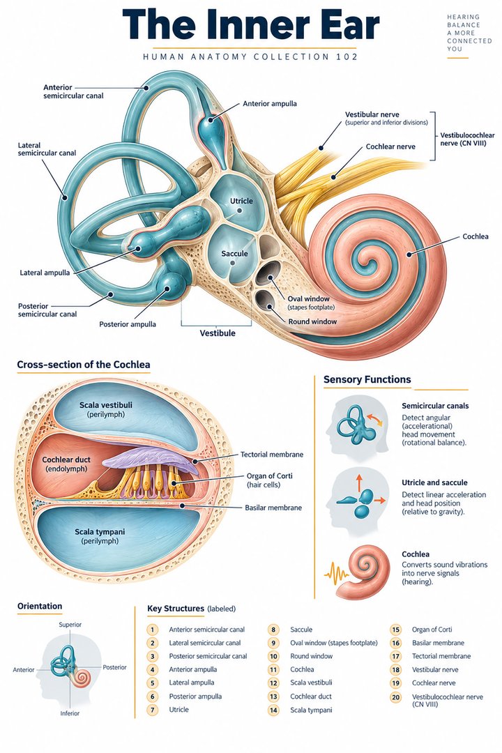
Within the cochlea, sound-related movement affects the basilar membrane and sensory hair-cell relationships. Bending stereocilia changes hair-cell electrical activity, which influences auditory nerve signaling. Higher frequencies preferentially engage more basal cochlear regions, while lower frequencies engage more apical regions. This spatial organization is a functional relationship within a coiled structure rather than a row of independent sound detectors. [NIDCD: how hearing works]
Compare an intact cochlear coil with an uncoiled teaching diagram. The uncoiled version makes the base-to-apex relationship easier to display but changes the shape deliberately. State that transformation in the caption. A frequency label on a drawing is an introductory map, not an individualized audiogram or proof of a person’s hearing ability at that frequency.
6. Vestibular structures perform a different sensory task
The three semicircular ducts participate in sensing rotational head movement. The utricle and saccule, the two otolith organs, contribute to sensing linear acceleration and orientation relative to gravity. These structures are part of the inner-ear sensory arrangement but are distinct from the cochlear auditory task. Their relationships explain why an ear anatomy guide must discuss more than hearing. [NIDCD: the balance system]
A useful revision sketch groups the rotation-related structures separately from the otolith organs. Label the relevant kind of movement rather than using the same generic “balance sensor” phrase for every part. The distinction adds precision without requiring a detailed treatment of every vestibular pathway. It also prevents the inner ear from becoming an automatic synonym for the cochlea.
7. Balance integrates information from several systems
The vestibular system contributes to equilibrium together with vision and somatosensory information from muscles, touch, and other body relationships. Balance should therefore not be explained as a function of the ear alone. A whole-body task can involve several sources of information even when one anatomical component is highlighted in a poster. [NIDCD: the balance system]
In eldercare and hospital teaching, this integrated framework helps explain why a general ear diagram cannot identify the cause of a person’s unsteadiness. Describe the observed difficulty and the established assessment separately from the mechanism being taught. The anatomical map can clarify terms in a care discussion, while clinical findings and the treating team’s plan determine the individual interpretation.
8. Hearing statistics need a population and definition
NIDCD reports disabling hearing loss in approximately five percent of US adults aged forty-five to fifty-four, ten percent aged fifty-five to sixty-four, twenty-two percent aged sixty-five to seventy-four, and fifty-five percent aged seventy-five and older. The estimates use 2015–2020 NHANES data and a definition of hearing loss of at least thirty-five decibels in the better ear. [NIDCD: hearing statistics and survey definitions]
These percentages concern the source’s specified definition, not every form of self-reported hearing difficulty or every population worldwide. The chart preserves the age groups and should not be read as a prediction for a particular person. Its value for healthcare teams is to connect anatomy with a clearly defined population statistic while retaining the need for an individual assessment.
9. Latin names help separate adjacent structures
Auricula names the auricle, meatus acusticus externus the external acoustic canal, and membrana tympani the tympanic membrane. Cavitas tympani identifies the middle-ear cavity. Malleus, incus, and stapes retain the ossicular names, while tuba auditiva refers to the auditory tube. Cochlea, utriculus, and sacculus identify distinct inner-ear structures. [FIPAT: Terminologia Anatomica, second edition]
Learn each term with a boundary or connection. A vocabulary table becomes more useful when it adds what lies before or after the named structure. For patient-facing discussion, translate the term into ordinary English while preserving whether it concerns the external canal, middle-ear space, auditory organ, or vestibular arrangement. This reduces ambiguity when several neighboring structures have unfamiliar names.
| English | Selected Latin |
|---|---|
| Auricle / external acoustic canal | Auricula / meatus acusticus externus |
| Tympanic membrane | Membrana tympani |
| Auditory ossicles | Ossicula auditus: malleus, incus, stapes |
| Auditory tube | Tuba auditiva |
10. A real external photograph establishes its own limits
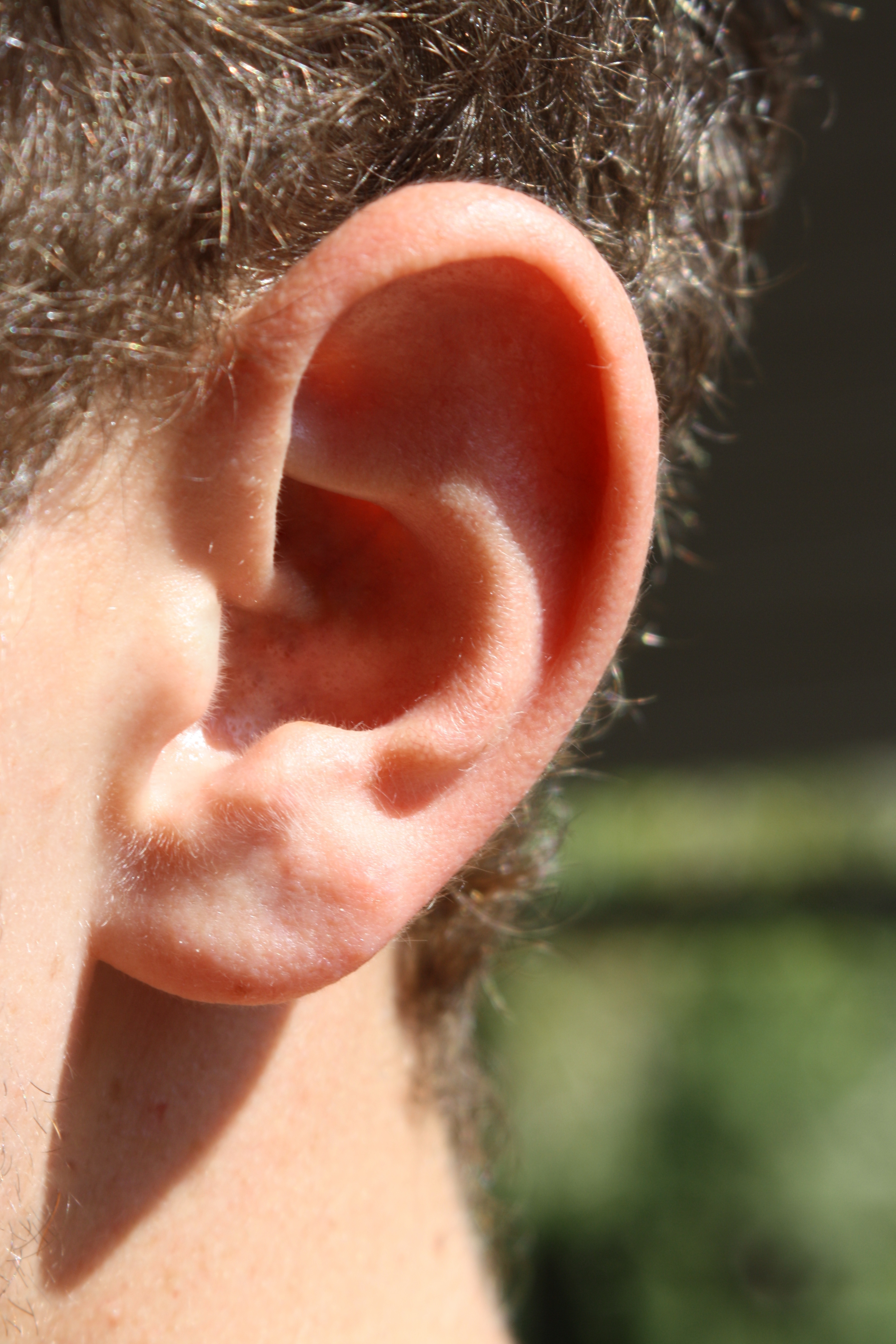
The real human ear photograph below shows external shape and surface relationships. It does not show the complete canal, ossicular chain, or inner-ear sensory compartments. Compare it with the featured opened-anatomy poster and identify which structures the illustration has exposed. The difference is intentional: the photograph records a visible surface, while the poster explains structures hidden beneath it.
For study, describe the view and one visible landmark before naming a concealed structure. This routine prevents a clean drawing from being treated as a view available in every ordinary examination. Retain the source and public-domain credit when sharing the photograph. Its real appearance supports orientation, while the cited anatomical sources support the internal relationships.
11. Use anatomy to support clear professional communication
Medical students can explain the auditory route and one vestibular distinction. Practice assistants and hospital staff can use the regional vocabulary to understand examination descriptions. Physicians can translate a report term into a location and function while distinguishing a population statistic from the person’s actual findings.
In eldercare, use established hearing and communication information when planning conversations and support. A general guide does not prescribe equipment or determine the cause of difficulty. End revision by explaining a mechanical interface, a neural connection, and a vestibular role. These three relationships make the ear’s anatomy more useful than a list of unfamiliar labels.
Sources and image credits
Sources checked on 8 October 2026. Numerical examples, anatomical classification counts, and research estimates are identified separately in the text.
- FIPAT: Terminologia Anatomica, second edition
- NIDCD: how hearing works
- NIDCD: the balance system
- NCBI Bookshelf: middle-ear anatomy and physiology
- NIDCD: hearing statistics and survey definitions
Explanatory prose and simplified SVG graphics are original. Photographs retain their stated Creative Commons licenses; creator and source links appear beside each image. Catalog artwork is educational and decorative; no clinical authorship or peer-review credential is claimed.