1. The eye combines optics with neural tissue
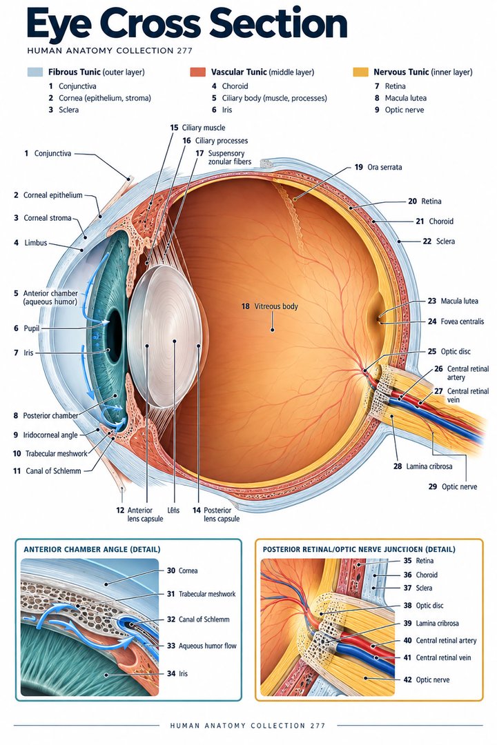
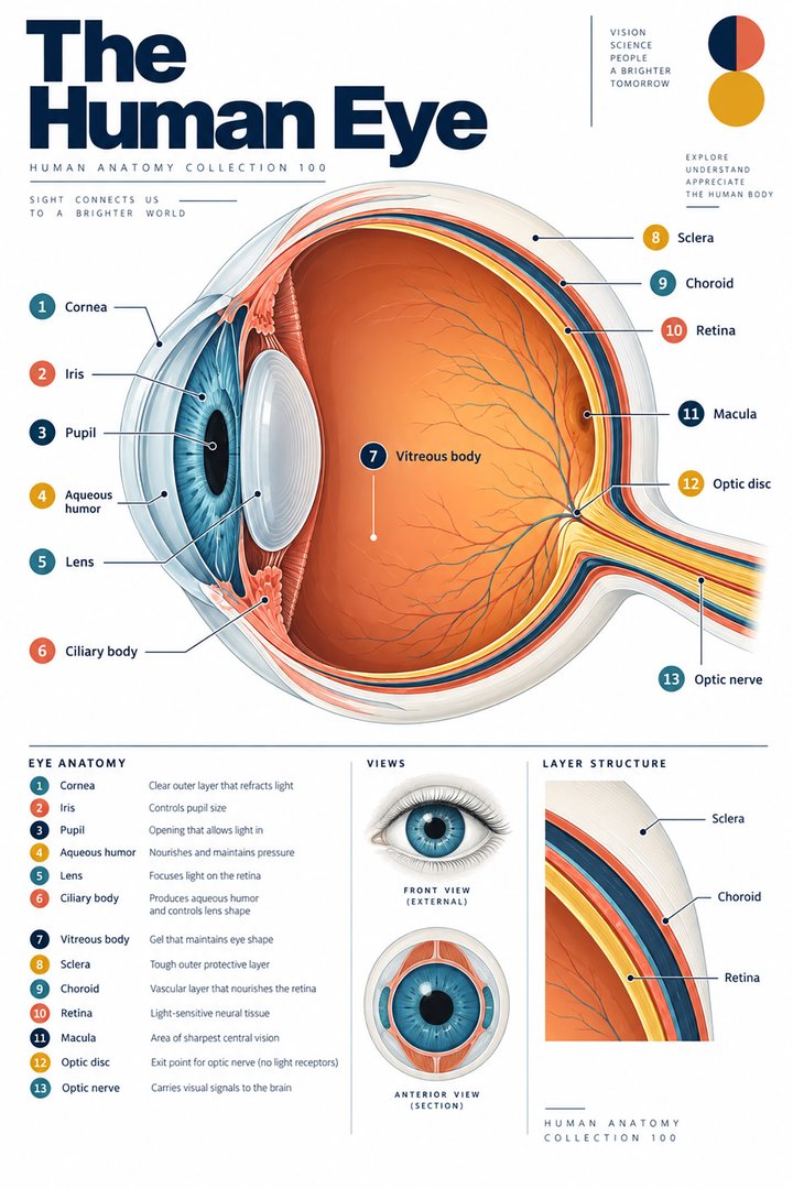
The human eye is an optical organ and a neural interface. Its transparent structures direct light, while retinal cells convert optical information into signals carried through connected visual pathways. A sectional poster is useful because it shows internal structures that cannot be seen in an ordinary external photograph. The two views complement each other rather than providing interchangeable evidence.
For medical students and healthcare teams, begin by separating the route followed by light from the route followed by neural information. Then add the tissues that maintain the optical environment and the structures supporting movement and protection. This sequence creates a coherent explanation of eye anatomy without reducing the organ to a camera with a passive film at the back.
2. Three tunics organize the globe wall
The outer fibrous tunic includes sclera and cornea. The middle vascular arrangement includes choroid, ciliary body, and iris. The inner neural arrangement includes retina. These groupings describe layers and associated regions, not three uniformly thick shells extending identically across the entire globe. A section must preserve transitions at the front of the eye. [Neuroscience: anatomy of the eye]
Use a layered drawing to trace each grouping around the globe. Ask where a structure changes its form or relationship rather than simply coloring three rings. The exercise explains why the cornea and sclera can belong to the same broad tunic while having very different visible properties. It also helps learners relate an external white surface to the deeper organization shown in a section.
3. Cornea, pupil, and lens have different optical roles
The cornea bends incoming light. The iris regulates the pupil’s aperture, while the lens contributes to focusing light toward the retina. The pupil is an opening, not a separate black tissue disk. A photograph can show its apparent outline, but it cannot directly reveal the internal lens or the full light path. [NEI: how eyes work]
For a diagram exercise, follow a ray through the front of the eye and label each boundary it encounters. Then describe what the iris changes without calling the iris a lens. This separates structures that can otherwise be merged by a simple camera analogy. Retain the limits of the analogy when explaining accommodation, optical focusing, or the reason a sectional view reveals structures hidden behind the iris.
4. Aqueous chambers differ from the vitreous chamber
The anterior chamber lies between cornea and iris. The posterior chamber lies behind the iris and in front of the lens and its associated supporting region. Both contain aqueous humor. The larger vitreous chamber lies behind the lens toward the retina and contains vitreous gel. The term posterior chamber must therefore not be used as a synonym for the entire posterior segment. [Webvision: gross anatomy of the eye]
Point to each chamber in a section and name its boundaries before naming its contents. This is a useful correction to diagrams that label all space behind the cornea as one compartment. It also illustrates a broader anatomical lesson: a directional word such as posterior gains meaning from the named compartment and should not be expanded to every structure lying farther back.
5. Aqueous circulation supports an optical environment
Ciliary processes contribute to aqueous-humor production. Aqueous passes from the posterior chamber through the pupil into the anterior chamber and toward drainage pathways. This fluid route should be distinguished from the route of light and from blood flow. Their arrows may cross in a two-dimensional drawing, but they represent different processes. [Neuroscience: anatomy of the eye]
Use separate line styles to trace the optical path and aqueous movement. Name the starting compartment and the junction through which the fluid passes. In clinical education, retain the anatomical route without setting a universal pressure threshold or inferring a diagnosis from a general diagram. A measured pressure requires the method, result, and established clinical interpretation.
6. The retina contains a layered processing network
Retinal photoreceptors include rods and cones. Rods are associated with high sensitivity under dim-light conditions, while cone systems contribute to color-related and detailed visual processing. Humans normally have three cone opsin types with overlapping spectral sensitivities. They should not be pictured as independent red, green, and blue camera pixels that each detect only one pure color. [OpenStax: sensory perception]
A retinal section reveals organization that an external eye photograph cannot show. Compare the photoreceptor layer with the route of information through additional retinal cells. The comparison explains why the retina is neural tissue with local processing rather than a passive screen. A simplified icon may identify a cell type, but it does not reproduce its full connections or physiological response.
7. Macula, fovea, and optic disc are different landmarks
The macular region and fovea provide important retinal landmarks associated with detailed central vision. The optic disc is the site where retinal output converges toward the optic nerve and where photoreceptors are absent. These landmarks should not be combined into one “center of vision” label. Their anatomical roles differ, even when a small poster places them close together. [OpenStax: sensory perception] [Webvision: gross anatomy of the eye]
Use a posterior retinal view alongside a globe section. Identify which landmark is highlighted and where it lies relative to the optical route. Then ask what the other view reveals. For healthcare communication, retain the specific name in an established report; replacing macula, retina, and optic nerve with the same broad term loses information about the structure discussed.
8. Extraocular muscles position the eye
Six principal extraocular muscles move each eye: four recti and two obliques. Their attachments and directions must be understood in three dimensions. A muscle’s name alone does not provide a complete account of its action in every gaze position. Movement depends on coordinated activity, and the whole orbit includes additional structures supporting the eye. [NCBI Bookshelf: extraocular muscles]
Compare a lateral muscle drawing with a superior view of the orbit. Explain how the same muscle changes appearance when the view changes. This reinforces the distinction between an attachment, an anatomical direction, and a resulting movement. A poster can support vocabulary during an examination discussion, while an individual movement finding belongs to the actual clinical assessment.
9. Quantitative anchors should retain their units
A commonly described adult globe has an axial length on the order of twenty-four to twenty-five millimeters. Individual eyes vary, and the number is an anatomical scale estimate rather than a universal clinical target. Three tunics and six extraocular muscles are classification counts. The categories should not be compared as though they measured the same quantity. [Webvision: gross anatomy of the eye] [NCBI Bookshelf: extraocular muscles]
The graphic below compares selected classification counts and deliberately excludes the dimensional estimate. Mixing millimeters with a number of muscles in a single unlabeled chart would be misleading. For student calculations, retain the unit and specify whether the value describes a length, a count, or a measured physiological variable. This habit becomes increasingly useful when moving from anatomy to clinical instrumentation.
10. Latin terms clarify structures that look close together
Bulbus oculi names the globe. Cornea, sclera, iris, retina, and lens are familiar anatomical expressions, while corpus ciliare and corpus vitreum identify the ciliary body and vitreous body. Macula lutea, fovea centralis, and nervus opticus retain important regional distinctions. Learn a term with its boundary and role rather than relying on its place in a colored legend. [FIPAT: Terminologia Anatomica, second edition]
Make a vocabulary card for the anterior and posterior chambers and draw their boundaries. Then add a separate card for the vitreous chamber. This is more effective than learning the word posterior without its compartment. When explaining a report to a patient, use ordinary English while preserving whether the finding concerns the front optical structures, retina, or neural output route.
| English | Selected Latin |
|---|---|
| Eyeball / globe | Bulbus oculi |
| Ciliary body / vitreous body | Corpus ciliare / corpus vitreum |
| Central fovea | Fovea centralis |
| Optic nerve | Nervus opticus |
11. A real eye photograph has a defined scope
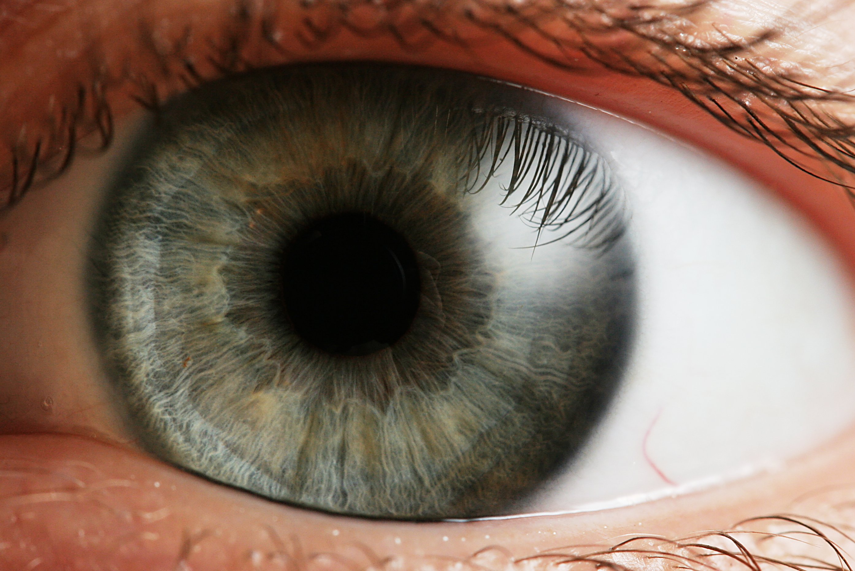
The close-up photograph below shows an actual external eye with iris, pupil, and surrounding tissues. It does not show a retinal section, optic nerve, or internal chamber boundaries. Reflections and lighting influence the appearance of transparent and glossy surfaces. Use the photograph to connect visible external anatomy with the hidden structures on the featured sectional poster.
Medical students can explain what the photograph reveals and what requires another view or method. Practice assistants, hospital staff, physicians, and eldercare teams can use the same distinction when clarifying established observations. End revision by tracing light, naming one fluid compartment, and identifying one neural structure. Those three explanations connect optics, anatomy, and information flow without overstating what an external image proves.
Sources and image credits
Sources checked on 8 October 2026. Numerical examples, anatomical classification counts, and research estimates are identified separately in the text.
- FIPAT: Terminologia Anatomica, second edition
- NEI: how eyes work
- Webvision: gross anatomy of the eye
- Neuroscience: anatomy of the eye
- OpenStax: sensory perception
- NCBI Bookshelf: extraocular muscles
Explanatory prose and simplified SVG graphics are original. Photographs retain their stated Creative Commons licenses; creator and source links appear beside each image. Catalog artwork is educational and decorative; no clinical authorship or peer-review credential is claimed.