1. Learn the system as several connected relationships
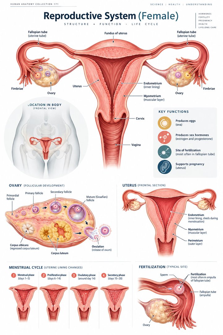
The female reproductive system includes ovarian tissue, uterine tubes, uterus, vagina, and external genital structures, together with hormonal regulation and supporting tissues. A single diagram can establish broad location, but it cannot capture all developmental stages, cyclical changes, and anatomical variations. Begin by separating the site of gamete development, the possible transport route, and the tissues that respond to hormonal change.
This guide uses the conventional organization of the ovarian reproductive system for anatomical teaching. Individual anatomy can differ through development, surgery, and other circumstances. Medical students and healthcare teams should discuss the structures relevant to the person and the established clinical information. Clear terminology supports respectful communication without assuming that an introductory diagram determines identity, symptoms, or reproductive function.
2. Ovarian follicles contain an oocyte and supporting cells
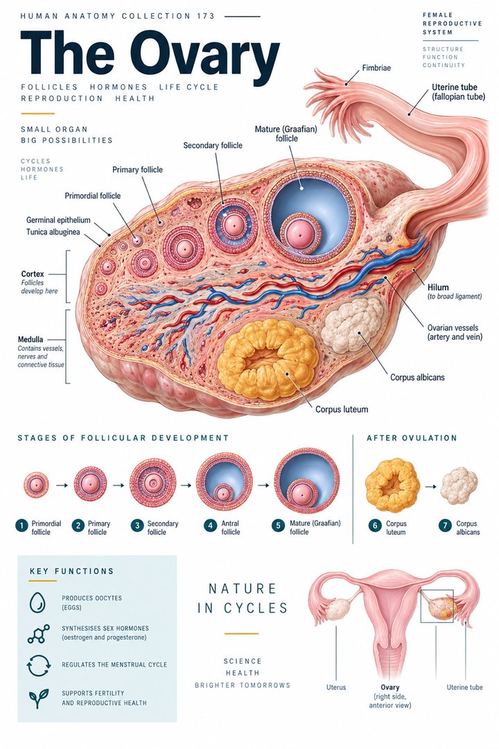
An ovarian follicle includes an oocyte and surrounding supporting cells. It is not simply another name for the oocyte. Follicles change during development, and the corpus luteum is a distinct post-ovulatory structure with endocrine activity. Keeping these entities separate makes a sectional ovarian illustration much easier to read. [OpenStax: ovarian reproductive system]
Cover the labels on a follicle drawing and identify which arrow points to the oocyte and which points to the surrounding follicular structure. Then compare another developmental stage. The exercise prevents the colored ring around a cell from being interpreted as a different gamete. It also illustrates how microscopic organization changes while the whole organ retains a broadly familiar external position.
3. The uterine tube is not sealed directly to the ovary
The uterine tube includes the infundibular region with fimbriae, ampulla, isthmus, and intramural portion. Its relationship to the ovary is close but not a sealed tube-to-organ junction. Fertilization commonly occurs in the ampullary region. The route must therefore be drawn with the ovary–tube relationship explained rather than as a continuous plumbing connection identical to a ureter leaving a kidney. [Human uterine tubes: anatomical and functional review]
Use a caption to name the small gap and the surrounding relationship in a simplified diagram. Ask learners what an arrow from the ovary toward the tube represents, since it does not indicate an enclosed duct running through the ovarian wall. This comparison with another organ system is useful precisely because similar-looking arrows can describe different anatomical arrangements.
4. The uterus has regions and a layered wall
The uterus lies between the bladder and rectum in the typical pelvic arrangement. Named regions include fundus, body, isthmus, and cervix. Its wall includes endometrium, myometrium, and perimetrium. The endometrium has functional and basal relationships that matter when explaining cyclical changes; the myometrium is smooth muscle rather than another mucosal lining. [NCBI Bookshelf: anatomy of the uterus] [NCBI Bookshelf: uterine histology]
Study a sagittal pelvic view and a uterine wall section together. The pelvic view explains neighboring organs; the section explains tissue organization. A wall layer is not a separate pelvic organ, so avoid transferring the large scale of a magnified section to the whole-body picture. State where the illustrator enlarged a detail and which relationship that enlargement is intended to teach.
5. Cervix, vagina, and vulva are different terms
The cervix is the lower uterine region connecting toward the vagina. The vagina is an internal canal, while vulva refers to the external genital structures. Using “vagina” for every external structure removes useful anatomical distinctions. Selected external structures include the labia and clitoris, each with its own organization and relationships. [OpenStax: ovarian reproductive system]
In teaching and clinical communication, clarify the term that best matches the actual site. A patient may use a broad everyday word without intending a precise anatomical distinction. Translate respectfully and document the clarified location. Do not allow a poster’s simplified view to substitute for an examination or the person’s description of what they are experiencing.
6. Ovarian and uterine changes are coordinated
Hormonal signaling links ovarian events with changes in uterine tissue. Follicular development, ovulation, and corpus-luteum activity belong to the ovarian sequence, while endometrial changes belong to the uterine sequence. These processes interact, but their named phases should not be treated as identical labels for the same tissue. [OpenStax: ovarian reproductive system]
Draw two parallel timelines labeled by organ. Add the regulatory relationship between them only after identifying which tissue each line describes. This makes a familiar cycle illustration more precise. It also prevents a “phase” label from being applied indiscriminately to an oocyte, follicle, hormone concentration, and uterine lining without stating the process under discussion.
7. Cycle numbers describe variation rather than a universal clock
NICHD describes an average menstrual cycle of about twenty-eight days, while emphasizing variation among people and between months. Its educational ranges include twenty-one to thirty-five days for adults and twenty-one to forty-five days for teenagers. The first day of bleeding marks the start of a cycle. These are descriptive ranges, not an individualized diagnostic rule. [NICHD: menstruation and cycle variability]
A twenty-eight-day drawing is an illustrative timeline, not evidence that every person ovulates on day fourteen. If teaching from such a graphic, label the assumptions and explain which events vary. A cycle history records the person’s actual observations. The numerical comparison below presents range endpoints from the cited educational source; it does not show a measured frequency distribution or probabilities.
8. Pelvic relationships matter beyond one organ
A reproductive diagram is easier to understand when neighboring structures are included. The uterus’s relationship to bladder and rectum helps orient a sagittal pelvic image. Supporting tissues and the pelvic floor contribute another layer of organization. A drawing that removes the neighbors to emphasize the reproductive tract should state that simplification rather than implying the organ occupies an otherwise empty space. [NCBI Bookshelf: anatomy of the uterus]
For study, first label the neighboring organs, then cover those labels and describe the reproductive structure’s position. This tests spatial understanding rather than color recognition. In professional discussion, anatomical proximity can help explain the language of a report, but it does not establish a symptom’s cause. Retain the actual clinical findings and the purpose of the investigation.
9. Latin names preserve distinctions at several scales
Ovarium, tuba uterina, uterus, cervix uteri, and vagina identify major structures. Fundus uteri and corpus uteri name uterine regions. Endometrium, myometrium, and perimetrium identify wall layers. Vulva and clitoris are also used in anatomical terminology. Learning terms at the organ, region, and layer levels prevents a familiar word from being used for everything within the same colored outline. [FIPAT: Terminologia Anatomica, second edition]
Build a vocabulary table that groups terms by scale. Add a one-sentence explanation for each group, then compare the groups on the featured poster. During patient-facing communication, translate the term while keeping the structural distinction. “Uterine lining” can be helpful ordinary language for endometrium; it should not become a synonym for the entire uterus or the muscular wall.
| English | Selected Latin |
|---|---|
| Ovary / uterine tube | Ovarium / tuba uterina |
| Uterine body / cervix | Corpus uteri / cervix uteri |
| Uterine wall layers | Endometrium / myometrium / perimetrium |
| External genital region | Vulva |
10. A museum photograph reveals a preserved representation
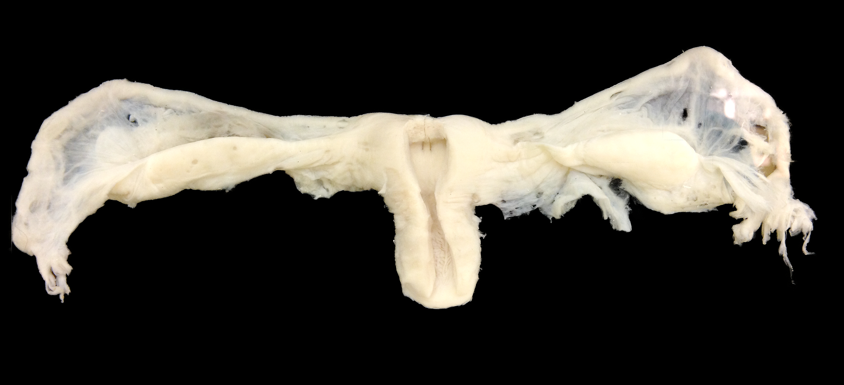
The real photograph below records a reproductive-anatomy display in the Anatomical Museum in Basel, including the ovarian and uterine region. It is a preserved museum representation, not a live surgical photograph or a view of normal tissue colors in a living person. Its preparation and viewpoint differ from the clean opened-organ drawings used in the catalog.
Compare the overall connections rather than assigning every visible detail a confident label. Ask what the museum preparation exposes and what the simplified poster removes. Retaining the caption’s context is essential when reusing the photograph: an image can be real while its subject is prepared, preserved, or arranged for teaching. That distinction improves both scientific accuracy and professional communication.
11. A useful study sequence for healthcare teams
Medical students can explain one ovarian structure, one tube relationship, and one uterine wall layer in sequence. Practice assistants and hospital staff can use the map to clarify documented sites and terminology. Physicians can use it when discussing established findings while distinguishing anatomy from cycle timing or reproductive function.
In eldercare, reproductive anatomy may remain relevant to a person’s documented history, pelvic concerns, prior procedures, and communication needs. Use current clinical information rather than assuming that a generic age category explains the individual’s anatomy. End a lesson by translating one report term into ordinary English and locating it on two different views. This turns vocabulary into a shared spatial explanation.
Sources and image credits
Sources checked on 8 October 2026. Numerical examples, anatomical classification counts, and research estimates are identified separately in the text.
- FIPAT: Terminologia Anatomica, second edition
- OpenStax: ovarian reproductive system
- NICHD: menstruation and cycle variability
- Human uterine tubes: anatomical and functional review
- NCBI Bookshelf: anatomy of the uterus
- NCBI Bookshelf: uterine histology
Explanatory prose and simplified SVG graphics are original. Photographs retain their stated Creative Commons licenses; creator and source links appear beside each image. Catalog artwork is educational and decorative; no clinical authorship or peer-review credential is claimed.