1. Separate production, transport, and hormone signaling
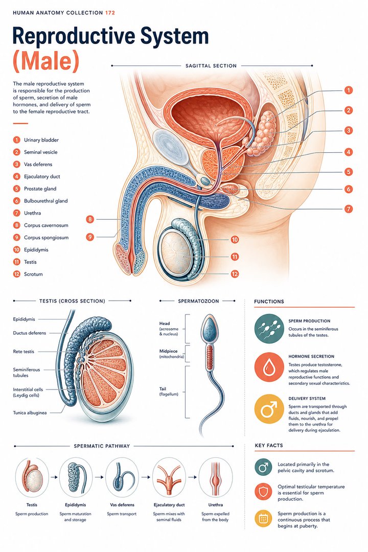
The male reproductive system combines gamete production, transport through a duct network, contributions from accessory glands, and endocrine signaling. A whole-system poster is useful because it displays these relationships together. It can also become misleading if every connected structure is assumed to be part of one continuous sperm-filled tube. Start by distinguishing the route followed by sperm from the places where additional secretions join that route.
This article uses the conventional anatomical organization of the testicular reproductive system. Individual anatomy, development, surgery, and other circumstances can differ from the introductory map. For medical students and healthcare staff, precise structure names are more useful than assumptions based on identity or outward appearance. Explain the documented anatomy relevant to the person and the purpose of the discussion.
2. Testes combine seminiferous tissue with endocrine cells
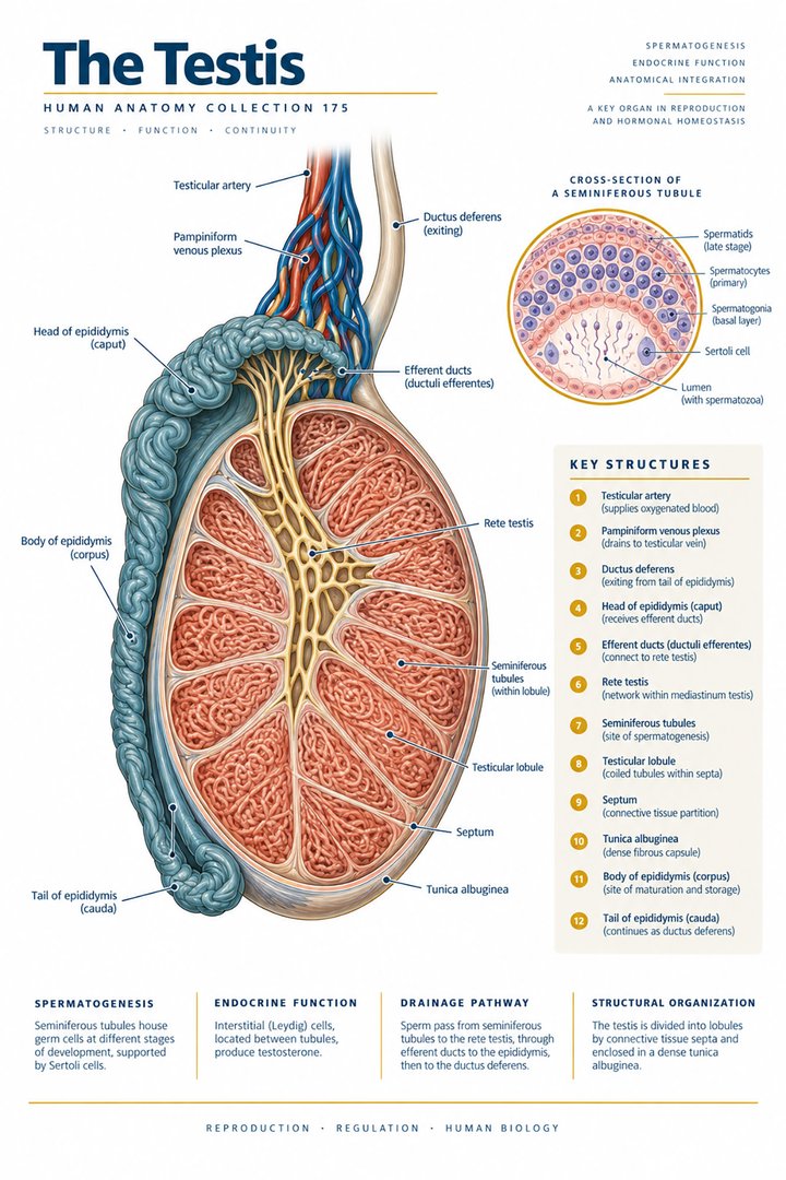
The testes contain seminiferous tubules where germ cells develop. Sertoli cells support the germ-cell environment, while Leydig cells in the interstitial tissue produce testosterone. A capsule and internal partitions organize the tissue. The scrotal arrangement supports a temperature environment different from core body temperature, but a poster cannot establish a person’s physiological or reproductive status. [NCI SEER: testes]
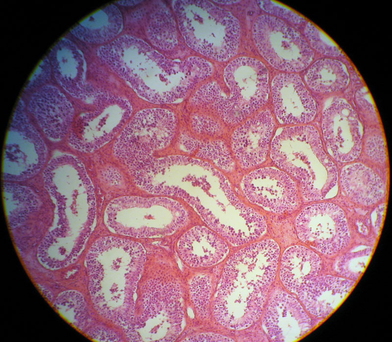
Compare a testicular section with a microscopic field. The section locates tubules and supporting tissue at a larger scale; the micrograph samples only part of that organization. Name the scale and preparation before interpreting the image. This prevents a cluster of tubule profiles from being mistaken for an entire duct route or a direct measure of sperm production.
3. Spermatogenesis includes meiosis and differentiation
The production of sperm involves germ-cell proliferation, meiotic division, and later differentiation. A primary spermatocyte normally undergoes two meiotic divisions that produce four spermatids, which then differentiate further. These stages should not be compressed into “one cell instantly becomes four sperm.” The nuclear chromosome complement and the morphology change through a sequence with distinct biological steps. [OpenStax: testicular reproductive system]
For revision, draw stage boxes with a label for what the box represents: a cell type, a division, or a differentiation process. A division arrow and a maturation arrow should not be treated as equivalent. This helps learners connect the chromosome-count graphic with the tissue rather than treating genetics and histology as separate topics that happen to share the same organ.
4. Trace the sperm route before adding glands
The seminiferous route connects through collecting channels and the rete testis toward efferent ductules and the epididymis. The epididymis supports maturation and storage. From there, the ductus deferens connects toward an ejaculatory duct and the urethral route. The ducts’ arrangement is a three-dimensional pathway, even when shown as a simple line on a poster. [NCI SEER: testes] [NCI SEER: male reproductive ducts]
Read the pathway aloud while following it with a finger. Then begin from the middle and identify both the preceding and following structure. This tests connectivity rather than memorization of one sentence. It also reveals where the illustration has shortened a long coiled duct to keep the whole system readable. Use the caption to clarify omitted branches or sections.
5. Accessory glands contribute secretions to semen
Seminal vesicles, the prostate, and bulbourethral glands contribute secretions associated with the reproductive route. Semen contains sperm together with fluid contributions; the words semen and sperm are therefore not interchangeable. The seminal vesicle’s duct joins the route, but a diagram should not suggest that sperm travel through the body of the gland as an obligatory intermediate stage. [OpenStax: testicular reproductive system]
Use two arrow colors when adding glands to the transport map: one for the sperm route and one for a gland’s contribution. Label the streams before they combine. This is an efficient way to correct a visually plausible but inaccurate pathway. In clinical communication, retain the precise term used for a specimen or analysis rather than replacing it with a less specific everyday synonym.
6. The penis contains organized erectile tissues
The penis contains paired corpora cavernosa and a corpus spongiosum associated with the urethral route. Their arrangement should be distinguished in a cross-section as well as a whole-organ picture. Erectile tissues, vessels, nerves, and the urethra have different structural roles, even when an introductory illustration uses one continuous outline for the organ. [OpenStax: testicular reproductive system]
For a teaching exercise, place a longitudinal view beside a cross-section and locate the same named tissue in both. Describe the axis of the section and which end of the organ is being viewed. This routine transfers the orientation principles used in other body regions to reproductive anatomy. Keep the explanation clinical and respectful, using the anatomical terms that best clarify the question.
7. Hormonal regulation links testes with the brain
Hypothalamic gonadotropin-releasing hormone supports pituitary gonadotropin signaling. Luteinizing hormone acts on Leydig cells, while follicle-stimulating hormone acts through Sertoli-cell-related processes. Testosterone participates in feedback. This regulatory arrangement connects tissue organization with endocrine physiology rather than leaving the testes as isolated production organs. [OpenStax: testicular reproductive system]
Map the source, responding cell population, and feedback separately. Do not make one arrow represent every action of a hormone. A whole-system teaching poster establishes the broad relationship, while laboratory interpretation requires the actual measurements, timing, and clinical context. The anatomy supports the vocabulary of the discussion without determining an individual’s hormone status or fertility from the picture alone.
8. Quantitative anchors describe structure and cell complements
The SEER teaching module describes a typical testis as roughly five centimeters long and three centimeters in diameter, with approximately two hundred and fifty lobules. These are broad descriptive estimates, not diagnostic thresholds. Coiling permits substantial tubular length within a small organ; a compact drawing should not be interpreted as a literal measure of every tube. [NCI SEER: testes]
Typical diploid human cell nuclei have forty-six chromosomes, while gametes have twenty-three. The comparison below describes chromosome complements rather than sperm concentration, fertility, or the amount of DNA at every developmental stage. Keeping the counted unit explicit prevents a useful genetic fact from becoming a claim about a person’s reproductive capacity. [NHGRI: diploid chromosomes]
9. Latin terms make adjacent structures easier to distinguish
Testis is singular and testes plural. Tubuli seminiferi names the seminiferous tubules; epididymis and ductus deferens identify later portions of the route. Funiculus spermaticus refers to the spermatic cord, which contains more than the duct alone. Corpora cavernosa and corpus spongiosum preserve the erectile-tissue distinction. Learn each expression with its relationship rather than merely its translation. [FIPAT: Terminologia Anatomica, second edition]
A practical revision card can pair a term with the preceding and following structure. For a cord or tissue group, add what the group contains or relates to. This prevents similar labels from collapsing into one generic “tube.” When explaining a report to a patient, translate the term into ordinary language while retaining enough detail to identify the actual structure discussed.
| English | Selected Latin |
|---|---|
| Testis / testes | Testis / testes |
| Seminiferous tubules | Tubuli seminiferi |
| Deferent duct | Ductus deferens |
| Spermatic cord | Funiculus spermaticus |
10. Real histology complements the transport map
The real testicular micrograph below shows a stained tissue preparation. Its multiple tubular profiles can be compared with a simplified organ section, but the image does not trace the entire sperm route. Tissue color comes from preparation and staining. No individual reproductive diagnosis is inferred from this educational image, and its source attribution remains beside the photograph.
Medical students can use the image to connect microscopic organization with the organ’s production role. Practice assistants, hospital staff, physicians, and eldercare teams can use the broader map to clarify established information and specimen terminology. End revision by explaining the sperm route, a gland’s separate contribution, and one hormonal feedback connection. Together, those explanations test structure, transport, and regulation.
Sources and image credits
Sources checked on 8 October 2026. Numerical examples, anatomical classification counts, and research estimates are identified separately in the text.
- FIPAT: Terminologia Anatomica, second edition
- NHGRI: diploid chromosomes
- NCI SEER: testes
- NCI SEER: male reproductive ducts
- OpenStax: testicular reproductive system
Explanatory prose and simplified SVG graphics are original. Photographs retain their stated Creative Commons licenses; creator and source links appear beside each image. Catalog artwork is educational and decorative; no clinical authorship or peer-review credential is claimed.