1. Lymphatic anatomy connects fluid return with defense
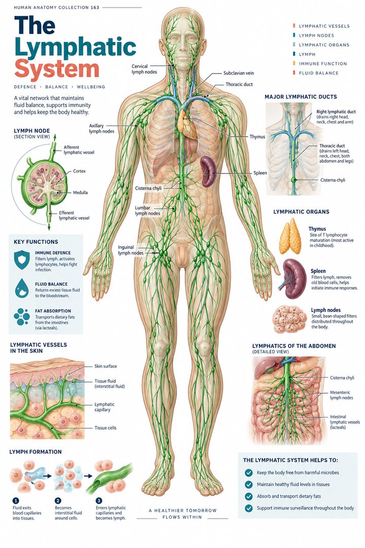
The human lymphatic system is easiest to understand through two connected roles: returning tissue fluid toward the circulation and supporting immune surveillance. Its vessels, nodes, and associated lymphoid organs form a distributed arrangement across the body. Unlike the blood circulation, this arrangement has no single central pump equivalent to the heart. A diagram should show both the direction of transport and the tissues participating along the route.
For healthcare learners, distinguish a lymphatic vessel from a lymph node, then distinguish lymph from blood and interstitial fluid. These terms describe different structures and compartments. They become particularly useful when reading a regional drainage map, discussing a node described in a report, or explaining why a whole-body illustration and a microscopic immune-cell photograph answer different questions.
2. Interstitial fluid enters an organized return route
Lymphatic capillaries take up fluid from the interstitial compartment. Collecting vessels, trunks, and ducts then provide a return route to venous circulation. The composition and movement of this fluid relate to the surrounding tissue and vascular exchange. The revised Starling framework gives a more nuanced account than the old universal illustration in which nearly all filtered fluid simply reenters the venous end of every capillary. [Levick and Michel: revised Starling principle]
In a learning diagram, keep the blood-vessel lumen, interstitium, and lymphatic lumen separate. An arrow between compartments should name what crosses and where it enters. Avoid placing a lymphatic channel directly inside a blood capillary unless the picture clearly concerns a connection at a different anatomical level. A few explicit boundaries make the fluid relationship easier to understand than a large number of unexplained arrows.
3. Lymph transport uses several mechanical influences
Valves in collecting vessels help limit backward flow. Intrinsic vessel contractions, surrounding skeletal movement, and respiratory pressure changes contribute to transport. These influences work within a living network whose flow varies with tissue conditions. The absence of a central heart-like pump does not mean that lymph simply sits motionless until a person deliberately moves. [NCI SEER: lymphatic vessels and drainage]
For study, compare a collecting lymphatic with a vein without treating them as identical. Both may be illustrated with valves, but they belong to different transport arrangements. Explain the direction before describing a supporting mechanism. For healthcare communication, a general account of movement should not be turned into individualized exercise or massage advice; those decisions depend on the person’s condition and the established clinical plan.
4. Major ducts organize broad drainage territories
The thoracic duct generally drains both lower limbs, the abdomen, and the left upper portion of the body. The right lymphatic duct generally serves the right upper quadrant. These are broad introductory territories, and variations in the terminal ducts and their connections occur. A colored drainage map should therefore present a usual pattern, not a promise about every vessel in every individual. [NCI SEER: lymphatic vessels and drainage]
When reviewing a diagram, name the territory before the duct. Then explain why a right-sided structure in the lower body does not automatically belong to the right lymphatic duct. This corrects a common inference based only on the word “right.” The duct’s name does not describe the entire right half of the body, so the regional map is essential.
5. A node is an organized tissue structure
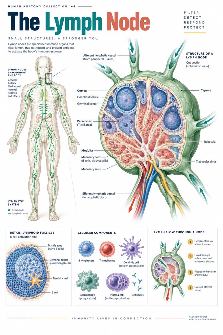
Lymph nodes are positioned along lymphatic routes and contain organized immune-cell populations. Afferent vessels bring lymph toward a node, while efferent vessels carry it away. A sectional drawing can reveal a capsule, cortex, and medullary organization that a whole-body chart cannot show. The node is not merely a swelling on a tube; its tissue organization supports interaction between transported material and immune cells. [NCI SEER: lymph-node organization]
Use a regional image and a node section together. First locate the group in the body, then explain what the enlarged sectional representation adds. State that the section is drawn at a different scale. This prevents a common visual error in which a magnified node is interpreted as being as large as an adjacent organ in the whole-body illustration.
6. Lymphoid organs are related but not interchangeable
Bone marrow and thymus are primary lymphoid organs associated with lymphocyte development. Nodes, spleen, and mucosal lymphoid tissues are secondary sites. The spleen interacts with blood rather than serving as a lymph-filtering station on the same route as a node. This distinction is important when a poster groups all lymphoid organs under one heading. [OpenStax: lymphatic and immune anatomy] [NCI SEER: spleen]
Create a table with a tissue or organ in the first column and the material or process it is associated with in the second. The table makes it harder to substitute “filters lymph” for every lymphoid organ. It also helps learners see that the lymphatic and immune systems overlap substantially without being identical concepts: one emphasizes transport anatomy, while the other emphasizes coordinated defensive and regulatory processes.
7. Intestinal lacteals connect absorption with transport
Lacteals are lymphatic channels within intestinal villi that participate in transporting absorbed dietary lipids. This is another example of an anatomical network supporting more than one broad function. The route links digestive processing with lymphatic transport rather than sending every absorbed substance directly into the same blood-vessel pathway. [OpenStax: lymphatic and immune anatomy]
Draw a villus with a separate blood-capillary network and lacteal. Label the two routes without assigning every nutrient to both indiscriminately. This small diagram connects digestive and lymphatic learning and explains why a whole-body drainage chart does not capture all of the network’s functions. It also makes a useful discussion point for medical students reviewing how microscopic tissue organization supports organ physiology.
8. Brain drainage requires careful compartment language
Research has established lymphatic vessels in the meninges. This should not be simplified into a claim that ordinary lymphatic vessels are distributed throughout the brain’s neural parenchyma in the same pattern as a limb. Meningeal lymphatics, cerebrospinal-fluid relationships, and glymphatic transport are related areas of study with distinct anatomical meanings. [NIH: meningeal lymphatic drainage research]
If a diagram labels a vessel near the brain, ask whether it lies in neural tissue, a meningeal compartment, or an extracranial drainage route. Retaining that location prevents an overgeneralized conclusion. Research findings in this area should also keep their experimental context. An anatomical learning guide can explain the compartments without presenting a research observation as a treatment or a complete explanation of a person’s neurological disease.
9. Numbers should identify the limits of the map
A commonly cited textbook estimate is approximately five hundred to six hundred lymph nodes in the body. It is an approximate anatomical estimate, not an exact number to apply to every person or count from a poster. The source’s range reflects the distributed system better than an illustration containing only a few prominently drawn node groups. [OpenStax: lymphatic and immune anatomy]
The count graphic below illustrates the reported range’s endpoints. It is not a distribution of measurements, does not show probabilities, and does not indicate how many nodes should be visible in a particular examination. Keep these distinctions beside any numerical display. A map that shows fewer nodes may simply have selected representative groups to preserve readability.
10. Latin vocabulary connects vessels, ducts, and organs
Lympha names lymph, vasa lymphatica the vessels, and nodi lymphoidei the lymph nodes. Ductus thoracicus and ductus lymphaticus dexter identify the major named ducts. Lien is a selected Latin term for the spleen, and thymus is used directly. Pair each expression with a location and a role so that similar-looking labels do not conceal different structures. [FIPAT: Terminologia Anatomica, second edition]
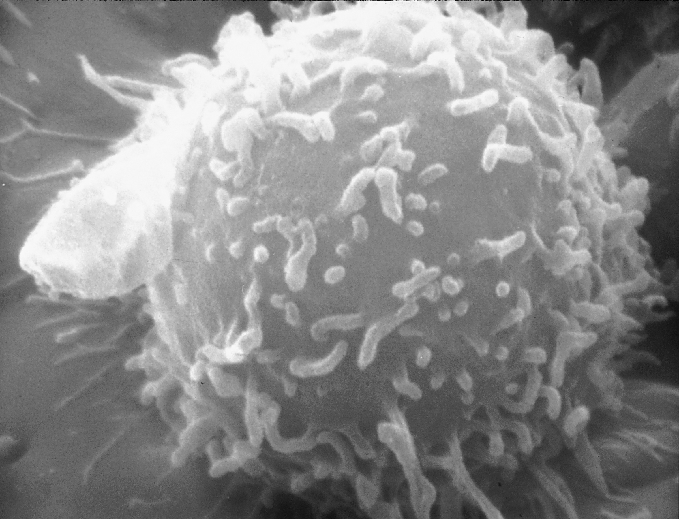
The real lymphocyte electron micrograph below adds a cellular view to the transport map. It does not identify the entire lymphatic network or establish whether the cell is a B or T lymphocyte. Use the image to introduce the difference between a cell and the tissue organization of a node. The image’s creator and public-domain source remain linked in the caption.
| English | Selected Latin |
|---|---|
| Lymph / lymphatic vessels | Lympha / vasa lymphatica |
| Lymph nodes | Nodi lymphoidei |
| Thoracic duct | Ductus thoracicus |
| Spleen | Lien |
11. Use drainage diagrams to support clear discussion
Medical students can trace a regional return route and then compare a node’s microscopic organization. Practice assistants, hospital staff, and physicians can use the same framework to clarify which structure a report names. Record a described location and established finding without inferring the cause of a node’s appearance from a general poster.
For eldercare teaching, explain the anatomy in relation to the person’s assessed condition and agreed care plan. A drainage illustration helps communicate a location and a route, but it does not establish treatment. Finish with three distinctions: lymph versus blood, node versus spleen, and usual drainage territory versus individual anatomy. These distinctions make the map useful across professional settings.
Sources and image credits
Sources checked on 8 October 2026. Numerical examples, anatomical classification counts, and research estimates are identified separately in the text.
- FIPAT: Terminologia Anatomica, second edition
- OpenStax: lymphatic and immune anatomy
- NCI SEER: lymphatic vessels and drainage
- NCI SEER: lymph-node organization
- NCI SEER: spleen
- Levick and Michel: revised Starling principle
- NIH: meningeal lymphatic drainage research
Explanatory prose and simplified SVG graphics are original. Photographs retain their stated Creative Commons licenses; creator and source links appear beside each image. Catalog artwork is educational and decorative; no clinical authorship or peer-review credential is claimed.