1. The urinary system manages an internal environment
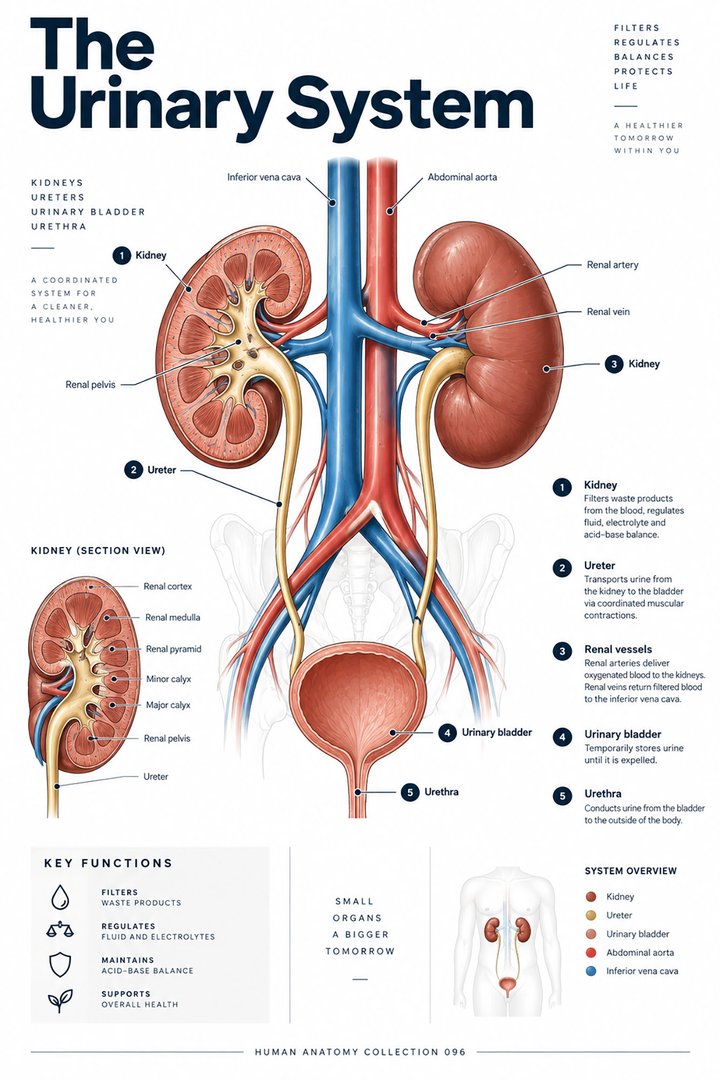
The human urinary system is often represented as two kidneys connected to a bladder, but its physiological role extends well beyond collecting waste. Kidney activity helps maintain water, electrolyte, and acid–base relationships while producing urine. The transport structures then move, store, and release that urine. Learning the system becomes easier when the renal processing route is distinguished from the downstream plumbing.
For healthcare learners, connect each structure with the question it can answer. A kidney section shows internal organization; a bladder image shows a reservoir; a urine measurement records an output under particular conditions. None provides all of the information needed to describe the entire system. This guide follows the anatomy from organ location to microscopic processing and then to the route out of the body.
2. Locate the kidneys before exploring their interior
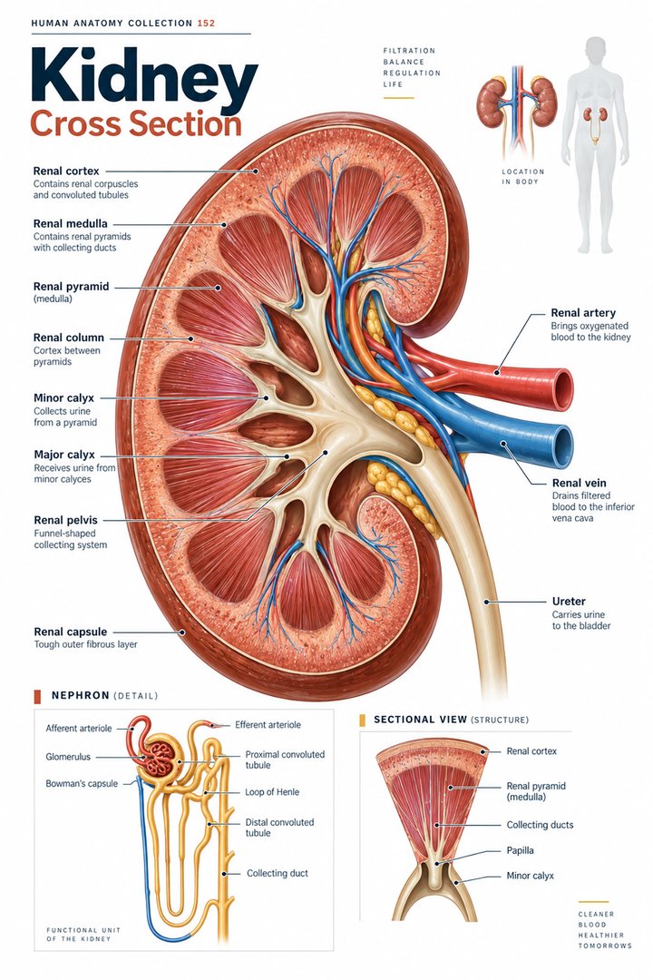
The kidneys are retroperitoneal organs on the posterior abdominal wall. Their medial hila provide routes for vessels and the urine-collecting outlet. A section distinguishes cortex, medulla, and a collecting arrangement that leads toward the renal pelvis. A whole-body poster can establish broad position but cannot show every relationship within these compartments. [OpenStax: gross kidney anatomy]
Compare an intact kidney with an opened section. Identify which surface has been removed and which internal areas become visible. Then trace a vessel separately from the urine outlet. This comparison prevents an oversimplified drawing from suggesting that blood and urine pass through the same central tube. It also helps learners understand why external shape alone says little about the functional organization inside the organ.
3. The nephron links filtration with tubular processing
A renal corpuscle combines the glomerulus with its surrounding capsule. The tubular route includes the proximal convoluted tubule, nephron loop, and distal convoluted tubule. Collecting ducts receive output from multiple nephrons and are technically distinct from the nephron itself in the usual structural definition. A simplified diagram should state where it changes from one nephron’s components to the shared collecting system. [OpenStax: microscopic kidney anatomy]
For revision, use two colors: one for the vascular route and another for the tubular fluid route. The color is a teaching convention, so add a legend. Follow both routes around the renal corpuscle before moving onward. This makes the relationship clearer than memorizing a list of segments without explaining how the blood-facing and fluid-facing compartments interact.
4. Filtration, reabsorption, and secretion describe directions
Filtration moves material from the glomerular circulation into the initial tubular compartment. Reabsorption returns substances from tubular fluid toward the circulation, while secretion moves substances into the tubular route. The final urine is therefore the result of multiple processes, rather than unchanged fluid that passed through a single passive filter. [OpenStax: microscopic kidney anatomy]
An effective study exercise adds arrows across a tubule wall and names the direction of each process. Include the starting and ending compartment. A learner who writes only “transport” has not yet specified what happened. The exercise also clarifies why the amount entering an early compartment is not identical to the amount finally excreted. Keep the same distinction when interpreting a numerical example of renal processing.
5. Kidney function extends to hormonal relationships
The kidneys help regulate water and mineral balance and remove waste. Their endocrine relationships include effects on blood pressure, red-cell production, and bone-related mineral physiology. This connects urinary anatomy with circulatory, endocrine, and skeletal learning. The kidney is therefore more than an excretory container attached to a ureter. [NIDDK: how the kidneys work]
Build an organ-system map with the kidney at the center and several labeled connections. Explain one connection without trying to compress every renal function into a slogan. For healthcare communication, distinguish an anatomical finding from a physiological measurement. The name of a renal structure on an image and a laboratory indicator of function may inform each other, but they are not interchangeable observations.
6. Ureters transport urine toward the bladder
Ureters carry urine from renal pelves to the urinary bladder. Their walls support peristaltic movement, so transport is not merely gravity-driven drainage through an inert tube. Their oblique passage through the bladder wall contributes to limiting reflux. The ureter should not be confused with the urethra, which carries urine from the bladder toward the outside. [OpenStax: urine transport]
Say the two names in a route sentence rather than learning them as unrelated definitions. “Renal pelvis to bladder” identifies a ureter; “bladder to outside” identifies the urethra. Then point to both on a poster. This small exercise is particularly useful in team teaching because a near-identical word can otherwise produce a description of the wrong anatomical connection.
7. The bladder combines a lining with a muscular wall
The bladder stores urine and changes its volume. Urothelium lines its internal surface, while the detrusor contributes the muscular wall involved in emptying. Its outlet and control mechanisms must be considered within the wider neural and pelvic arrangement. A bladder diagram that shows only an empty sac omits important tissue and coordination. [OpenStax: urine transport]
Compare the storage concept with a wall section. Ask what must change as a reservoir fills and how tissue organization supports that change. Keep the explanation anatomical rather than setting a universal capacity for every person. Patient history, neurological status, prior procedures, medications, and actual measurements all influence clinical interpretation; a standard poster cannot substitute for those details.
8. Quantitative facts require units and context
NIDDK describes approximately one million nephrons in each kidney. This is a useful scale estimate rather than an exact count in every individual. It also describes usual adult urine production on the order of one to two US quarts daily, approximately 0.95 to 1.9 liters. Output varies with intake, losses, and physiological circumstances; the range is not an individualized care target. [NIDDK: how the kidneys work]
The length comparison below uses common adult teaching approximations of around four centimeters for the female urethra and twenty centimeters for the male urethra. Individual anatomy varies, and procedures must use the person’s actual anatomy and clinical protocol. The values explain a broad structural distinction, not a universal measurement or a prediction of the right equipment for a particular patient. [OpenStax: urine transport]
9. Latin names preserve the route’s distinctions
Ren is the singular term for kidney and renes the plural. Cortex renalis, medulla renalis, and pelvis renalis identify internal regions. Vesica urinaria names the urinary bladder. Ureter and urethra remain distinct named routes. Corpusculum renale refers to the renal corpuscle. Learn the expression with the relevant compartment rather than translating only the word “renal.” [FIPAT: Terminologia Anatomica, second edition]
A practical vocabulary exercise begins with a whole-body route and then adds one microscopic structure. This moves between scales without losing the organ context. For patient-facing explanations, use “kidney,” “tube to the bladder,” and “outlet from the bladder” as needed, then connect those phrases to the technical terms appearing in the report. The objective is shared understanding, not a display of terminology.
| English | Selected Latin |
|---|---|
| Kidney / kidneys | Ren / renes |
| Renal cortex / medulla | Cortex renalis / medulla renalis |
| Renal pelvis | Pelvis renalis |
| Urinary bladder | Vesica urinaria |
10. Real renal tissue adds a microscopic perspective
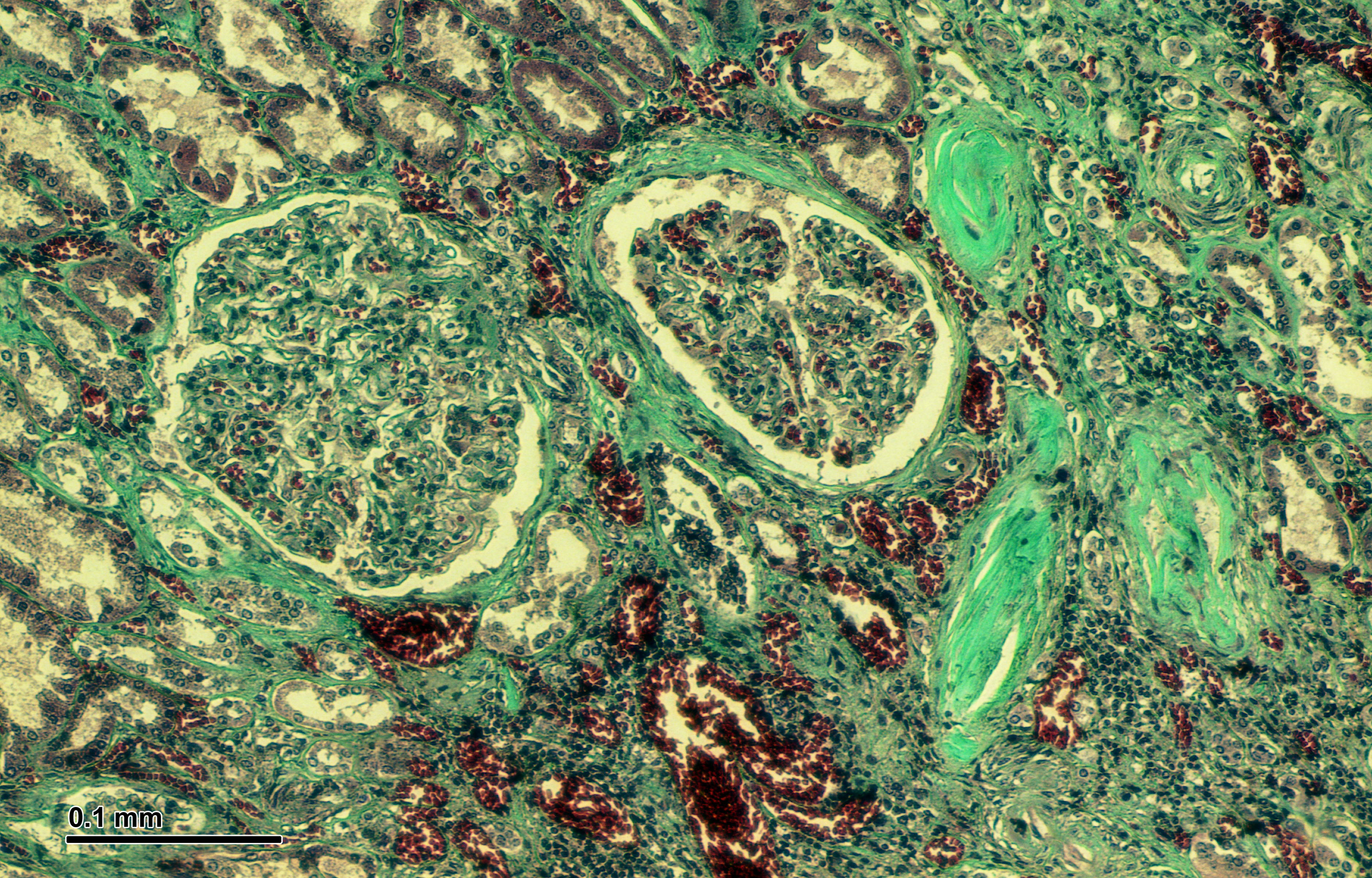
The real human kidney micrograph below shows tissue under bright-field microscopy. Display resizing changes its apparent scale, so a fixed optical magnification is not claimed here. Tubular profiles and renal tissue organization can be compared with the simplified nephron drawing, but a single preparation does not reproduce an entire nephron as a continuous colored line.
Medical students can use this difference to separate structural evidence from schematic explanation. Practice assistants, hospital staff, physicians, and eldercare teams can use the route map to understand established findings and documentation. End revision by explaining what happens before urine enters a ureter, where it is stored, and how it leaves the bladder. Those distinctions make the system’s anatomy usable in everyday professional communication.
Sources and image credits
Sources checked on 8 October 2026. Numerical examples, anatomical classification counts, and research estimates are identified separately in the text.
- FIPAT: Terminologia Anatomica, second edition
- NIDDK: how the kidneys work
- OpenStax: urine transport
- OpenStax: gross kidney anatomy
- OpenStax: microscopic kidney anatomy
Explanatory prose and simplified SVG graphics are original. Photographs retain their stated Creative Commons licenses; creator and source links appear beside each image. Catalog artwork is educational and decorative; no clinical authorship or peer-review credential is claimed.