Two complementary questions about one body
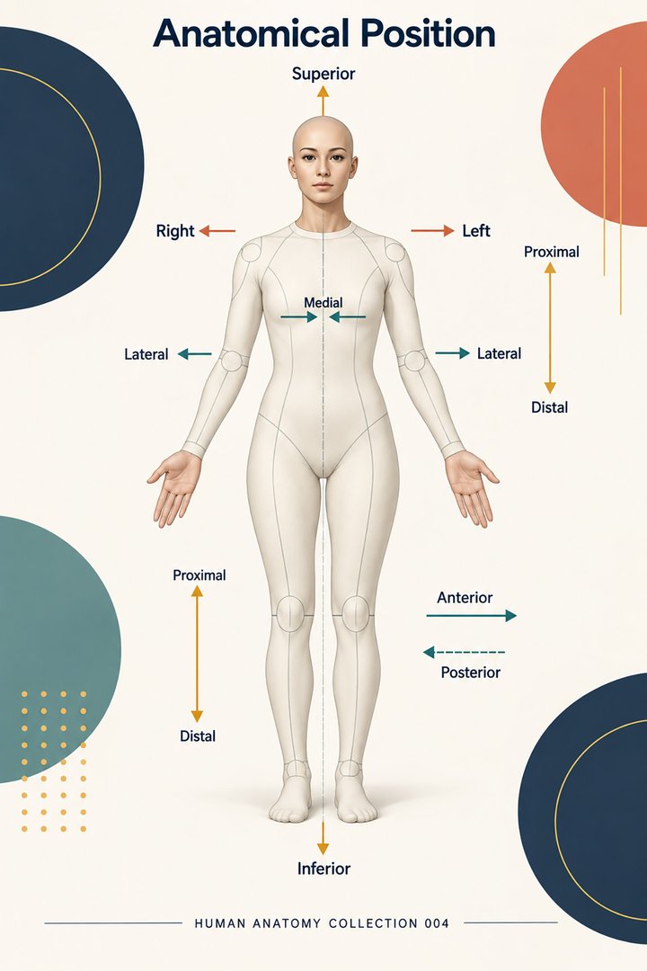
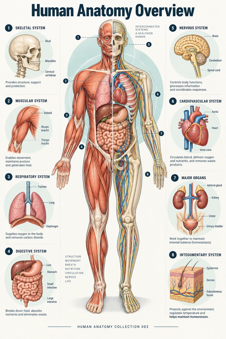
Anatomy asks how the body is organized; physiology asks how its organized parts work. Neither subject becomes fully understandable in isolation. Naming a structure without explaining its role produces a static inventory. Describing a process without identifying the structures involved produces an abstract sequence with nowhere to happen. Students and healthcare teams need both kinds of knowledge when moving between an atlas, a real image, a physical observation, and an explanation to another person. OpenStax: Overview of anatomy and physiology
Begin with two questions: “What is here?” and “What changes here?” The first might identify a chamber, membrane, muscle, or tissue layer. The second might identify movement, exchange, signaling, or regulation. Add a third question: “How would I know?” This introduces the method used to observe structure or function. A sectioned specimen, a moving image, and a recorded pressure trace may all concern the same organ, yet each answers a different part of the problem. Combining the answers is more powerful than asking one method to prove everything.
A scale ladder helps connect disciplines
The body can be studied at molecular, cellular, tissue, organ, system, and organism levels. These levels organize questions; they do not imply that the body is assembled from independent boxes. A mechanism at one level may require an explanation from another. OpenStax: Structural organization
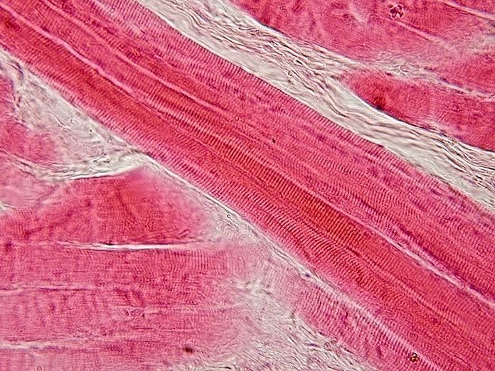
Consider an educational discussion of a contracting muscle. A photograph shows gross shape and attachment. A microscope shows tissue arrangement. A molecular explanation concerns interactions too small for that photograph to display. A movement assessment concerns the result at the level of a joint and the whole person. If the discussion stalls, identify the scale of the unanswered question. “Where does it attach?” and “How does it generate force?” are related but require different evidence. This simple distinction prevents a student from trying to answer a molecular question by staring harder at a gross anatomical illustration.
Latin terms can anchor that ladder: cellula for cell, textus for tissue, organum for organ, and named regional structures for larger arrangements. The value of terminology is that it lets several observers refer to the same object precisely. Terms should support explanations rather than substitute for them. A learner who can pronounce a long name but cannot identify the relevant layer or pathway needs another kind of practice, not merely another vocabulary list.
Shape creates opportunities and constraints
Every anatomical arrangement offers possibilities and imposes limits. A broad exchange surface permits more contact than a small one. A thick wall can support different mechanical demands from a very thin wall. An opening allows passage but also defines what can pass and where. These general relationships help learners construct questions before examining detailed organ physiology. They should not be mistaken for a claim that shape alone determines every outcome; tissue properties, regulation, timing, and surrounding conditions also matter.
An original teaching experiment uses two paper models: a flat sheet and the same sheet folded into a compact space. Neither model is living tissue, but they show how a large surface can occupy a small volume. The next step is to ask what the model leaves out: blood supply, cell behavior, selective transport, structural strength, and adaptation. A useful analogy ends by identifying its limits. Otherwise students may remember the paper model more vividly than the mechanism it was intended to introduce.
Barriers are active interfaces
An anatomical boundary can separate spaces while still supporting regulated transfer. The distinction between inside and outside is therefore not just a matter of drawing a line. In the digestive tract, material in the lumen becomes available to the internal environment through absorption across an epithelial interface. The mucosa, underlying support, vessels, and lymphatic pathways make that interface a structured part of an organ. OpenStax: Digestive system overview
To explain a barrier, trace one hypothetical particle from its starting compartment to its destination. Name each boundary it must cross, then ask what kind of evidence would demonstrate crossing. Seeing material on one side does not establish the rate of transfer. Finding it on the other side does not by itself identify the route. This exercise helps distinguish an anatomical map from a physiological measurement. It can also clarify why the word “permeable” needs a subject: permeable to which substance, under which conditions, through which mechanism, and over what period?
Movement requires an arrangement and a sequence
Movement is easier to understand when the structures and their timing are considered together. A valve, a chamber, and a vessel define a pathway, but the direction of flow also depends on changing pressure. A muscle's position is important, yet the pattern of contraction determines what actually moves. A static diagram can describe available routes; it cannot, on its own, show the state of every structure throughout a cycle.
In the cardiac cycle, filling and ejection occur through coordinated changes rather than continuous opening of all routes at once. Valve states follow pressure relationships. A diagram of one moment should therefore identify the phase it represents. OpenStax: Cardiac cycle
For practice, take a familiar flow diagram and redraw it as three panels: before movement, during movement, and after movement. Label what remains constant and what changes. The exercise is deliberately more demanding than copying arrows because it requires temporal reasoning. It also makes a common misunderstanding visible: a structure that permits passage is not necessarily a structure that supplies the driving energy for passage. Direction, force, and timing are separate parts of a complete explanation.
Homeostasis describes regulation, not immobility
Homeostasis concerns maintaining suitable internal conditions despite changing demands. Negative feedback opposes a deviation; positive feedback amplifies a process until an endpoint or other limiting event occurs. These descriptions refer to the direction of a feedback effect, not whether an effect is beneficial or harmful. OpenStax: Homeostasis
A classroom thermostat offers a limited model of negative feedback. If the measured temperature falls below a selected value, heating increases; when the deviation decreases, the response subsides. The model is useful because the direction of the correction is visible. The human body is more complicated: multiple signals, delays, competing priorities, and different time scales interact. Rather than memorizing “negative means good,” identify the variable, the sensor information, the response, and the expected direction of change. Then state what evidence would show that the response actually corrected the deviation.
A physiological value always has conditions
Measurements acquire meaning from their conditions. A number recorded during activity answers a different question from the same number recorded during quiet rest. A reading obtained at one site may not be interchangeable with a reading at another. A value estimated from an image may depend on the method used to define its borders. The conditions are part of the physiological information, not optional notes added after the science is complete.
Suppose two students record the same variable ten minutes apart. One observes a seated person who has been resting; the other observes that person after walking upstairs. If their values differ, the first task is not to choose which student is wrong. The first task is to compare the contexts and the procedure. A complete learning record includes the time, posture, recent activity, method, units, and relevant circumstances. These details let an instructor judge whether the difference reflects a changing body, a changing technique, or both. They are also useful habits in ordinary healthcare documentation.
Units reveal the kind of quantity being measured
Length, volume, pressure, concentration, and flow are different quantities. Their units should remain attached to the number from the moment it is recorded to the moment it is explained. A flow expresses an amount per unit time; a concentration expresses an amount relative to a volume; a ratio compares two quantities. Losing those distinctions can make a correct numerical calculation answer the wrong physiological question.
Take an explicitly hypothetical cardiac calculation: 70 milliliters per beat multiplied by 75 beats per minute equals 5,250 milliliters per minute, or 5.25 liters per minute. This illustrates how stroke volume and rate combine in cardiac output. It does not establish either input for a real patient, and it does not justify adding right and left ventricular output into a single doubled systemic flow. OpenStax: Cardiac physiology
The educational value of the example lies in the cancellation of “per beat” with “beats.” Ask students to say the units aloud while doing the multiplication. Then replace one input and predict the change before calculating it. Numerical reasoning becomes more robust when learners can explain both what the result means and which assumptions the calculation depends on.
Integrating systems through a meal
An ordinary meal provides an approachable example of integrated physiology. Food is transformed and moved through a digestive pathway, secretions join that pathway, and absorption supplies material to internal transport systems. Hormonal and neural signals influence the process. The liver, pancreas, and gallbladder contribute without food passing through those organs as if they were segments of the alimentary tube. NIDDK: Digestive system and how it works
For a teaching exercise, ask different learners to follow different objects: a bite of food, an absorbed molecule, a signal, and the blood supplying an intestinal segment. Their routes will overlap but will not be identical. This separation is important because a diagram can unintentionally place all arrows into one undifferentiated flow. The food pathway is luminal; the vascular pathway lies within tissues; signaling pathways communicate across additional boundaries. A learner who can distinguish these routes has moved beyond naming the organs toward explaining their cooperation.
Integrating mechanics through a breath
The diaphragm separates thoracic and abdominal regions while contributing to ventilation. Its contraction changes thoracic dimensions, and the surrounding mechanical relationships permit lung expansion. This links gross anatomy with changing volume and pressure. NCBI Bookshelf: Diaphragm anatomy
A useful observation exercise begins with a torso model and a simple drawing of the diaphragm. Ask which surface faces the thorax and which faces the abdomen, then predict what happens to the model's cavity dimensions if the dome descends. The prediction should be stated before arrows are added. Next ask what remains unrepresented: pleural coupling, airway resistance, tissue elasticity, and active control. This makes the transition from an anatomical landmark to a physiological mechanism explicit. It also avoids treating a single muscle as an isolated piston that explains every aspect of breathing.
Different evidence answers different questions
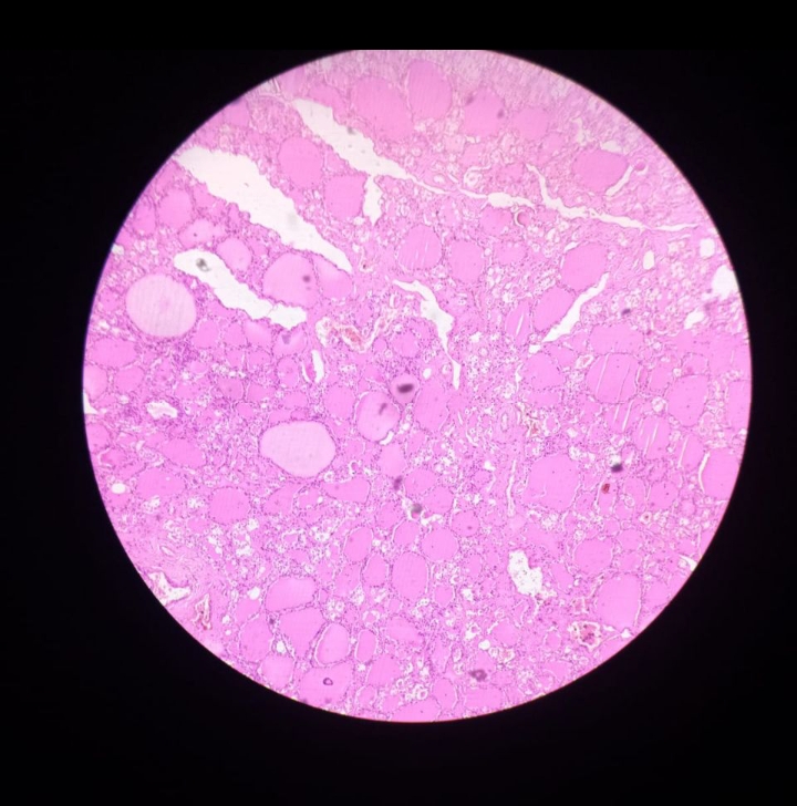
Histology, gross examination, imaging, and physiological recording all contribute to understanding the body, but their outputs should not be merged carelessly. A micrograph establishes local tissue appearance. A scan establishes a spatial representation using a particular acquisition method. A recorded trace shows change over time. A patient description communicates experience. None is a universal replacement for the others, and agreement between them often requires careful alignment of location, timing, and definition.
Imagine a learner presenting a single static image to explain movement. The instructor can ask what the image directly shows and what has been inferred. Then the learner can identify the additional observation needed: another phase, a continuous recording, or a measurement of the relevant variable. This question is constructive rather than punitive. It teaches students to separate observation from interpretation, which makes their explanations more precise. In a healthcare team, the same distinction helps colleagues understand whether a statement is a visible finding, a reported experience, a numerical result, or a proposed mechanism.
A practical structure-function worksheet
Choose one organ and write five connected sentences. First identify its location and the relevant anatomical part. Second identify a tissue feature that helps its role. Third describe a process occurring there. Fourth name a measurable quantity associated with that process. Fifth state a limitation of the available evidence. Keep the sentences linked: the tissue feature should concern the named part, and the measurement should concern the described process. This prevents a worksheet from becoming five unrelated facts about the same organ.
For example, a learner studying a hollow organ might describe its wall, its lumen, movement of contents, a change over time, and what cannot be established from an unlabeled photograph. Another learner studying a barrier might focus on two compartments, the separating tissue, transfer, a concentration difference, and uncertainty about the transfer rate. The framework adapts to many topics without forcing every organ into the same mechanistic story. It emphasizes questions that can be investigated, rather than claims that merely sound technical.
Connecting knowledge to respectful explanation
Healthcare learners often need to explain anatomy and physiology to people who have not studied either subject. A clear explanation can begin with the person's question, identify one relevant structure, and describe one process in ordinary language before introducing terminology. More detail is useful only if it answers the question. A long list of Latin names may demonstrate recall while leaving the listener uncertain about what happens in the body.
Practice explaining the same mechanism at three levels: a short conversation, a student tutorial, and a detailed professional discussion. The underlying science should stay consistent, while the vocabulary and depth change. Avoid using certainty to compensate for an incomplete explanation. If a measurement is an estimate, say so; if a diagram is simplified, explain what it omits. Structure and function become most valuable when they support accurate, understandable communication. The goal is a connected account of the body that a listener can follow and a colleague can assess against evidence.
Anatomical Latin: quick reference
| Latin term | English meaning |
|---|---|
| Cellula | Cell |
| Textus | Tissue |
| Organum | Organ |
| Cor | Heart |
Sources and image credits
Sources reviewed on 9 October 2026. Counts, estimates, study statistics and hypothetical calculations retain their context in the text.
- OpenStax: Overview of anatomy and physiology
- OpenStax: Structural organization
- OpenStax: Digestive system overview
- OpenStax: Cardiac cycle
- OpenStax: Homeostasis
- OpenStax: Cardiac physiology
- NIDDK: Digestive system and how it works
- NCBI Bookshelf: Diaphragm anatomy
Explanatory prose and SVG graphics are original. Photograph authors, source records and licenses are credited beside each image. Catalog illustrations provide educational context; anatomical facts are supported by the linked sources.