A useful fact explains its own limits
Human anatomy attracts impressive numbers: hundreds of bones, trillions of cells, millions of microscopic air spaces, and a heart that works without an ordinary rest break. These statements can help learners remember scale. They become more useful when we ask what was counted, whose body was studied, which method produced the estimate, and whether the result describes a population or an individual. For a medical student or healthcare colleague, those questions turn trivia into scientific literacy. A confident number without a definition can create more confusion than a carefully explained approximation.
Think of a fact as a small record with four fields: the structure, the unit, the conditions, and the source. “Four chambers” names a standard structural organization of the heart. “Five liters per minute” describes a flow under particular circumstances. “Thirty trillion cells” is an estimate assembled from evidence about many tissues. These are different kinds of information. The first is a categorical count, the second changes over time, and the third depends on a model. Reading them as though they have equal certainty hides the biology that makes them interesting.
The adult skeleton: a count with a convention
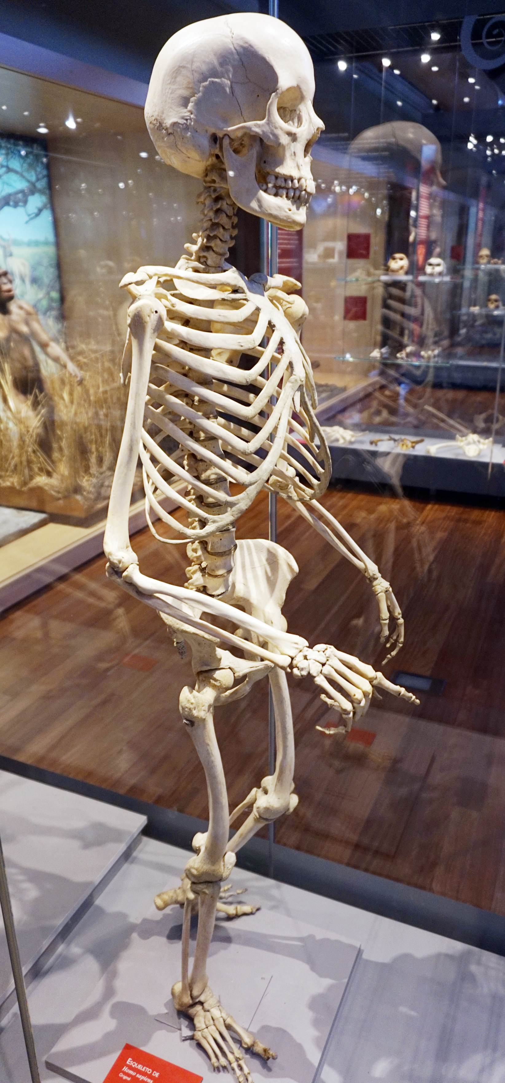
The conventional adult skeleton contains 206 bones. The number is a teaching framework, not an inventory guaranteed for every person. Developmental fusion changes how structures are counted, and accessory or sesamoid bones introduce variation. The skeleton is also divided into axial and appendicular components; its role includes support, movement, protection, mineral storage, and blood-cell production. OpenStax: Skeletal divisions
An original classroom exercise is to compare three ways of counting a hand. One student counts named bones; another counts visible pieces on an image; a third counts joints. Their totals answer different questions. A joint is an articulation, not a bone. A shadow on a radiograph can overlap another shadow. An immature skeletal element may have more than one ossification center. Before correcting a total, identify the object being counted. This habit transfers directly to everyday work with anatomical images, where a label and a visible outline may represent different stages of development.
The Latin os means bone; its plural is ossa. Skeleton axiale and skeleton appendiculare identify organizational divisions. These terms help learners recognize related vocabulary without mistaking a linguistic category for a separate physical layer. An index can organize the same bone by region, shape, development, and function. Multiple valid descriptions do not mean that the bone has multiplied; they mean that anatomy can be examined through several useful questions.
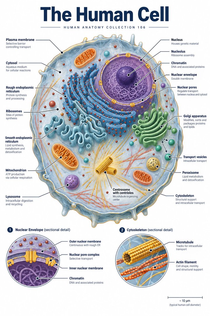
Trillions of cells do not mean trillions of identical units
A 2023 synthesis estimated about 36 trillion cells for its reference adult male, 28 trillion for its reference adult female, and 17 trillion for its reference child. These are modeled bodies, not fixed biological quotas. Cell size varies enormously; small circulating cells contribute strongly to counts, whereas much larger cells contribute differently to tissue mass. Hatton et al.: Human cell count and size distribution, 2023
Imagine two containers with equal volume: one holds fine sand and the other holds large stones. The sand container has many more objects, although its outside dimensions do not tell you that. Cell inventories pose a more complex version of this problem. A change in body mass does not translate neatly into an equal proportional change in every cell population. Neither can a count of nuclei be treated automatically as a count of cells, because cell structures and nuclear arrangements differ. The scientifically useful lesson is scale and heterogeneity, rather than a single spectacular total.
In clinical education, distinguish cellula, tissue, and organ. A whole organ contains more than its most familiar working cell. Supporting connective tissue, vascular endothelium, nerves, immune cells, and extracellular material contribute to the organ's organization. A photograph of one cell type therefore cannot stand for the complete cellular census of that organ. This distinction is especially valuable when teaching why a tissue section contains structures with several appearances rather than a uniform field of identical cells.
The lung demonstrates why sampling matters
Ochs and colleagues used a stereological method to investigate alveolar number in six adult human lungs. They reported a mean of 480 million and a range of 274–790 million. This is a result from a small anatomical study, not a reference interval defining every healthy person. Its value is partly methodological: it demonstrates that apparently simple counting requires a strategy for reconstructing three-dimensional structure from sampled material. Ochs et al.: Number of alveoli in the human lung, 2004
A drawing of ten alveoli, alveoli pulmonis, makes a pattern understandable but does not provide a scale model. A microscopic field shows only part of a sample, and its empty spaces may change appearance with the plane of section. When a learner sees several round profiles, it is tempting to equate profiles with separate three-dimensional objects. The temptation is precisely why counting methods matter. A diagram should state that it is schematic; a study should state the sampling approach; a classroom discussion should separate what is visible from what is inferred.
An instructive comparison is to slice a bunch of irregular balloons at different heights. One slice might show many small circles, another a few broad outlines, and another no profile from a balloon that still exists above it. Real alveolar geometry is not a bunch of isolated balloons, but the analogy illustrates a measurement problem. The point is not to replace histology with a toy; it is to make the hidden assumptions of a two-dimensional image explicit before studying the real tissue.
Four heart chambers and one connected circulation
The standard human heart has two atria and two ventricles. The atria receive returning blood; the ventricles direct it into pulmonary and systemic pathways. A septum separates the right and left sides. These structural counts remain a useful starting point even though the chambers differ in wall structure, shape, connections, and mechanical work. NHLBI: Heart anatomy
The phrase “two pumps” can be helpful when discussing pulmonary and systemic flow, but it should not suggest two independent devices that can deliver unrelated volumes indefinitely. A circulation diagram needs a closed route. If a hypothetical right-sided pump delivered substantially more volume than the left side for a prolonged period, the difference would have to accumulate somewhere. Conservation of volume makes the chambers easier to understand than memorizing arrows alone. This is a reasoning exercise, not a diagnosis based on any one patient's measurements.
A daily heartbeat total is a calculation, not an observation
Choose a hypothetical steady heart rate of 70 beats per minute. Multiplying 70 by 60 minutes and then by 24 hours gives 100,800 beats in one day. At 80 beats per minute the same calculation gives 115,200. The difference illustrates how sensitive a daily total is to the assumed rate. Nobody has established a person's actual daily total simply by taking one brief pulse measurement and multiplying it by a day.
A transparent infographic can label these as hypothetical scenarios and display the arithmetic. It should not announce that every heart beats exactly 100,000 times daily. Sleep, activity, emotional state, illness, medication, and measurement context can alter the observed rate. The educational goal is to connect units and time, then distinguish an estimate from a recorded series. Healthcare staff encounter this distinction whenever a short measurement is used to summarize a changing process. Retaining the original timestamp and circumstances keeps that summary interpretable.
Body systems are useful maps, not sealed compartments
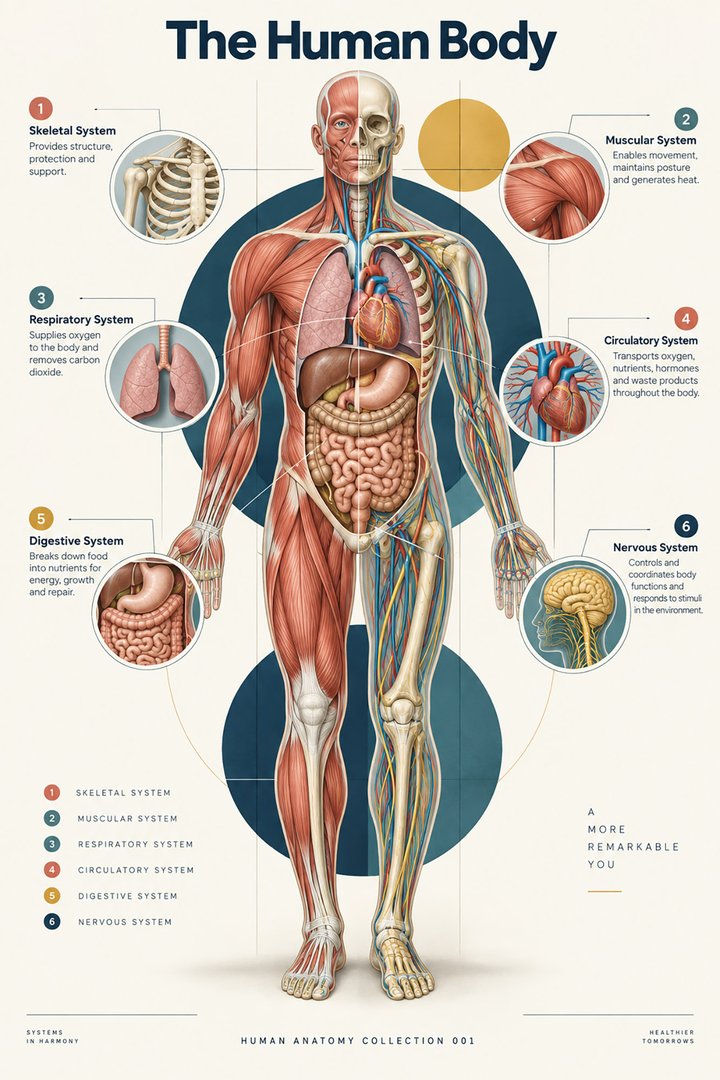
Textbooks commonly organize the body into systems because related structures can be learned together. The hierarchy from cells through tissues and organs to organ systems helps connect different scales of study. A system is a functional and educational grouping; the boundaries of a textbook chapter do not form impermeable walls in a living person. OpenStax: Structural organization
Consider the pancreas as an exercise in classification. One discussion concerns secretions reaching the digestive tract; another concerns hormones communicating through blood. A learner who insists that each organ belongs to exactly one chapter will struggle to connect the two discussions. Instead, ask which pathway and product are being described. The same strategy works for skin, bone, kidney, and many other organs. A label can be correct yet incomplete. Healthcare teaching improves when an organ's roles are explained as relationships rather than as membership in a single exclusive list.
Symmetry is a starting pattern, not a promise
The external body offers many paired structures, and right-left comparison is a powerful learning tool. Internal anatomy is less simply mirrored. The heart, liver, stomach, and spleen contribute to a strongly asymmetric arrangement. Anatomical right and left refer to the subject, not the viewer. Standard orientation therefore matters even in an apparently straightforward photograph. OpenStax: Anatomical terminology
An original observation exercise is to place a frontal anatomy diagram beside a photograph of a torso model. Before naming an organ, identify the subject's right side, the viewpoint, and any removed surface. Ask whether the model shows an anterior dissection, an internal view, or a transverse section. Many apparent contradictions disappear at that stage. A label that looks reversed might reflect a different viewing direction; a missing structure might have been removed to expose another. The reasoning protects against errors more effectively than memorizing that red always belongs on one side of a poster.
“Normal” has more than one meaning
In an anatomy atlas, normal often describes a representative structural pattern. In a laboratory report, a reference interval describes a distribution under specified measurement conditions. In a discussion of function, normal may refer to an appropriate response to a situation. These meanings overlap but are not interchangeable. A healthy person's pulse during activity should not be expected to match the pulse recorded during quiet rest. A population average height should not become a template for judging the proportions of every body.
For teaching, separate “common,” “average,” “reference,” and “required.” Common anatomy occurs frequently; an average summarizes measured values; a reference supports comparison; a required feature defines a particular classification. A small numerical difference may represent measurement variation rather than a meaningful biological change. Conversely, a structural variation can be important for an individual procedure even when it is harmless in ordinary life. Good anatomical literacy leaves room for both possibilities without forcing every observation into a binary list of perfect and defective bodies.
A real image is evidence of one specimen
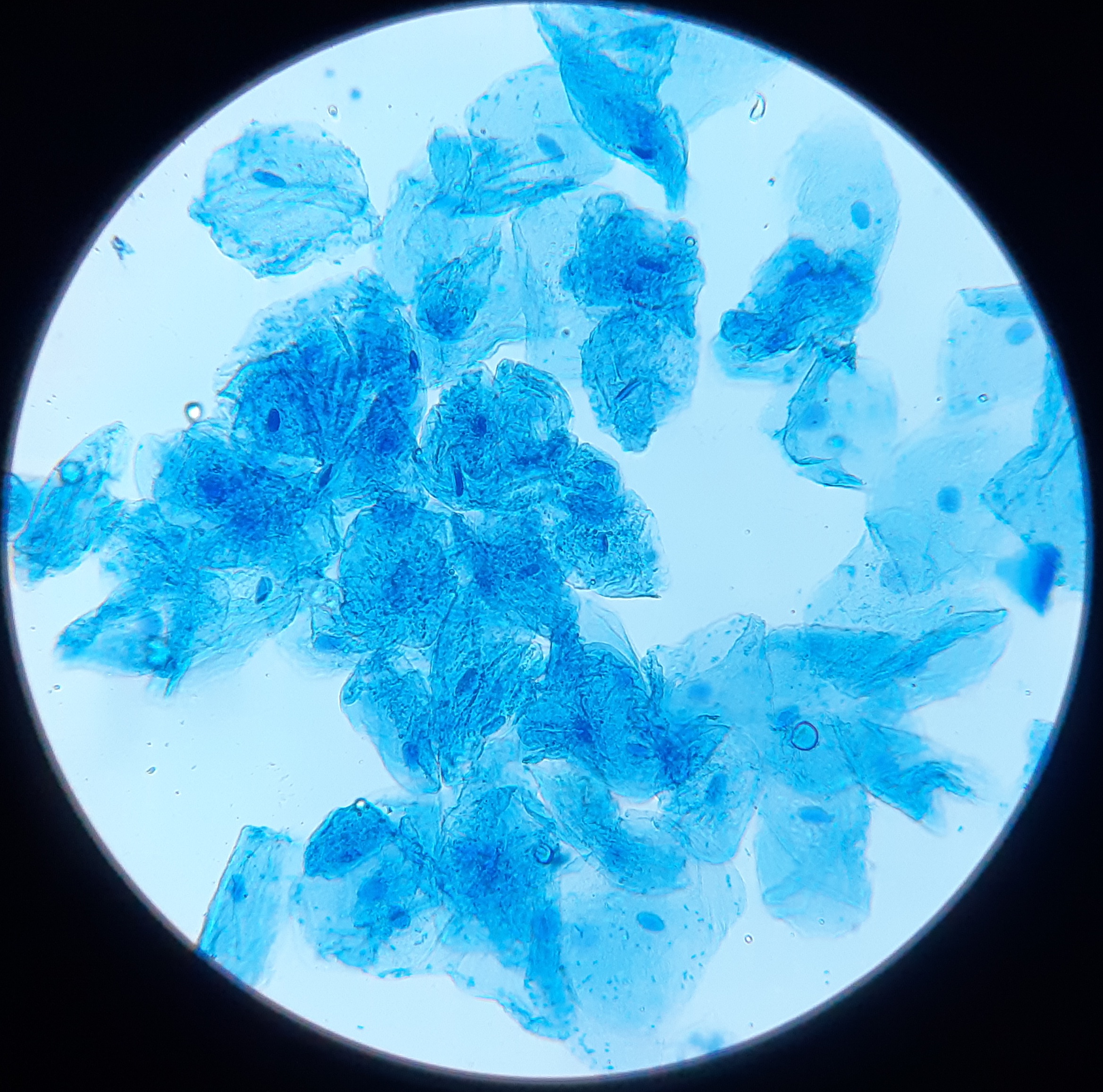
Real photography and microscopy provide something a simplified drawing cannot: texture, irregularity, overlapping structures, and the consequences of preparation. They also bring limits. A specimen may have been fixed, sectioned, stained, enlarged, or photographed under conditions that change its appearance. A chest radiograph is a projection, not a surface photograph. A micrograph displays a particular field, not a complete organ. Understanding those limits makes real images more useful rather than less trustworthy.
When presenting an image to colleagues, identify the method, the orientation, and the scale if available. Preserve the original author and license credit. Describe only the structures the image and its source support. For example, “cardiac muscle micrograph” is safer than “healthy human heart tissue” if the source does not identify species or health status. A caption should be a factual bridge between the image and the lesson, not a decorative claim that the photograph cannot establish. This is a practical skill for anyone preparing educational material in a practice or hospital.
How to investigate a surprising body fact
Begin with the exact claim rather than its popular summary. Does a statement about “the lung” refer to a pair, one organ, a volume of tissue, or a model? Does a bone count include accessory elements? Does “cells replaced each day” include every tissue or selected populations? Identify the denominator before comparing two numbers. Then examine the study design, the sample, and the time period. An apparent disagreement may reflect different questions rather than a scientific mistake.
Create an evidence note in plain language. For a modeled cell count, write “estimated for reference bodies from a literature synthesis.” For alveolar data, write “study of six adult lungs using stereology.” For the hypothetical pulse calculation, write “arithmetic assuming constant rate.” These notes are short enough to fit below a graphic and strong enough to prevent a misleading impression. Students can use the same technique when preparing presentations: every numerical label should retain its unit, its context, and its explanation of what kind of number it represents.
Facts that stay useful in healthcare work
Useful body facts improve observation, explanation, or communication. Knowing that an image has a defined plane helps you describe it. Knowing that an organ contains multiple tissues helps you interpret a specimen. Knowing that an average is not a personal target helps you speak respectfully about variation. The practical benefit is not winning a trivia competition; it is making fewer unsupported assumptions while working with complicated biological information.
For a final teaching exercise, select one structural count, one measured variable, and one modeled estimate. Explain each without using the word “always.” Then state what would be needed to verify it in an individual. The structural count may require imaging or direct inspection, the changing variable may need repeated observations, and the modeled estimate may not be directly measurable in routine care at all. That difference is itself a memorable human body fact. Anatomy becomes more interesting when impressive numbers are accompanied by honest reasoning about how we know them.
Anatomical Latin: quick reference
| Latin term | English meaning |
|---|---|
| Cellula | Cell |
| Os / ossa | Bone / bones |
| Cor | Heart |
| Pulmo | Lung |
Sources and image credits
Sources reviewed on 9 October 2026. Counts, estimates, study statistics and hypothetical calculations retain their context in the text.
- OpenStax: Skeletal divisions
- Hatton et al.: Human cell count and size distribution, 2023
- Ochs et al.: Number of alveoli in the human lung, 2004
- NHLBI: Heart anatomy
- OpenStax: Structural organization
- OpenStax: Anatomical terminology
Explanatory prose and SVG graphics are original. Photograph authors, source records and licenses are credited beside each image. Catalog illustrations provide educational context; anatomical facts are supported by the linked sources.