Why a cavity is more than an empty space
A body cavity is an anatomical space defined by surrounding structures, but the word does not mean that the space is empty. Some large compartments contain organs, vessels, nerves, connective tissue, and fluid. Other named cavities are thin potential spaces between surfaces that normally lie close together. A third concept, the lumen of a hollow organ, describes the interior of that organ. These distinctions are essential when reading a report or explaining where something is located. “Inside the chest,” “inside a lung,” and “within the pleural space” are not interchangeable statements.
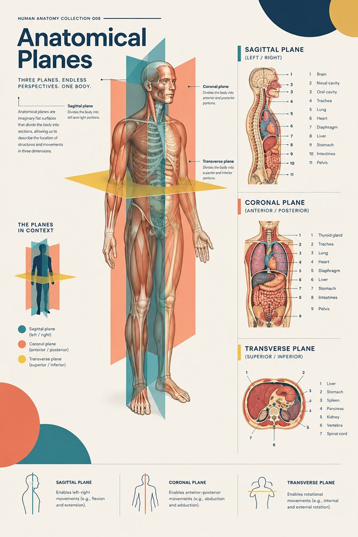
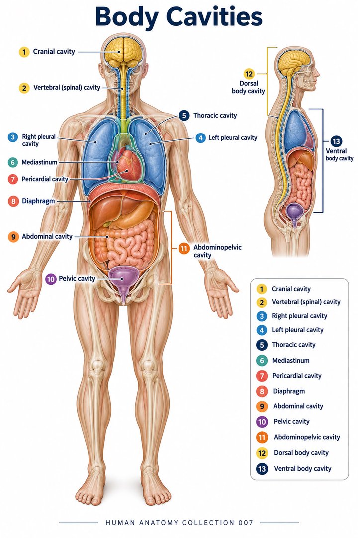
An introductory map divides the major cavities into posterior and anterior groups. Cranial and vertebral cavities form the posterior group; thoracic and abdominopelvic regions form the anterior group. This map is a first level of organization, not a claim that all smaller spaces share the same lining or mechanical behavior. OpenStax: Anatomical terminology
For healthcare learners, a useful description names the boundary before the contents. If someone says that a structure is “within a cavity,” ask which wall or membrane defines that cavity. This approach makes subsequent details easier to place. It also exposes ambiguous diagrams: a colored shape may represent a region containing organs, a membrane itself, or the narrow space between two membranes. The legend must tell the reader which interpretation is intended.
The cranial cavity and its internal supports
The cranial cavity contains the brain and associated protective structures within the skull. Its floor is organized into anterior, middle, and posterior cranial fossae. These are depressions in the cranial base, not three disconnected containers. Their contours support different parts of the brain and provide a spatial framework for identifying openings, ridges, and relationships. The three meninges are dura mater, arachnoid mater, and pia mater. NCBI Neuroscience: The meninges
The terminology invites a layer-by-layer mental picture. The tough outer covering, the arachnoid, and the closely applied pia are not simply three copies of the same material. Their relationships define spaces with different contents and significance. When a drawing separates the layers widely so that labels fit, the separation is an explanatory exaggeration. A learner should not infer that the living cranial cavity contains large open gaps arranged exactly like the diagram. The purpose of a magnified illustration is to reveal relationships that would otherwise be difficult to distinguish.
The vertebral canal connects the posterior map
The vertebral canal follows the column of aligned vertebral openings. It houses neural structures and their coverings; the contents change with the level being examined. A canal is therefore not described completely by one transverse drawing. A section through one region may show the spinal cord, whereas a more inferior section can show descending roots within the continuing dural sac. The important anatomical question is the level and the exact structure being labeled, rather than simply whether a circular space appears on a scan.
The spinal epidural space lies outside the dura within the vertebral canal. It differs from the subarachnoid space, which lies between arachnoid and pia. The name identifies a relationship to a covering; it does not refer to a compartment in the chest or abdomen. Littrell: Epidural space anatomy, 1991
This distinction matters in communication about regional anatomy even without discussing a procedure. If a colleague uses the word “spinal,” clarify whether they mean the vertebral column, the spinal cord, a nerve root, or a particular meningeal space. Each term refers to a different object. Accurate vocabulary lets a learner describe an image while recognizing that the image may not show every boundary equally well.
The thoracic cavity includes several different compartments
The thoracic region extends between the root of the neck and the diaphragm. Its walls include the sternum anteriorly, the vertebral column posteriorly, and the ribs with their associated tissues around the sides. It contains paired pulmonary regions and a central mediastinal compartment. The shape of this region matters because its boundaries participate in breathing rather than acting as a rigid, motionless box. NCBI Bookshelf: Anatomy of the thorax
Latin cavitas thoracis identifies the thoracic cavity. The term should not be reduced to “the space around the heart.” The heart is part of a central arrangement that also relates to vessels, airways, the esophagus, nerves, and lymphatic pathways. A frontal drawing compresses this organization into a single plane. Studying a transverse view restores the depth that the frontal view hides: an anterior wall, pulmonary tissue on the sides, central structures, and posterior structures can all occupy the same vertical level without touching the same surface.
Pleural cavities are interfaces around the lungs
Each lung has a visceral pleural covering; the parietal pleura relates to the thoracic wall, diaphragm, and mediastinum. The pleural cavity is the potential space between these surfaces. A thin fluid film permits movement with limited friction. The right and left pleural arrangements are separated by the mediastinum, so the thorax should not be imagined as one uninterrupted pleural chamber. OpenStax: Lungs and pleura
Distinguish three nested concepts: the lung tissue, the air spaces within the lung, and the pleural space outside the lung surface. An airway connects to air-containing respiratory spaces; it does not normally open into the pleural cavity. The pleural interface contributes to mechanics while remaining anatomically separate from the route followed by inhaled air. This nested picture makes the term “pleural” more precise than a loose description such as “around the breathing tubes.” It also explains why an educational diagram should use separate colors and labels for air, tissue, membranes, and the potential space.
The mediastinum is a region, not a serous cavity
The mediastinum runs between the pleural sacs. A traditional anatomical model divides it into superior and inferior portions; the inferior portion is described as anterior, middle, and posterior. These subdivisions organize structures and relationships without requiring complete physical walls between every compartment. The heart and pericardial arrangement occupy the middle mediastinal region, while other central structures occupy different or continuing routes. NCBI Bookshelf: Mediastinum
Students sometimes add up these divisions as though the word “cavity” means the same thing in every label. It does not. A region may be defined by reference planes and neighboring structures, while a cavity may be defined by opposed membrane surfaces. A simple numerical fact is that the traditional inferior mediastinum has three named subdivisions. This is a classification count, not a measurement of three fluid volumes. Keeping the type of number explicit prevents a chart from implying anatomical equivalence where there is only an organizational convention.
The pericardial cavity surrounds a moving organ
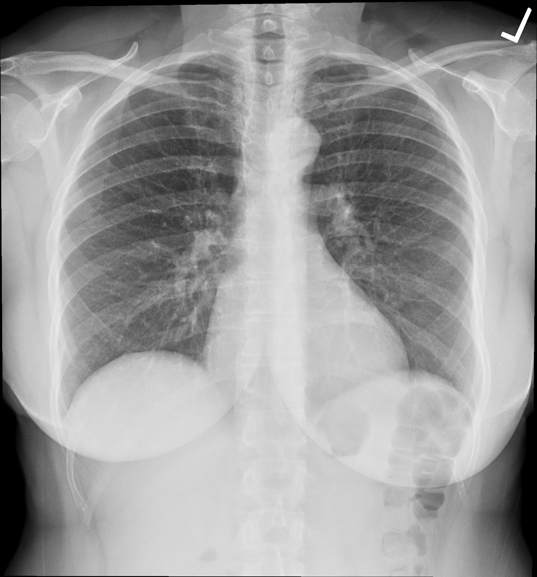
The pericardial sac surrounds the heart and contributes to its protection and position. Fluid between its serous surfaces reduces friction during movement. The cardiac chambers, by contrast, contain the blood moving through the heart. A blood-filled atrium or ventricle is not a subdivision of the pericardial cavity. NHLBI: Heart anatomy
This distinction is particularly useful in a cross-sectional illustration. Begin at the interior of a chamber, move through the heart wall, then identify the relevant external coverings and surrounding interface. The sequence traverses several anatomical objects before reaching the neighboring thoracic region. A diagram that labels only “heart” and “chest” loses these distinctions. Healthcare learners benefit from tracing the path outward because it encourages exact descriptions: in the chamber, within the wall, on the surface, between serous surfaces, or in an adjacent compartment. These phrases locate observations far more clearly than an isolated organ name.
The diaphragm separates regions while allowing passage
The diaphragm forms the floor of the thoracic region and the roof of the abdominal region. It is a muscular and tendinous sheet with openings and pathways for structures that travel between the two regions. Its dome-shaped organization and movement contribute to ventilation, so its position is not a fixed horizontal shelf. NCBI Bookshelf: Diaphragm anatomy
This creates an important spatial relationship: the same boundary participates in two neighboring compartments. A change in its position alters the shapes of those compartments even though the named anatomical regions remain recognizable. When interpreting an image, note the phase and posture rather than treating a textbook outline as an unchanging contour. The diaphragm is also a reminder that anatomical separation does not mean functional isolation. Vessels, nerves, and the esophagus connect regions across the boundary. A complete cavity map should show these connections without implying that the compartments are one undivided space.
Abdominal cavity and peritoneal cavity are different terms
The abdominal region contains organs and tissues arranged in relation to the peritoneum. The peritoneal cavity specifically identifies the potential space between parietal and visceral peritoneal surfaces. Parietal peritoneum lines the wall; visceral peritoneum covers many organ surfaces. The membrane also forms folds associated with support and routes for vessels, lymphatics, and nerves. NCBI Bookshelf: Peritoneum
An organ commonly described as intraperitoneal is not freely floating inside an empty bag. The description concerns its peritoneal relationships. The organ has tissue substance, attachments, and connected structures. A useful conceptual drawing shows a wall, a reflected membrane, an organ surface, and the thin intervening space. If the drawing uses a wide gap for clarity, its caption should say so. Otherwise the learner may imagine the potential space as a large preexisting chamber occupied by the organ itself. The distinction is subtle in everyday language and fundamental in anatomical reasoning.
Retroperitoneal anatomy adds a posterior layer
The retroperitoneum lies posterior to the parietal peritoneum. It includes structures from urinary, endocrine, digestive, and vascular systems. Kidneys and major posterior vascular routes help anchor this layer in the mental map. Its subdivisions include perirenal and pararenal spaces, demonstrating that the posterior abdominal arrangement is more detailed than a single label suggests. NCBI Bookshelf: Retroperitoneum
In an anterior torso diagram, posterior structures may be moved sideways or exposed after other organs have been removed. That arrangement can be excellent for teaching names yet misleading for teaching depth. A learner should ask whether the figure preserves the original spatial arrangement or deliberately separates structures. Following the posterior abdominal wall on a sectional view helps restore the relationship. The idea is not to discard schematic posters; it is to use them for one purpose and then add a depth-oriented representation for another purpose. Both become stronger when their limitations are stated.
The pelvic region continues the abdominopelvic arrangement
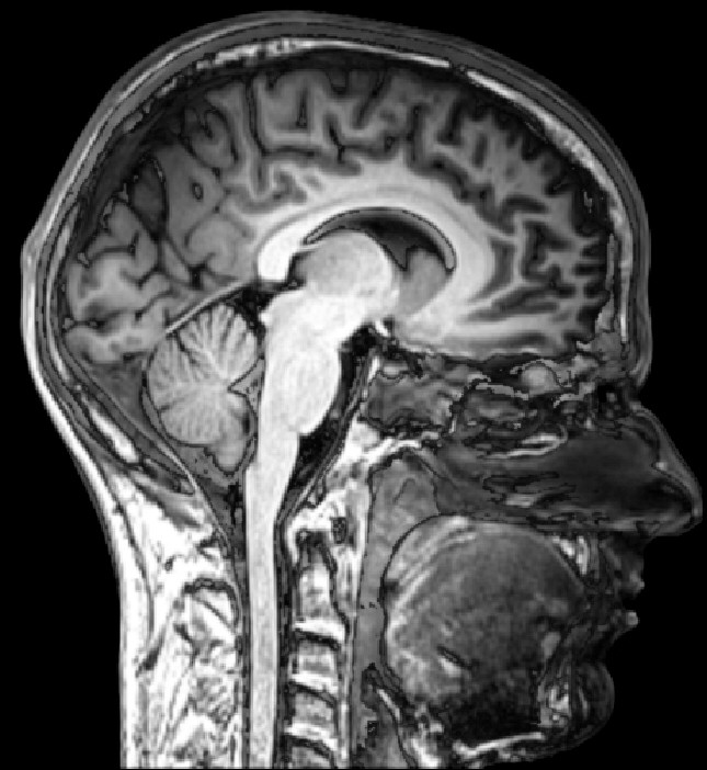
Abdominal and pelvic regions are continuous rather than separated by a complete diaphragm. The pelvis provides a bony framework and an inferior musculofascial support. The pelvic floor includes levator ani and coccygeus components, contributes to support of viscera, and participates in continence and coordinated evacuation. Its relationships concern urinary, reproductive, and anorectal structures. NCBI Bookshelf: Pelvic floor
The rectum lies posteriorly in the pelvic visceral arrangement, with neighboring relationships varying according to reproductive anatomy. Its continuation from the sigmoid colon and transition toward the anal canal make it part of a pathway crossing regional boundaries. NCBI Bookshelf: Rectum anatomy
The regional distinction therefore serves orientation rather than dividing every organ into an exclusive box. A hollow organ can span more than one region, and an organ's size or filling can change how much of it extends into a neighboring region. The terms abdomen, pelvis, peritoneal, and extraperitoneal answer different questions. Combining them accurately gives more information than using any one of them alone.
Lumens and body cavities should never share an unlabeled arrow
The lumen of a hollow organ is the space inside its wall. A digestive lumen, a urinary lumen, and a cardiac chamber differ in connections and contents. A serous cavity lies between membrane surfaces outside an organ's working interior. A regional compartment can contain several organs and other tissues. These are three distinct spatial categories. A teaching graphic should identify which category each colored region represents before asking the learner to trace any route through it.
For practice, imagine following a swallowed item, a molecule in circulating blood, and a thin film between serous surfaces. The routes do not merge simply because all three may occupy the same broad torso region. One remains luminal until absorption, one travels through vascular and cardiac interiors, and one belongs to a membrane interface. This exercise tests whether a learner understands location rather than merely remembering names. It is also a useful check when preparing diagrams for mixed healthcare audiences, whose experience with specific anatomical terms may differ considerably.
A systematic way to read a cavity diagram
Start with the viewpoint and subject orientation. Identify the external body wall, then the major boundary of the region, then the membranes, then the organ surfaces and internal lumens. Move from large-scale organization to smaller interfaces rather than jumping directly to a familiar organ. For each label, state whether it names tissue, a compartment, or a potential space. Finally, identify which important relationships have been omitted for clarity. A schematic becomes a trustworthy guide when the viewer knows exactly what it represents.
In healthcare communication, a complete location statement might include a region, a relation to a membrane, and a neighboring structure. The precise wording depends on the observation, but the principle is stable: locate before interpreting. Avoid letting a diagram create certainty about an individual's exact anatomy when it is only a representative model. Cavities provide the framework for understanding how structures share space, move against one another, and remain connected across boundaries. Learning that framework turns a flat organ list into a three-dimensional account of the human body. An additional check is to explain a label without pointing at the page. If “between two membranes” and “inside an organ” become confused in that explanation, revisit the boundaries before moving to more detailed terminology. Spatial clarity should survive the removal of a familiar picture.
Anatomical Latin: quick reference
| Latin term | English meaning |
|---|---|
| Cavitas thoracis | Thoracic cavity |
| Cavitas abdominis | Abdominal cavity |
| Cavitas pelvis | Pelvic cavity |
| Cavitas peritonealis | Peritoneal cavity |
Sources and image credits
Sources reviewed on 9 October 2026. Counts, estimates, study statistics and hypothetical calculations retain their context in the text.
- OpenStax: Anatomical terminology
- NCBI Neuroscience: The meninges
- Littrell: Epidural space anatomy, 1991
- NCBI Bookshelf: Anatomy of the thorax
- OpenStax: Lungs and pleura
- NCBI Bookshelf: Mediastinum
- NHLBI: Heart anatomy
- NCBI Bookshelf: Diaphragm anatomy
- NCBI Bookshelf: Peritoneum
- NCBI Bookshelf: Retroperitoneum
- NCBI Bookshelf: Pelvic floor
- NCBI Bookshelf: Rectum anatomy
Explanatory prose and SVG graphics are original. Photograph authors, source records and licenses are credited beside each image. Catalog illustrations provide educational context; anatomical facts are supported by the linked sources.