The torso joins support, protection, and passage
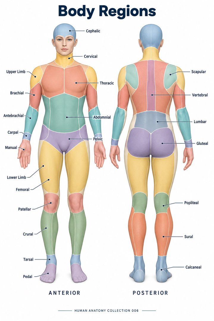
The torso, or trunk, is the central region linking the head and neck with the limbs. It includes the thorax, abdomen, pelvis, and back. Learning it as a stack of organ names misses an essential feature: the walls and their attachments matter as much as the contents. The trunk supports posture and movement, surrounds vital structures, and provides routes for air, food, blood, lymph, neural signals, and urinary or reproductive pathways. Several of these routes cross more than one region, so no single regional diagram tells the whole story.
The most useful mental model has depth. At the surface are skin and superficial tissues; beneath them are muscles, connective tissues, and skeletal structures; deeper still are compartments and organs. The arrangement changes from one part of the trunk to another. An anterior chest wall is not built exactly like an anterior abdominal wall, and neither is built exactly like the back. A layer-by-layer approach reveals those differences while preserving the connections that make the torso function as a whole.
The thoracic cage provides a moving framework
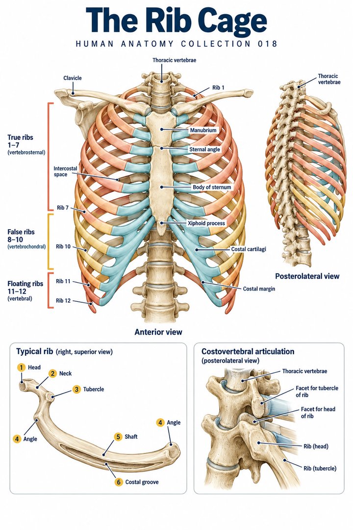
The standard thoracic cage has 12 pairs of ribs associated posteriorly with 12 thoracic vertebrae. Anteriorly, the sternum is divided into manubrium, body, and xiphoid process. Ribs one through seven have direct cartilaginous connections to the sternum; eight through ten connect indirectly through the costal margin; eleven and twelve lack an anterior sternal connection. These categories describe attachments, not a difference between real and unreal bones. OpenStax: Thoracic cage
Latin costa means rib, sternum names the breastbone, and processus xiphoideus names its inferior process. The costal margin is a boundary of great educational importance because structures below its surface projection may still lie partly protected by the lower cage. The lower edge of the visible chest is not a complete boundary between all thoracic and abdominal organs. A learner should distinguish an external landmark from the internal dome of the diaphragm and from the three-dimensional extent of an organ.
Intercostal layers help explain chest movement
The intercostal spaces contain layered muscles, connective tissues, vessels, and nerves. External, internal, and innermost intercostal components contribute to the wall's organization. During inspiration, thoracic dimensions can increase vertically, from front to back, and from side to side. Pump-handle and bucket-handle descriptions illustrate aspects of rib movement; they are mechanical analogies rather than statements that all ribs behave identically. NCBI Bookshelf: Thoracic wall movements
These relationships connect a skeletal poster with a breathing diagram. A rib is not only a protective arch; it is part of a moving framework linked to vertebrae, cartilage, muscle, and connective tissue. In a classroom model, a rigid chest cage may make attachments easy to see while failing to demonstrate motion. The lesson is to supplement it with a schematic that shows changing dimensions. When describing observed chest movement in healthcare settings, specify the area and the observation rather than assuming that one visible movement reveals the activity of every deep muscle.
The diaphragm is both boundary and mechanism
The diaphragm is a muscular and tendinous partition with a dome-shaped arrangement. It separates the thoracic and abdominal regions and supplies a major mechanical contribution to inspiration. Its descent changes thoracic volume while affecting the neighboring abdominal arrangement. The right and left domes do not have perfectly identical relationships; the liver contributes to the higher right-sided contour. Structures traveling between thorax and abdomen pass through or behind defined diaphragmatic routes. NCBI Bookshelf: Diaphragm anatomy
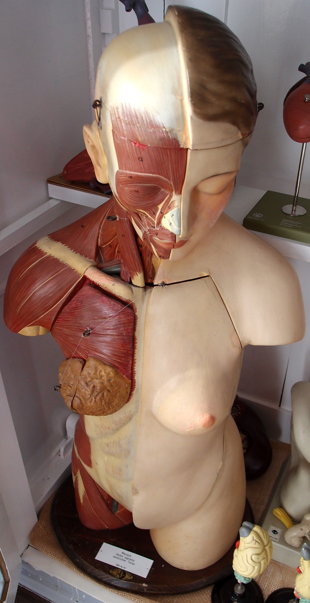
The Latin diaphragma helps identify the same structure across anatomical and physiological discussions. A sectioned torso model may show a flat cut edge where the diaphragm actually forms a curved surface. If a learner interprets the cut edge as the whole muscle, the dome becomes difficult to visualize. Following the peripheral attachment and central region in more than one view restores the shape. This is a recurring principle in torso anatomy: a plane through a structure reveals one slice of its geometry, not its entire form.
The mediastinum organizes the center of the chest
Between the pulmonary regions lies the mediastinum. A traditional anatomical division identifies superior mediastinum and an inferior region subdivided into anterior, middle, and posterior portions. The heart and pericardium occupy the middle region, while major vessels, airways, the esophagus, nerves, and lymphatic routes form a connected central arrangement. These subdivisions are useful reference regions rather than fully sealed boxes. NCBI Bookshelf: Mediastinum
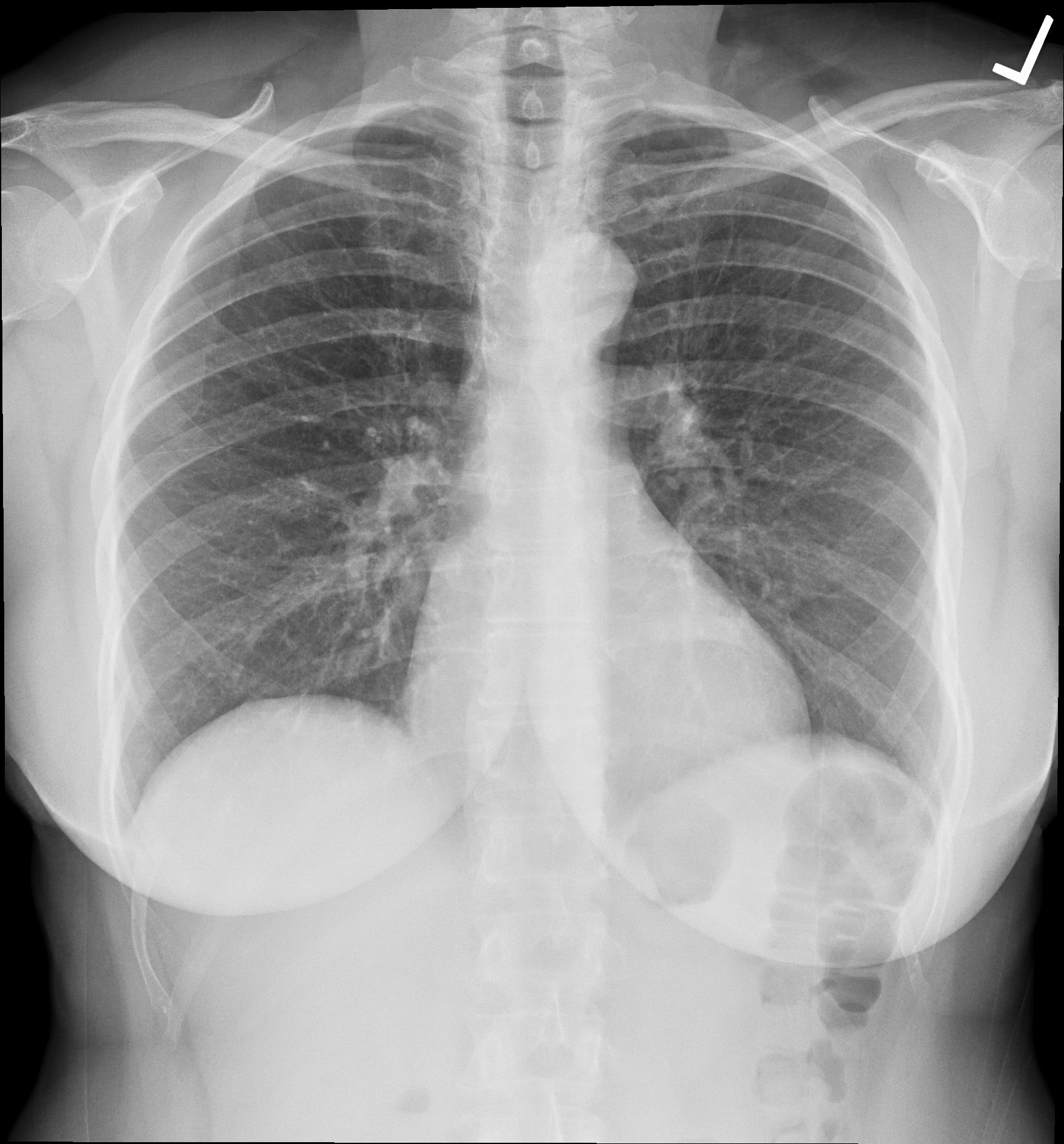
The central chest therefore contains pathways as well as organs. A frontal diagram may place the esophagus and trachea side by side for clarity, but the real depth relationship is crucial. A learner should ask which structure is anterior, which is posterior, and which continues beyond the displayed level. The same questions help interpret a chest image in which overlapping structures produce composite shadows. The familiar outlines of heart and lungs are a starting point; understanding the central routes requires a layered view rather than a single silhouette.
Pulmonary surfaces meet the wall through pleural relationships
The lungs have costal surfaces related to the cage, diaphragmatic bases, and medial relationships to central structures. Visceral pleura covers lung surfaces and extends into fissures; parietal pleura relates to the wall and neighboring boundaries. The potential pleural space lies between these coverings, separate from the airways and alveolar spaces inside the lung. OpenStax: Lungs and pleura
This is an important torso distinction because a diagram can unintentionally collapse tissue, air, and membrane interfaces into one colored area. A well-designed graphic shows an airway entering lung tissue, a lung surface, a membrane covering, and the opposing wall-related membrane. The actual interface is thin, even if the educational drawing separates it widely. A caption should identify the exaggeration. Spatial accuracy allows learners to connect breathing mechanics with regional anatomy without imagining that inhaled air normally enters a large chamber outside the lung.
The anterior abdominal wall has intersecting muscle directions
The three broad lateral muscle layers are external oblique, internal oblique, and transversus abdominis. Their differing fiber directions support the wall and contribute to movement and pressure generation. Rectus abdominis forms a paired longitudinal arrangement near the midline. Its tendinous intersections create visible subdivisions in some bodies, but these subdivisions do not turn the muscle into a collection of separate organs. NCBI Bookshelf: Abdominal wall muscles
The Latin names remain informative: musculus rectus abdominis, musculus obliquus externus abdominis, musculus obliquus internus abdominis, and musculus transversus abdominis. Reading the names beside a layer diagram is more productive than treating them as an isolated vocabulary exercise. “External” and “internal” refer to relative position, while “transverse” describes an orientation. The wall operates through coordinated tissue relationships, so a simplified exercise that highlights one muscle should not imply that the remaining layers become irrelevant during that movement.
Aponeuroses and fascia give the wall continuity
The broad muscle layers continue into aponeuroses that contribute to the rectus sheath and the midline linea alba. The arrangement of the sheath changes inferiorly at the arcuate line: the posterior aponeurotic covering does not continue in exactly the same form throughout the lower wall. Deep to the muscular arrangement lies transversalis fascia, with further tissues separating it from parietal peritoneum. NCBI Bookshelf: Anterolateral abdominal wall
These terms identify different materials and boundaries. Muscle fibers, an aponeurotic sheet, a fascial layer, and a serous membrane should not all be drawn as interchangeable strips. Their distinctions explain why a torso can be described both as a surface of named muscles and as a layered wall enclosing an internal region. Fascia also continues between regions, so an anatomical illustration cropped at the abdomen may hide connections beyond its border. NCBI Bookshelf: Abdominal wall fascia
For an original learning exercise, draw the same region first as a list of layers and then as a continuous folded arrangement. The second drawing reveals why a wall cannot be understood entirely by memorizing a top-to-bottom list. Layers change position, merge into related connective structures, and form routes around other structures. This is regional anatomy rather than a stack of identical sheets.
The abdomen contains anterior and posterior arrangements
The digestive tract and its associated organs occupy much of the abdominal region, but not all structures share the same relationship to peritoneum. Peritoneal coverings and folds support mobile portions and provide routes for vessels, lymphatics, and nerves. The narrow peritoneal cavity is a potential space between serous surfaces, not a hollow storage area in which organs float freely. NCBI Bookshelf: Peritoneum
Behind the parietal peritoneum lies the retroperitoneal region, including kidneys and other structures belonging to several systems. This posterior layer remains important even when a model presents abdominal organs in a single frontal display. NCBI Bookshelf: Retroperitoneum
The contrast gives healthcare learners a way to understand why a surface location and an internal relationship may both be correct while referring to different aspects of anatomy. “Upper abdomen” provides a broad region; “posterior to the peritoneum” provides depth. Neither alone tells the full arrangement. Combining regional and relational descriptions creates a more precise statement and a more useful mental map.
Digestive and vascular routes cross torso regions
The alimentary pathway links mouth, pharynx, esophagus, stomach, small intestine, and large intestine to the anal outlet. The esophagus travels through the torso to connect with the stomach; the digestive tract then changes direction and arrangement within the abdomen and pelvis. Liver, pancreas, and gallbladder contribute through associated secretions without becoming luminal segments through which a swallowed item passes. NIDDK: Digestive system and how it works
Venous return from lower body regions travels through the inferior vena cava toward the right atrium. Its relation to the diaphragm and posterior abdominal arrangement helps connect regional anatomy with the direction of blood flow. NCBI Bookshelf: Inferior vena cava
On a torso diagram, keep the food pathway and vascular pathway visually distinct. Both may cross the same broad region, but their contents occupy different interiors. A graphic can use separate arrow styles and a legend rather than relying entirely on color. The distinction is particularly helpful for staff explaining anatomy to people who may otherwise interpret every arrow as showing where food travels.
The back provides a different view of the same trunk
The vertebral column supplies a central posterior framework with regional changes in shape and joint relationships. Thoracic vertebrae relate to ribs, while lumbar structures contribute to the lower trunk's load-bearing arrangement. Vertebral bodies, arches, processes, discs, and articulations describe different components; the whole column should not be reduced to a rigid line. OpenStax: Vertebral column
The posterior view also changes how internal structures are imagined. An organ visible in an anterior diagram may be concealed behind other tissues from the back. The same organ can have an anterior surface, a posterior relation, and a projection near a lateral landmark. Asking learners to translate among these descriptions builds three-dimensional understanding. A correct translation does not require pretending that every organ touches the external landmark used to describe its position; surface projection is a map, not direct contact.
Pelvic support completes the lower torso
The pelvis links the central trunk with the lower limbs and surrounds lower visceral structures. Its floor is a musculofascial complex that supports organs while permitting necessary passage. Levator ani and coccygeus components participate in this support, with connective tissue and coordinated activity contributing to continence and evacuation. Urinary, reproductive, and anorectal relationships occupy the region rather than separate, isolated chambers. NCBI Bookshelf: Pelvic floor
This lower boundary must be understood alongside the diaphragm and abdominal wall. A simplified diagram of pressure generation can show those three components as related surfaces, while acknowledging that real activity is coordinated and variable. The same trunk that supports a posture also accommodates breathing, organ filling, and movement. An anatomy guide should therefore resist depicting the pelvis as a motionless bowl with contents glued in position. A stable structural map can coexist with a dynamic account of how tissues and organs behave.
Superficial structures belong to torso anatomy too
Breast tissue lies in the superficial pectoral region and contains glandular and supporting components, distinct from the deeper chest musculature. Its relation to superficial fascia, skin, vessels, and lymphatic drainage is part of regional anatomy. A model that removes superficial tissues to expose ribs or organs is useful, but it is showing a deliberately incomplete surface. NCBI Bookshelf: Mammary gland
The same principle applies to the entire torso. Skin, subcutaneous tissue, superficial veins, scars, and other visible landmarks do not reproduce the deep anatomy directly. They provide a surface context that must be interpreted with the person's posture and individual variation. Healthcare observation should describe what is actually seen before inferring a deep structure. This does not make surface anatomy unimportant; it makes it a distinct layer of evidence that deserves the same care as an internal image or a specimen.
Using torso anatomy in clear communication
When describing a torso location, combine a region with a side, a depth relation, and an appropriate landmark. Use the subject's right and left, and identify the image plane if working from a scan. A statement such as “in the lower anterior wall” communicates something different from “within a posterior abdominal compartment.” Both may concern the same broad torso but different tissues. Precise wording helps a team compare observations without assuming that everyone has imagined the same view.
A final integration exercise uses three drawings of the same torso: frontal, sagittal, and transverse. Follow one structure across the views, then identify which parts have been removed or simplified. Name a supporting wall, a neighboring compartment, and a route connecting it to another region. This exercise makes the torso more than a collection of labels. It becomes a connected arrangement of boundaries, movement, support, and passage, which is the regional understanding needed to interpret anatomy in a living person.
Anatomical Latin: quick reference
| Latin term | English meaning |
|---|---|
| Thorax | Chest region |
| Abdomen | Abdominal region |
| Pelvis | Pelvic framework / region |
| Diaphragma | Diaphragm |
Sources and image credits
Sources reviewed on 9 October 2026. Counts, estimates, study statistics and hypothetical calculations retain their context in the text.
- OpenStax: Thoracic cage
- NCBI Bookshelf: Thoracic wall movements
- NCBI Bookshelf: Diaphragm anatomy
- NCBI Bookshelf: Mediastinum
- OpenStax: Lungs and pleura
- NCBI Bookshelf: Abdominal wall muscles
- NCBI Bookshelf: Anterolateral abdominal wall
- NCBI Bookshelf: Abdominal wall fascia
- NCBI Bookshelf: Peritoneum
- NCBI Bookshelf: Retroperitoneum
- NIDDK: Digestive system and how it works
- NCBI Bookshelf: Inferior vena cava
- OpenStax: Vertebral column
- NCBI Bookshelf: Pelvic floor
- NCBI Bookshelf: Mammary gland
Explanatory prose and SVG graphics are original. Photograph authors, source records and licenses are credited beside each image. Catalog illustrations provide educational context; anatomical facts are supported by the linked sources.