One route, many regional tasks
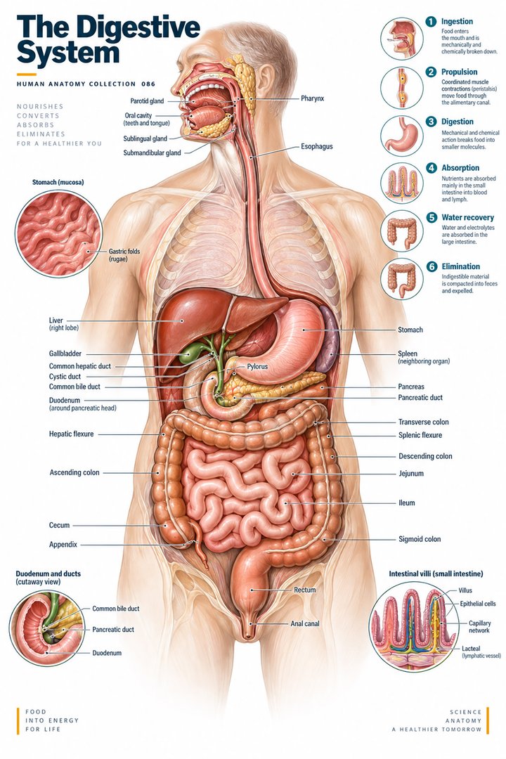
The digestive tract is the continuous luminal route from the oral opening to the anal outlet. It is also called the alimentary canal or gastrointestinal tract. Its regions do not all perform the same task: entry, propulsion, storage, mixing, chemical processing, absorption, and elimination are distributed across a connected sequence. The digestive system is a broader concept that includes organs contributing secretions and regulation without belonging to the route followed by a swallowed item. Recognizing that distinction is the foundation of an accurate anatomy diagram.
For a medical student or healthcare colleague, studying the tract means learning both the route and the wall. A familiar outline of the intestines can conceal the layers and interfaces that perform the work. Conversely, a beautiful tissue section can be difficult to interpret if the learner cannot place it along the route. This guide joins the two approaches: move from region to region, then consider what the regional arrangement permits and how it connects with other pathways. A swallowed item, an absorbed molecule, a vessel, and a neural signal should not all be represented by the same arrow.
Oral preparation begins before the esophagus
The oral cavity contains structures that prepare material for swallowing, including teeth and tongue, while saliva joins the contents. The tongue helps position and move the prepared bolus; the pharynx links the oral region with the esophageal route. Swallowing requires coordinated changes that direct the bolus while protecting the airway. The epiglottis is part of that coordination, not a stand-alone lid whose position explains the entire process. OpenStax: Mouth, pharynx, and esophagus
Latin cavitas oris names the oral cavity and lingua the tongue. These labels identify structures whose appearance varies across gross anatomy, clinical observation, and sectional images. A surface view of the mouth does not show the whole pharyngeal arrangement, and a sagittal diagram may remove one side to reveal deep relationships. When explaining swallowing, state the viewing plane and separate the food route from the nearby respiratory route. That clarification helps listeners understand why structures sharing a region can still lead to different destinations.
The esophagus is a transport segment with changing muscle
The esophagus connects the pharyngeal route to the stomach through neck, thoracic, and short abdominal portions. Its wall produces coordinated propulsion rather than relying simply on gravity. The upper portion contains predominantly skeletal muscle, the lower portion predominantly smooth muscle, and the intervening region includes a transition. Once the swallow passes its voluntary oral stage, subsequent coordinated phases are largely reflexive. NCBI Bookshelf: Gastrointestinal physiology
This arrangement explains why “a tube that drops food downward” is an incomplete description. A diagram should show the esophagus as an active wall enclosing a lumen, with neighboring structures relevant to orientation. The lower esophageal junction also requires careful terminology: a physiological sphincter region is not identical to a separate round object placed around an otherwise passive pipe. Learners can compare a gross diagram with a schematic of coordinated contraction, then identify which features each representation establishes. The first locates the route; the second illustrates a mechanism operating along it.
The stomach transforms the incoming material
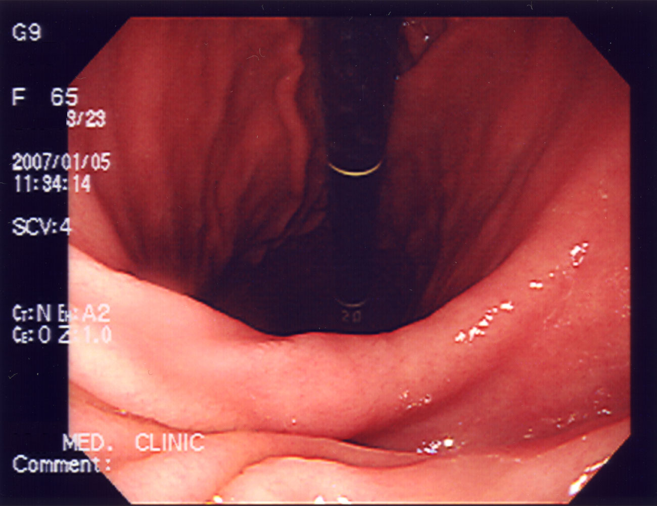
The stomach's regions include cardia, fundus, body, and pyloric arrangement. Its wall adds an oblique muscle orientation to circular and longitudinal components, supporting vigorous mixing. Gastric pits connect to glands, and the mucosa has specialized secretory cells rather than the villous arrangement associated with small-intestinal absorption. The pyloric region connects the gastric lumen with the duodenum. OpenStax: Stomach
The term chyme describes the mixed gastric contents passing onward, not a new organ or a separate anatomical compartment. A diagram that changes the appearance of the contents at the stomach can help communicate processing, provided the caption states that it is a schematic. Real material does not move through the tract as a single unchanged lump with a convenient label attached. Keeping content transformation separate from wall structure makes the explanation clearer: the organ supplies conditions and movement, while the luminal material changes during its passage.
The duodenum is a receiving and processing junction
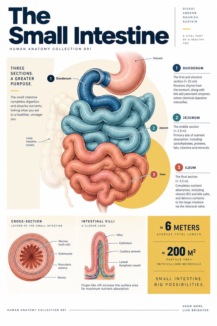
The proximal small intestine is the duodenum, followed by jejunum and ileum. The duodenum receives material from the stomach and joins it with associated digestive secretions. Its relationship to the pancreatic region and to posterior abdominal structures makes its shape and position different from the more freely arranged distal loops. The transition toward jejunum occurs at the duodenojejunal flexure. NCBI Bookshelf: Small intestine anatomy
This junction illustrates why proximity and continuity are different anatomical relationships. The pancreas contributes to the digestive process through a duct pathway, but a swallowed item does not pass through pancreatic tissue. Similarly, a vessel near a bowel segment supplies or drains its wall rather than carrying the unabsorbed contents of its lumen. A well-designed illustration keeps all three routes visible without joining them incorrectly. The educational question is not just “what is next to the duodenum?” but “how is it connected, what passes through that connection, and where does the material go afterward?”
Jejunum and ileum continue a long, adaptable surface
The small intestine has three named regions, but the transitions involve changing patterns rather than three detachable tubes with identical walls. Circular folds and villous arrangements contribute to the mucosal surface, with differences along the tract. Jejunal folds tend to be more prominent than ileal folds, and the terminal ileum joins the large-intestinal route. The length reported for bowel varies with individual anatomy and the way it is measured. NCBI Bookshelf: Small bowel anatomy
Latin intestinum tenue refers to the small intestine. “Small” does not mean that this is the shorter intestinal component; it refers to caliber in the familiar comparison. This distinction is worth stating because a learner may otherwise infer the wrong spatial scale from the everyday word. Measurements obtained in living, relaxed, fixed, or surgically handled tissue should not be treated as automatically interchangeable. A numeric length needs a method and context. For educational graphics, the relative order of the regions is often more defensible than pretending that one drawn loop count represents every person's bowel.
Folds, villi, and microvilli belong to different scales
Circular folds are gross mucosal-submucosal structures; villi are smaller mucosal projections; microvilli are features of the epithelial cell surface. They should not be labeled as three synonyms for the same structure. Together they illustrate how an organ can place a large functional surface within a compact region. Intestinal glands and associated cells contribute additional regional functions. OpenStax: Small and large intestines
A magnification diagram should therefore use three linked frames rather than one image with mixed-scale labels. The first frame shows the bowel segment, the second a local surface arrangement, and the third an epithelial boundary. This is not merely a design preference. It prevents a student from imagining a microvillus as something visible on a routine whole-organ photograph. It also helps colleagues explain why a histological image and a gross anatomy image can look so different while both concern the same segment. Scale is part of the anatomical information, not a decorative detail in the caption.
The common wall plan has important local differences
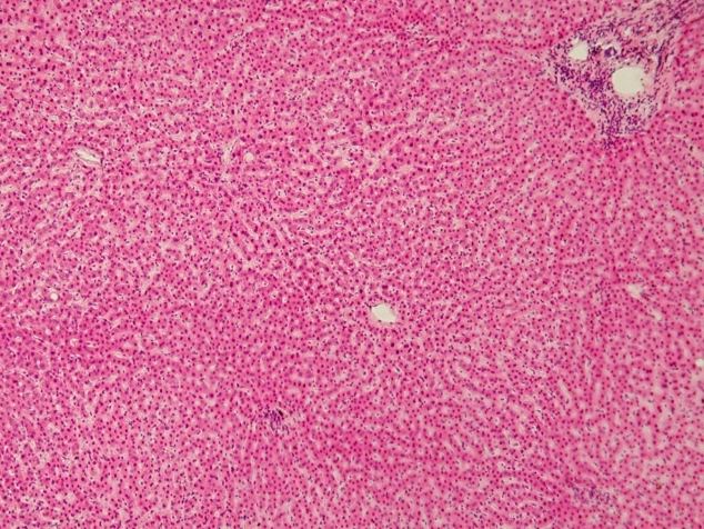
From the lumen outward, a common gastrointestinal wall plan identifies mucosa, submucosa, muscularis externa, and an outer covering. The mucosa itself includes epithelium, lamina propria, and muscularis mucosae. The outer covering varies according to the segment's relationships, with serosal or adventitial arrangements. The thin muscularis mucosae should not be confused with the larger muscle layers responsible for major movement of the wall. OpenStax: Digestive system overview
These distinctions matter when comparing tissue images. A thick connective area is not necessarily a muscular layer; a thin muscle strip near glands does not necessarily represent the outer propulsive layer. A learner should orient the section from lumen to outer surface and then examine texture and organization. When the section is tangential or incomplete, some layers may not be displayed cleanly. The correct response is to describe the limitation rather than force every expected layer into a poorly oriented field. Real specimens teach variation in visibility as well as textbook structure.
Movement is divided between propulsion and mixing
Peristalsis and segmentation illustrate different patterns of wall activity. Propulsive movement advances contents; mixing activity increases their interaction with secretions and surfaces. The balance changes by region and circumstances. Digestion, absorption, and motility are linked but distinct processes: breaking down material does not itself prove that the resulting molecules have crossed into the internal environment. OpenStax: Digestive processes and regulation
An original flow exercise places three labels beneath a drawing: movement, chemical processing, and absorption. At each region, students identify what the drawing actually shows and which label would require additional evidence. An arrow along the lumen demonstrates direction in the schematic; it does not quantify transport across the wall. A colored secretion entering the lumen shows a source relationship; it does not establish a rate of digestion. This exercise encourages precise explanations while remaining closely tied to gastrointestinal anatomy, where multiple processes occur in neighboring structures at the same time.
The large intestine has a recognizable external pattern
The large-intestinal route begins at the cecal arrangement and proceeds through ascending, transverse, descending, and sigmoid portions of the colon before the rectum and anal canal. Haustra form sacculations, and three longitudinal bands, the taeniae coli, distinguish much of the colon's external organization. Fat-containing omental appendices provide another gross feature. The appendix connects with the cecum rather than forming the next obligatory segment through which all contents must pass. NCBI Bookshelf: Large intestine anatomy
The numerical facts “three small-intestinal regions” and “three colonic longitudinal bands” count different anatomical categories. A comparison graphic should keep separate labels rather than placing them on a shared quantitative axis. Counts can be memorable without pretending that they measure equivalent amounts of tissue. The colon's outline also changes with filling and individual arrangement, so a loop drawn across the upper abdomen is a representative route rather than a fixed survey of every person's organ placement.
Colonic function continues processing rather than merely storing waste
The colon participates in absorption of water and electrolytes and in movement of contents toward the rectum. Haustral contractions and stronger mass movements describe different motility patterns. The microbial environment contributes to luminal processing, but the presence of microorganisms should not be reduced to a claim that every microbe or every product has the same effect. NCBI Bookshelf: Large intestine physiology
The anatomical distinction between a narrow lumen, a wall, and a surrounding vascular route remains important here. Changes in content consistency concern the lumen; absorption concerns crossing the epithelial interface; propulsion concerns coordinated wall activity. These processes occur together but can be described separately. A healthcare explanation becomes more understandable when it states which process is under discussion. Calling the colon a passive disposal pipe hides the active interfaces and movement that its regional structure supports.
The rectum and anal canal complete a coordinated outlet
The rectum follows the sigmoid route within the pelvis and transitions toward the anal canal. Its posterior pelvic position and neighboring relationships differ with reproductive anatomy. The arrangement supports temporary storage and coordinated evacuation, while continence depends on more than a single circular muscle label. NCBI Bookshelf: Rectum anatomy
The pelvic floor contributes muscular and connective support around anorectal, urinary, and reproductive pathways. Its activity must be considered alongside the terminal digestive route rather than as a separate platform unrelated to that route. NCBI Bookshelf: Pelvic floor
This organization is useful for learners working in hospitals or older-person care, where clear language about function and location matters. A report of a bowel movement describes an event, not an anatomical diagnosis. Information about timing, content, effort, and the person's experience can be documented distinctly without inferring a specific deep structural problem from one observation. Anatomical knowledge supports communication; it does not replace assessment of an individual.
Local neural networks connect the wall with regulation
The myenteric plexus lies between the circular and longitudinal muscle arrangements and is closely associated with motility control. Submucosal networks occupy a different wall location and contribute to regulation nearer the mucosa. Enteric organization includes local circuits while receiving communication from broader autonomic pathways. NCBI Bookshelf: Myenteric plexus
Parasympathetic and sympathetic pathways influence gastrointestinal activity, and the effects involve communication with enteric networks. The familiar “rest and digest” phrase is a broad teaching shorthand; it does not identify every regional effect or every timing pattern. NCBI Bookshelf: Gastrointestinal nervous control
An anatomy graphic should locate the networks rather than placing an unlabeled nerve outside the tract with an arrow to everything. Locating the myenteric and submucosal arrangements makes their different relationships visible. It also explains why the tract's wall contains more than muscle and glands: local sensing and coordination are built into the structure. The digestive tract is an organized responsive tissue pathway, not a sequence controlled only by commands traveling from the brain.
Absorption follows destinations beyond the lumen
Many absorbed nutrients enter vascular routes, while lipid transport also involves intestinal lymphatic pathways. Vitamin B12 illustrates a particularly useful regional connection: intrinsic factor originates from gastric parietal cells, and the complex is absorbed in the distal ileum. The location of production and the location of uptake therefore differ. NIH ODS: Vitamin B12
This is a powerful final route exercise. Begin with the luminal item, identify a contributing secretion, locate the epithelial crossing, and follow the destination beyond the tract. Do not skip from “food is digested” directly to “every molecule goes everywhere.” Regional anatomy supplies interfaces and connections that make each transition meaningful. For healthcare education, the strongest digestive-tract diagram combines continuity, local specialization, distinct wall layers, and separate transport routes. It shows how the same continuous tube changes its organization to perform different tasks while remaining linked to the rest of the body.
Anatomical Latin: quick reference
| Latin term | English meaning |
|---|---|
| Oesophagus | Esophagus |
| Gaster | Stomach |
| Intestinum tenue | Small intestine |
| Intestinum crassum | Large intestine |
Sources and image credits
Sources reviewed on 9 October 2026. Counts, estimates, study statistics and hypothetical calculations retain their context in the text.
- OpenStax: Mouth, pharynx, and esophagus
- NCBI Bookshelf: Gastrointestinal physiology
- OpenStax: Stomach
- NCBI Bookshelf: Small intestine anatomy
- NCBI Bookshelf: Small bowel anatomy
- OpenStax: Small and large intestines
- OpenStax: Digestive system overview
- OpenStax: Digestive processes and regulation
- NCBI Bookshelf: Large intestine anatomy
- NCBI Bookshelf: Large intestine physiology
- NCBI Bookshelf: Rectum anatomy
- NCBI Bookshelf: Pelvic floor
- NCBI Bookshelf: Myenteric plexus
- NCBI Bookshelf: Gastrointestinal nervous control
- NIH ODS: Vitamin B12
Explanatory prose and SVG graphics are original. Photograph authors, source records and licenses are credited beside each image. Catalog illustrations provide educational context; anatomical facts are supported by the linked sources.