Four chambers form a connected sequence
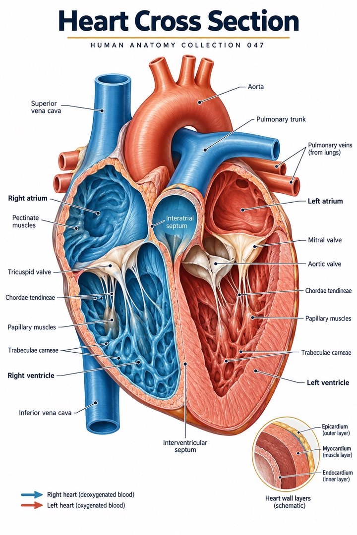
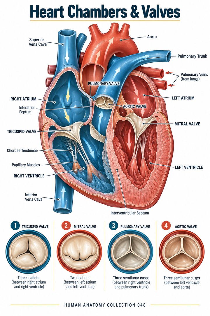
The human heart's standard chamber arrangement includes right atrium, right ventricle, left atrium, and left ventricle. The Latin labels are atrium dextrum, ventriculus dexter, atrium sinistrum, and ventriculus sinister. Atria receive returning blood, while ventricles supply the principal ejection into pulmonary and systemic pathways. Their organization is connected: the four chambers do not behave as unrelated containers. NHLBI: Heart anatomy
For healthcare learners, the best way to remember a chamber is to identify its inflow, its outflow, its internal landmarks, and its relation to neighboring structures. These four questions connect shape with function. They also help distinguish a simple circulation diagram from a real anatomical image. A diagram can arrange chambers into a neat square to make the route easy to follow. The living heart is rotated in the thorax, has uneven chamber shapes, and contains structures that cannot all be seen from one viewing direction. A correct route can therefore coexist with a deliberately simplified drawing.
The right atrium receives systemic and coronary return
The superior and inferior venae cavae supply major returning systemic pathways to the right atrium. The coronary sinus contributes return from much of the heart's own venous network. From this receiving chamber, blood passes through the right atrioventricular connection toward the right ventricle. The sequence distinguishes the route of blood through cardiac interiors from the separate coronary vessels supplying cardiac tissue. NHLBI: Blood flow through the heart
This distinction is easy to overlook in a poster showing many red and blue lines. A chamber contains circulating blood, but the myocardium still needs its own vascular supply. Drawing a coronary vessel on the external surface does not make that vessel an alternative entrance directly into every chamber. Learners should trace the luminal circuit first, then trace the tissue-supply circuit separately. Each trace answers a different question: where blood is being pumped, or how the pump's tissue receives support. Maintaining separate paths prevents a familiar color scheme from replacing anatomical reasoning.
The right atrium contains identifiable regions
The crista terminalis separates a smoother atrial region from the pectinate muscular arrangement, which is especially apparent toward the right atrial appendage. The fossa ovalis is a depression seen from the right side of the interatrial septum and relates to the developmental atrial communication. The fossa is a landmark, not an ordinary open passage through which adult blood normally moves from one atrium to the other. NCBI Bookshelf: Fossa ovalis
Latin musculi pectinati names the comb-like muscular ridges, while septum interatriale names the partition between atria. The names describe recognizable organization, yet a specimen's orientation affects what is visible. A section may omit the ridge or remove part of the appendage. A learner should not conclude that the structure is absent simply because it is not displayed in one image. Compare the cutting plane, the exposed surface, and the original caption before treating an educational photograph as a complete inventory of the chamber.
The right ventricle has an inflow and an outflow arrangement
The right ventricle lies prominently toward the anterior cardiac surface and has a complex internal geometry. The tricuspid connection defines inflow from the right atrium; the outflow continues toward the pulmonary valve and trunk. Trabeculated regions and a smoother outflow region make its interior different from a simple smooth-walled sphere. The tricuspid apparatus, septal relationships, and prominent muscular structures support identification across views. Anatomy for right ventricular lead implantation: Anatomical review, 2022
Thinking in terms of inflow and outflow is useful because a single chamber name can hide directional organization. A fluid route enters at one opening and exits at another, while the surrounding wall changes shape and pressure through time. A gross specimen can show the openings and muscular structures. A dynamic image can show changing geometry. A pressure record can show a physiological variable. None of these methods alone fully describes every aspect of the chamber, but their relationship provides a connected explanation of how the right ventricular arrangement serves the pulmonary circulation.
Trabeculae and the moderator band need precise labels
Trabeculae carneae are muscular ridges within ventricular interiors, not valve leaflets or blood vessels. The right ventricular moderator band is a named muscular connection associated with the conduction arrangement. A morphological study examined selected right ventricular conduction structures in fetal, child, and adult specimens, illustrating that these landmarks can be studied directly rather than merely inferred from a schematic. Morphological study of right ventricular conduction structures, 2012
In an educational image, a prominent band can appear to divide an interior into additional spaces. That appearance should not automatically alter the basic four-chamber count. A chamber classification is not produced by counting every recess or ridge on a cut surface. The learner should identify the relevant anatomical definition before adding a new label. This is especially important when a magnified specimen contains more internal detail than a basic textbook diagram. Increased detail expands understanding of the chamber; it does not make the simple diagram's count inherently wrong.
The left atrium receives pulmonary venous return
Pulmonary veins return blood from the lungs to the left atrium. A common anatomical pattern has four principal pulmonary veins, with superior and inferior routes from each lung, but venous ostia and branching show variation. The left atrium is a posterior cardiac structure, so its openings are not all exposed in a routine anterior view. Its relation to pulmonary venous connections helps distinguish it from the right atrium. NCBI Bookshelf: Pulmonary veins
The quantity four is therefore best presented as a common configuration, not a guarantee for every individual. In a diagram, an artist may combine venous openings to avoid visual crowding. The reader needs to know whether that choice represents a simplified route or a documented variant. A careful caption can say that the figure depicts a representative pattern and that individual anatomy differs. This small qualification improves accuracy without making the basic blood route difficult to understand. The central connection remains pulmonary return to left atrium, followed by passage toward the left ventricle.
The left ventricle supplies systemic ejection
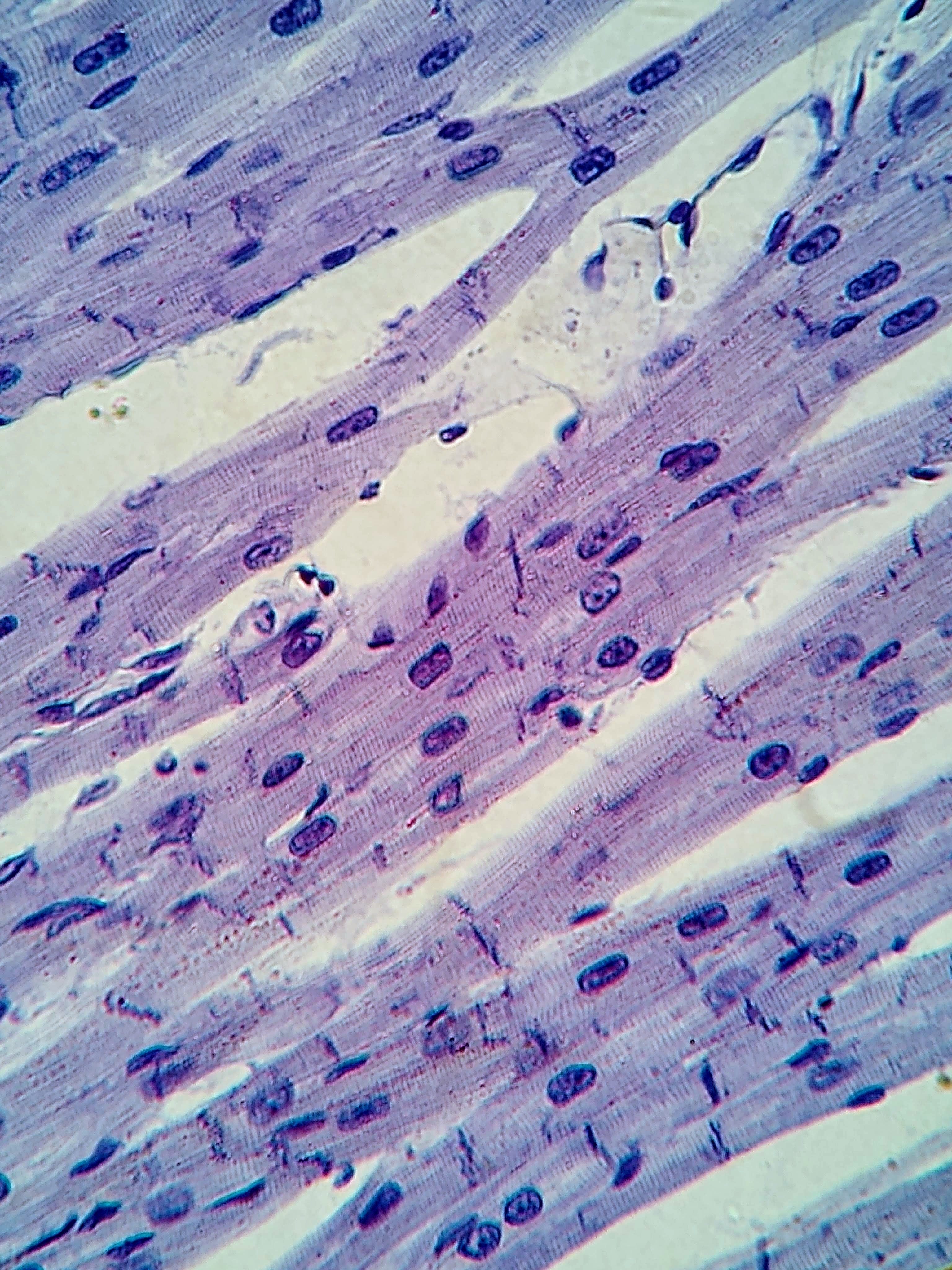
The left ventricle has a substantially thicker muscular wall than the right ventricle in the usual adult arrangement, reflecting the pressure demands of the systemic pathway. Its cavity, septal relationship, and contribution to the apex help distinguish it in a section. The wall includes endocardial lining, muscular myocardium, and an outer epicardial component; the surrounding pericardial arrangement is related but should not be substituted for the chamber's lumen. OpenStax: Detailed heart anatomy
Wall thickness is not the same measurement as chamber volume, and neither is the same as ejected volume. A thick wall surrounds a cavity whose dimensions change during the cycle. A photographic section can make one cavity appear larger or smaller depending on plane and state. When comparing ventricles, state which property is being compared. “Thicker wall,” “larger cavity in this view,” and “higher generated pressure” are different statements. This precision lets a learner connect anatomical appearance with function without assuming that one visible dimension proves every physiological feature.
The mitral apparatus joins several structures
The mitral valve lies between left atrium and left ventricle. Its apparatus includes leaflets, annular relationships, chordae tendineae, papillary muscles, and the ventricular wall supporting them. The familiar two-leaflet description is a useful gross framework, but the apparatus is more than two loose flaps. Chordal connections and surrounding geometry contribute to maintaining the relationship during changing chamber pressure. NCBI Bookshelf: Mitral valve anatomy
The Latin chordae tendineae identifies connective cords, and musculi papillares identifies ventricular muscle projections associated with them. Papillary muscles develop tension in the chordal arrangement during ventricular contraction; they do not act as strings pulled to open the valve. The leaflets respond to pressure relationships while the supporting apparatus limits inappropriate displacement. NCBI Bookshelf: Papillary muscles
An original model can illustrate this by using a flexible flap and supporting cords. First allow the flap to move with a pressure difference, then show how cords limit excessive motion in one direction. The model should explicitly state that it simplifies tissue properties and geometry. Its purpose is to correct a mechanism misconception, not to imitate the full valve apparatus.
Four principal valves separate different transitions
The tricuspid and mitral valves govern atrioventricular transitions; pulmonary and aortic valves govern ventricular outflow. The right-sided route proceeds through tricuspid then pulmonary connections; the left-sided route proceeds through mitral then aortic connections. A circulation map should place the lungs between right ventricular output and left atrial return rather than joining the two atria directly. This preserves the pathway that gives the chamber names their functional meaning.
The count of four principal valves and the count of four chambers refer to different categories. Neither should be inferred from the other. A valve is associated with a connection, not an additional chamber. An educational schematic can label each chamber with a shape and each valve with a distinct symbol. This visual separation is particularly useful for mixed healthcare audiences, where a first-time reader may otherwise treat every labeled object as one more compartment. The number of cusps or leaflets also describes a different level of structure from the number of whole valves.
Chamber filling and ejection happen in phases
During filling, blood enters the ventricles through atrioventricular pathways. During ejection, blood passes through outflow pathways into pulmonary trunk and aorta. Isovolumetric phases refer to intervals when ventricular pressure changes while volume remains approximately constant because the principal inlet and outlet valves are closed. These phases link changing chamber pressure, valve state, and chamber volume. OpenStax: Cardiac cycle
The word systole identifies contraction-related phases, while diastole identifies relaxation-related phases, but the chamber or phase being discussed should be specified. Atria and ventricles do not all contract and relax in an undifferentiated single event. A cardiac-cycle diagram should therefore state whether it is describing atrial or ventricular events. A time sequence is also different from a route diagram: one shows when structures change state, the other shows where blood travels. Placing the two beside each other helps students avoid trying to read time directly from a static set of anatomical arrows.
Electrical coordination is not the same as a mechanical image
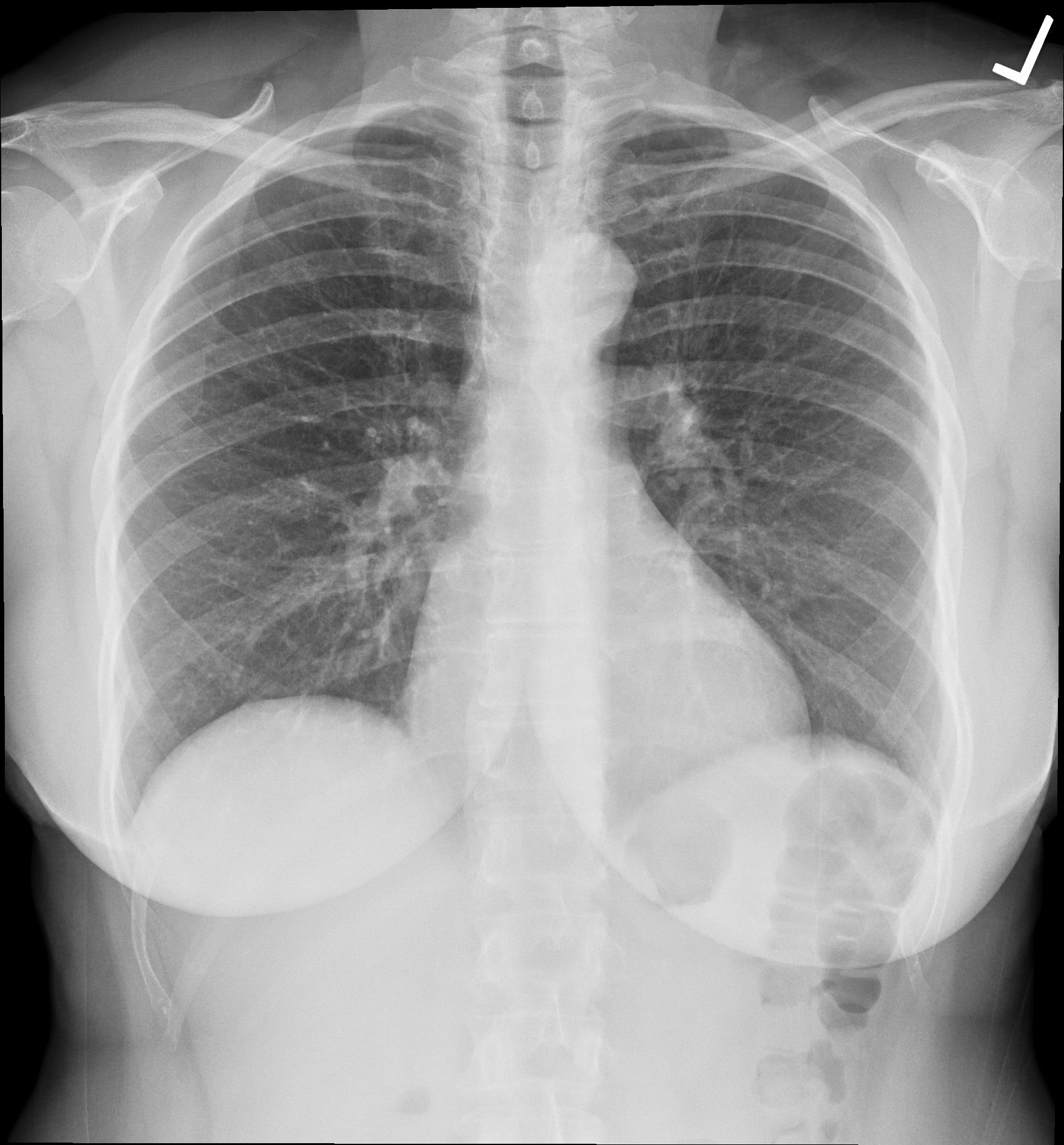
The sinoatrial node initiates the usual coordinated rhythm, with conduction through atrial and atrioventricular arrangements toward ventricular pathways. The sequence allows atrial and ventricular events to be coordinated rather than occurring as unrelated muscle contractions. Heart sounds relate to valve-closure events and associated vibrations; they do not provide a photograph of the chambers. NHLBI: How the heart beats
An electrical recording, a sound recording, and a dynamic image concern related events but do not measure the same thing. The distinction matters when interpreting an educational graphic that places them on one timeline. Their alignment illustrates a relationship, but a visible electrical deflection should not be labeled as the exact instant when every ventricular cell has finished a mechanical action. Learners should identify the recorded signal and then describe the associated event carefully. This approach keeps useful diagrams from implying greater precision than their simplified design actually supports.
Volume calculations make chamber function concrete
Stroke volume is the difference between end-diastolic and end-systolic ventricular volume. Ejection fraction compares that ejected amount with end-diastolic volume. Cardiac output combines stroke volume with heart rate. These quantities answer different questions and require their units or denominator to remain visible. OpenStax: Cardiac physiology
Use an explicitly hypothetical example: end-diastolic volume 120 milliliters and end-systolic volume 50 milliliters give stroke volume 70 milliliters. Seventy divided by 120 gives about 58 percent. At 75 beats per minute, 70 milliliters per beat gives 5.25 liters per minute. These are arithmetic teaching values, not measurements from a patient or a universal target. The example also shows why ejection fraction does not mean the chamber becomes completely empty: 50 milliliters remain in the hypothetical end-systolic state. A clear graphic should show the residual as well as the ejected amount.
Connected circuits impose a conservation relationship
In a closed circulation over time, right and left outputs must remain closely matched apart from small physiological pathway differences and transient changes. The left ventricular wall's greater thickness does not mean that it normally supplies twice the flow of the right side. Pressure demand and volume flow are different aspects of the work. Two pumps in series can move broadly comparable amounts while generating different pressures across different downstream conditions.
This relationship can be understood without introducing a clinical diagnosis. Imagine two reservoirs connected by pumps in a loop. If one pump transfers more volume than the other for an extended period, material accumulates in one reservoir and falls in the other. The thought experiment makes a hidden constraint visible. It is not a model of every cardiovascular mechanism, but it explains why “larger muscle means proportionally greater ongoing flow” is an unsafe inference. Anatomy supports the explanation, while conservation supplies the logical connection.
Reading chamber images and explaining their limits
Begin with orientation: anterior, posterior, sagittal, transverse, or another defined view. Identify a recognizable connection, then follow it to the chamber, valve apparatus, and adjacent partition. A real image may expose only part of a chamber and may have removed tissue to reveal its interior. A model may separate structures or change scale. These choices should be acknowledged rather than mistaken for the complete living arrangement. A clear anatomical description tells the reader which relationships are visible and which are supplied by a schematic explanation.
For practical learning, compare one route drawing with one specimen or imaging view. Trace the right-sided and left-sided sequences, then identify one internal landmark that the simple drawing omits. Explain why that landmark improves identification without changing the basic circuit. Finally, distinguish structure from a functional measurement: chamber shape, valve state, volume, and flow each contribute information, but none replaces all the others. The four heart chambers become memorable when they are understood as distinct anatomical arrangements working together in one continuous, coordinated circulation.
Anatomical Latin: quick reference
| Latin term | English meaning |
|---|---|
| Atrium dextrum | Right atrium |
| Atrium sinistrum | Left atrium |
| Ventriculus dexter | Right ventricle |
| Ventriculus sinister | Left ventricle |
Sources and image credits
Sources reviewed on 9 October 2026. Counts, estimates, study statistics and hypothetical calculations retain their context in the text.
- NHLBI: Heart anatomy
- NHLBI: Blood flow through the heart
- NCBI Bookshelf: Fossa ovalis
- Anatomy for right ventricular lead implantation: Anatomical review, 2022
- Morphological study of right ventricular conduction structures, 2012
- NCBI Bookshelf: Pulmonary veins
- OpenStax: Detailed heart anatomy
- NCBI Bookshelf: Mitral valve anatomy
- NCBI Bookshelf: Papillary muscles
- OpenStax: Cardiac cycle
- NHLBI: How the heart beats
- OpenStax: Cardiac physiology
Explanatory prose and SVG graphics are original. Photograph authors, source records and licenses are credited beside each image. Catalog illustrations provide educational context; anatomical facts are supported by the linked sources.