1. Lobes organize a complicated surface
The cerebral lobes provide a practical starting map for learning the brain. Their names divide a folded surface into recognizable regions, making it easier to describe where a structure lies and how it relates to neighboring tissue. For healthcare students and staff, this map supports the language used in teaching, imaging reports, and clinical explanations. It is most useful when treated as a spatial framework rather than a collection of isolated boxes.
A lobe is not a separate miniature brain. Connections cross regional boundaries and link the cortex with deeper structures. Begin each study session by identifying the view, hemisphere, and surface shown. Then establish the major landmarks before adding functional associations. This sequence makes the same names usable across a poster, a specimen photograph, and a section through the head.
2. Four surface lobes and a concealed insula
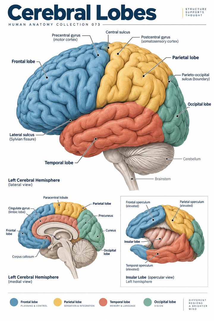
The four familiar lobes visible on the external cerebral surface are frontal, parietal, temporal, and occipital. The insula lies deeper within the lateral sulcus and is often described as a fifth lobe. Some anatomical classifications also discuss a limbic lobe on the medial surface. State which classification a diagram uses instead of treating different counts as necessarily contradictory. [National Academies: Major Structures and Functions of the Brain] [NCBI Bookshelf: The Anatomy of the Cerebral Cortex]
The brain has two cerebral hemispheres, and the same broad regional names apply on both sides. A single lateral picture therefore shows only one side of the arrangement. When teaching a group, label the hemisphere before asking anyone to name a lobe. A left-side surface view and a right-side surface view can look similar after rotation, while carrying different laterality information.
3. Use sulci to establish boundaries
A gyrus is a ridge of cortex; a sulcus is a groove. The central sulcus separates the frontal and parietal lobes. The lateral sulcus helps separate the temporal lobe from the frontal and parietal regions above it. The parieto-occipital sulcus is especially helpful on the medial surface, while the full lateral boundary of the occipital lobe also uses conventional anatomical landmarks. [OpenStax: central nervous system]
These landmarks are more dependable for study than the colors chosen by an illustrator. A poster may use bright green for one lobe, but another atlas may use green elsewhere. Trace the groove and explain the neighboring regions without relying on color. Then compare a smoother schematic with an actual specimen, where folds can appear irregular and the chosen section may hide an expected landmark.
4. The frontal lobe connects action with planning
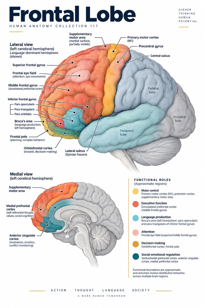
The frontal lobe lies anterior to the central sulcus. Its posterior precentral gyrus contains the primary motor cortex, while more anterior regions contribute to complex cognitive processes. Introductory descriptions often associate frontal regions with planning, judgment, and aspects of behavioral regulation. These broad associations are a starting point; they do not mean that every such process belongs to one small place. [OpenStax: central nervous system] [NIMH: Get to Know Your Brain]
For a practical explanation, distinguish a motor landmark from a general cognitive association. Point to the precentral region when describing the cortical motor map, then explain why planning an action also involves connected systems. This prevents an anatomical chart from implying that movement is produced by a single independent command center. It also helps learners separate a region’s location from the much larger question of how a task is performed.
5. The parietal lobe integrates sensory relationships
The parietal lobe lies posterior to the central sulcus. The postcentral gyrus contains primary somatosensory cortex. Other parietal regions contribute to integrating sensory information and relationships involving the body and surrounding space. A basic map therefore links this lobe with sensation while also recognizing that its contribution extends beyond a simple receiving station. [OpenStax: central nervous system]
Compare the precentral and postcentral gyri together. Their names encode a relationship to the same sulcus: one is before it and the other behind it. This pairing is an efficient memory aid because it retains the landmark that makes the distinction meaningful. In a handover or report, however, use the precise term provided by the clinical assessment rather than inferring a cortical location from a single symptom.
6. The temporal lobe supports hearing and memory networks
Temporal regions lie below the lateral sulcus. Their functions include auditory processing, while medial temporal structures participate in memory-related networks. The hippocampus is a deeper medial temporal structure, not a visible label on every external surface view. A diagram showing only the outer cortex cannot be assumed to reveal the internal organization of the entire lobe. [OpenStax: central nervous system] [Neuroscience: internal anatomy of the cerebral hemispheres]
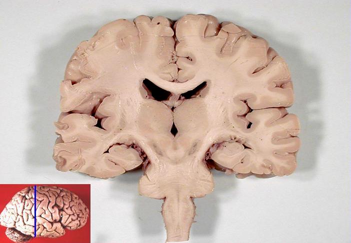
This distinction is useful when comparing a lateral poster with a coronal specimen. The poster establishes broad external location; the section reveals tissue that was concealed in that view. Ask learners to name what has changed in the representation. They should describe the cut surface and depth before transferring an external label to a structure that happens to appear nearby in two dimensions.
7. The occipital lobe contributes to visual processing
The occipital lobe occupies the posterior cerebral region. Primary visual cortex is associated with the medial occipital surface around the calcarine sulcus. Processing visual information also involves connections with other cortical areas. An introductory association between the occipital lobe and vision is therefore accurate as a starting point, but incomplete as an explanation of recognizing an object or navigating a room. [OpenStax: central nervous system]
A useful exercise is to place a lateral image beside a medial view and ask where a familiar functional label has moved. Nothing has physically changed in the brain; the representation has exposed a different surface. This exercise reinforces a general anatomical principle: a structure may be important even when it is hidden in the most commonly used illustration.
8. The insula illustrates the importance of depth
The insula is concealed by surrounding opercular regions in an ordinary external view. Opening the lateral sulcus exposes it. Its position makes it an excellent example of why a lobe map should distinguish visible surface regions from deeper cortex. Descriptions of its functions include contributions to internal bodily sensation and taste-related processing within wider networks. [NCBI Bookshelf: The Anatomy of the Cerebral Cortex] [OpenStax: central nervous system]
Do not draw the insula as another patch added onto an intact lateral surface without explaining the exposure. Use a caption such as “surrounding tissue moved aside” if that is the representation intended. In a teaching discussion, ask what the illustrator removed or displaced to make a label visible. The answer connects the colored shape with a three-dimensional structure.
9. Language and cognition cross simple lobe labels
Language assessment considers several abilities, including comprehension, naming, repetition, and production. Introductory maps often emphasize left-hemisphere frontal and temporal language regions, but language depends on connected pathways and must be assessed in context. The popular claim that people divide into a logical left-brain type and a creative right-brain type is not a useful clinical classification. [OpenStax: mental status examination]
Likewise, a reported difficulty does not permit an unqualified anatomical conclusion from a teaching chart. The same observed task can draw on sensory input, attention, language, memory, and motor output. A professional explanation should name the observed difficulty and the established clinical findings. Use lobe associations to understand the vocabulary, while keeping diagnosis tied to the actual assessment and investigations.
10. Latin terms make the map portable
The Latin forms lobus frontalis, lobus parietalis, lobus temporalis, and lobus occipitalis correspond to the familiar English lobe names. Insula is used directly. Gyrus precentralis and gyrus postcentralis describe ridges on either side of sulcus centralis. Learning these expressions as complete terms helps preserve both the structure and its relationship to an anatomical landmark. [FIPAT: Terminologia Anatomica, second edition]
Use the vocabulary in a short description: identify a hemisphere, locate a sulcus, and name the region beside it. Then translate the description into ordinary English for a patient-facing explanation. This practice is useful across medical school, hospital departments, practices, and eldercare because the same region can appear in a technical report and in a conversation with someone unfamiliar with anatomical terminology.
| English | Selected Latin |
|---|---|
| Frontal lobe | Lobus frontalis |
| Parietal lobe | Lobus parietalis |
| Temporal lobe | Lobus temporalis |
| Occipital lobe | Lobus occipitalis |
| Central sulcus | Sulcus centralis |
| Precentral / postcentral gyrus | Gyrus precentralis / gyrus postcentralis |
11. Compare a real scan with a specimen photograph
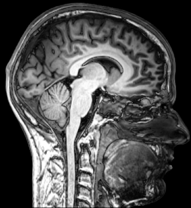
The sagittal MRI below shows an approximately midline section of a living human head. The face points toward the image’s right. The coronal specimen photograph shows a different plane and includes an original orientation inset. Its visible cut surface should not be mistaken for an intact external view. These are real images with different subjects, preparation methods, and purposes.
Begin by explaining what each view reveals and conceals. Look for broad relationships between the cerebrum and deeper structures rather than claiming to identify every colored region from a poster. A lobe map helps orient the viewer, while the actual images demonstrate why anatomy cannot be reduced to one idealized perspective. Preserve the credits and captions when sharing either image for teaching.
12. Build a study routine around relationships
Choose one surface landmark and explain two relationships around it. Change to another view and identify which relationship remains visible. Finish by describing one function as a contribution within a network. This three-step routine combines location, representation, and function without requiring a long recitation of labels.
For team education, let one colleague describe the view while another identifies a region. Discuss disagreements about side, depth, or terminology before discussing function. In eldercare and other patient-facing settings, use the guide to clarify established information about the person’s condition and communication needs. The strongest learning outcome is the ability to explain the map accurately while recognizing what the map alone cannot establish.
Sources and image credits
Sources checked on 8 October 2026. Numerical examples, anatomical classification counts, and research estimates are identified separately in the text.
- OpenStax: central nervous system
- OpenStax: mental status examination
- NCBI Bookshelf: The Anatomy of the Cerebral Cortex
- National Academies: Major Structures and Functions of the Brain
- Neuroscience: internal anatomy of the cerebral hemispheres
- NIMH: Get to Know Your Brain
- FIPAT: Terminologia Anatomica, second edition
Explanatory prose and simplified SVG graphics are original. Photographs retain their stated Creative Commons licenses; creator and source links appear beside each image. Catalog artwork is educational and decorative; no clinical authorship or peer-review credential is claimed.