1. A cell diagram is a model of organization
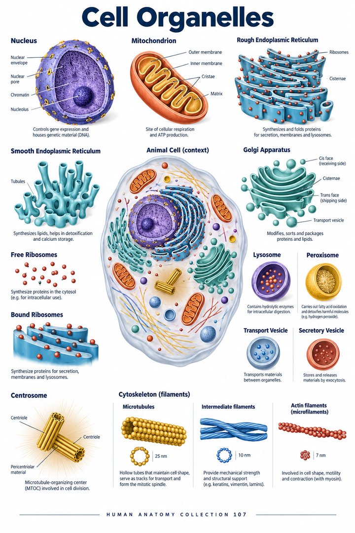
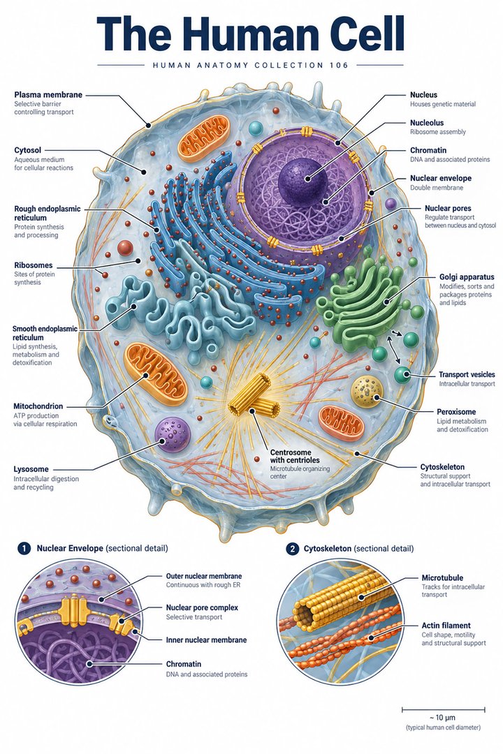
Human cell structure provides a bridge between visible anatomy and the processes that sustain tissues. A typical cell diagram shows a membrane, nucleus, and several organelles, but it combines features into a deliberately readable model. Real cells differ in shape, size, contents, and specialization. A neuron, epithelial cell, skeletal muscle fiber, and mature red blood cell should not be imagined as identical spheres with different labels.
For healthcare learners, the most useful question is how a structure supports a process. Relate the membrane to exchange, the nucleus to genetic information, and intracellular compartments to specific tasks. Then connect those tasks with the tissue in which the cell operates. This approach supports later study of organs without claiming that a clean illustration reproduces everything visible in an actual microscopic preparation.
2. The plasma membrane establishes an active boundary
The plasma membrane contains a phospholipid bilayer and associated proteins. Its organization permits selective exchange and supports signaling and attachment. The membrane is dynamic rather than a rigid wall. Channels, carriers, and pumps provide different transport mechanisms, while receptors help cells respond to particular signals. A membrane drawing should distinguish the lipid boundary from the proteins operating within it. [OpenStax: cell membrane]
When teaching membrane transport, specify the substance, route, and direction. An arrow crossing a cell outline does not explain whether movement occurs through the lipid layer, a channel, or another mechanism. Add those details before discussing energy requirements. This habit makes a diagram informative without making it excessively crowded and helps learners avoid using “diffusion” as a name for every movement into or out of a cell.
3. Cytoplasm and cytosol are related but different
Cytoplasm describes cellular contents between the plasma membrane and the nucleus, including organelles and cytosol. Cytosol is the fluid component. The cytoskeleton includes protein systems that contribute to shape, intracellular transport, and movement. These structures connect apparently separate compartments into an organized working environment. An organelle suspended in a textbook picture is therefore part of a coordinated cell rather than an independent machine. [OpenStax: cellular organelles]
Compare a diagram’s labels for cytoplasm and cytosol. If both point to the same empty background without explanation, ask what distinction the illustration intends. In a practical lesson, list a structure that belongs to the cytoplasm but is not merely cytosol. This tests understanding of categories, which becomes important when a report refers to a change in the cytoplasm rather than a specific organelle.
4. The nucleus stores and organizes genetic information
The nuclear envelope separates nuclear contents from the cytoplasm, with pores supporting regulated exchange. DNA is organized with proteins as chromatin; chromosomes become particularly conspicuous during cell division. The nucleolus participates in ribosome assembly. A dark nuclear profile in a stained light micrograph does not show every chromosome as an individually countable object. The preparation method determines which details can be seen. [OpenStax: nucleus and DNA]
For study, distinguish the location of information from its use. Nuclear genetic information supports RNA production, while protein synthesis occurs on ribosomes. Avoid drawing a protein emerging directly from a chromosome into the bloodstream. A sequence diagram can preserve the intermediate steps and show how nuclear activity connects with the cytoplasm, membranes, and eventual destination of a protein.
5. Ribosomes, endoplasmic reticulum, and Golgi apparatus
Ribosomes are sites of protein synthesis and are not membrane-bound organelles. Rough endoplasmic reticulum has associated ribosomes and participates in processing proteins destined for secretion or membranes. Smooth endoplasmic reticulum supports other tasks, including lipid-related processes. The Golgi apparatus modifies and sorts cargo. These labels describe relationships in a trafficking system, not merely differently shaped structures. [OpenStax: cellular organelles]
Trace a simplified secreted-protein route from a ribosome associated with rough endoplasmic reticulum toward Golgi processing and a secretory vesicle. State that the drawing concerns one selected route, since proteins have many destinations. This limitation makes the schematic more accurate. The exercise also helps explain why a cell specialized for secretion can have a different appearance from a cell whose main function is contraction.
6. Energy production and intracellular recycling
Mitochondria participate in producing ATP through cellular respiration and have their own genetic material. Lysosomes support digestion of material within the cell, while peroxisomes contain enzymes involved in oxidative reactions. Calling mitochondria “powerhouses” can provide a memory aid, but a useful explanation names ATP and connects energy availability with cellular work. Recycling and quality control are similarly active processes rather than passive disposal. [OpenStax: cellular organelles]
In a teaching exercise, pair a task with its supporting structures instead of assigning each organelle a single slogan. Consider a cell that transports substances against a concentration gradient or repeatedly secretes proteins. Ask which processes require energy and which compartments participate. The answer should connect several structures. This prevents a vocabulary list from becoming a misleading set of one-to-one equivalences.
7. Chromosome counts need an explicit cellular context
Most human somatic cells are diploid, with forty-six chromosomes arranged in twenty-three pairs. Gametes normally contain twenty-three chromosomes. These conventional counts must not be expanded into “every human cell has forty-six chromosomes.” Mature erythrocytes lack a nucleus, and other specialized cellular arrangements also complicate that statement. Always specify the cell type and the biological context of a count. [NHGRI: diploid chromosomes] [Molecular Biology of the Cell: chromosome organization]
The graphic below compares chromosome counts, not the amount of total genetic material in every cell at every stage. DNA replication and division require additional context, and mitochondria introduce another genetic compartment. When discussing a numerical result with students, first name what is being counted. A chromosome, chromosome pair, nucleus, and cell are different units that should not be substituted for one another.
8. Real microscopy reveals only selected levels of detail
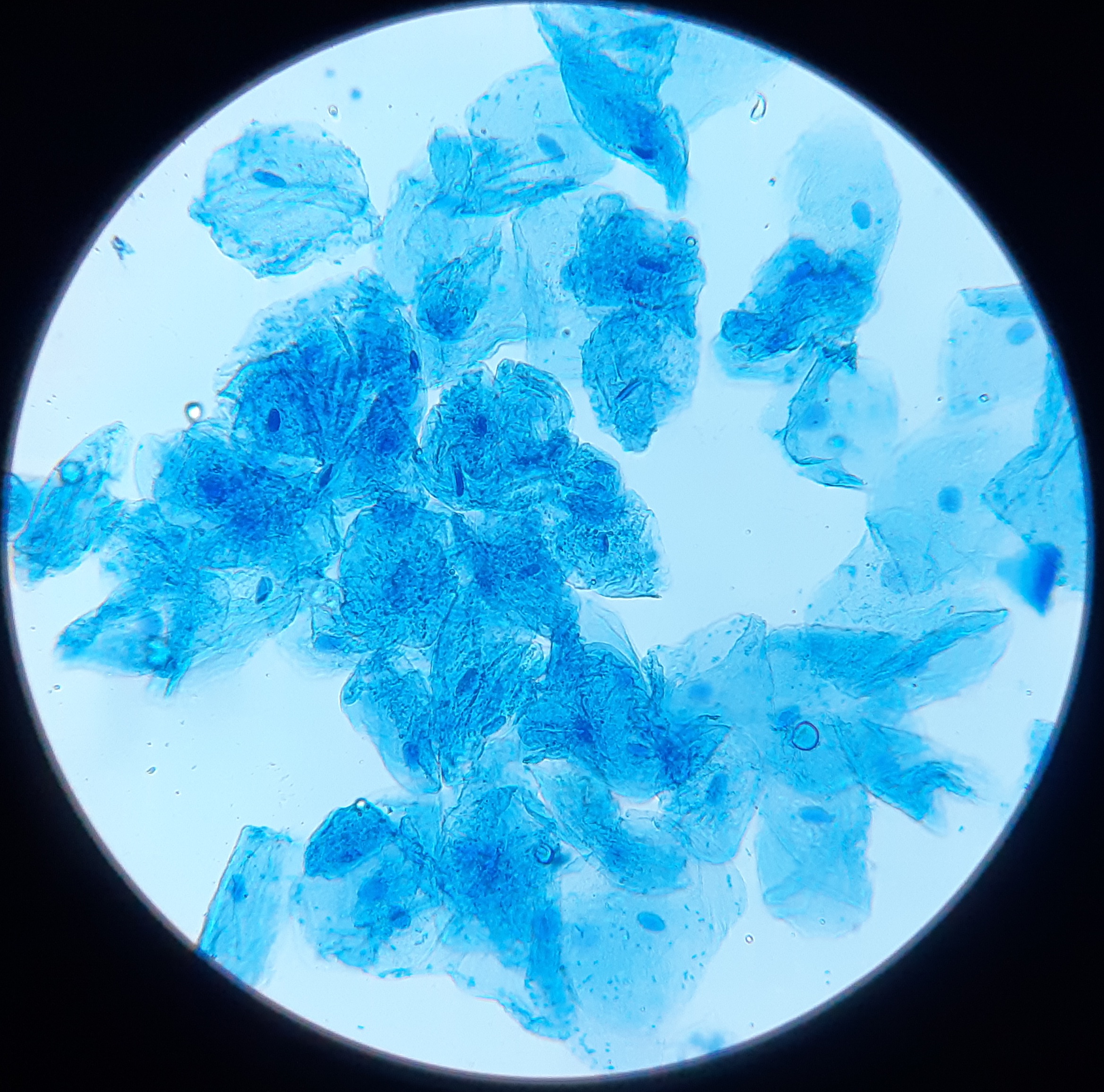
The cheek-cell photograph below is a real light-microscope image. Its source does not specify a stain or scale, so no magnification or exact cell diameter is inferred here. A recognizable cell profile and nuclear region do not make every organelle on the featured poster visible. Resolution, contrast, sample preparation, and the section or focal plane determine the available information.
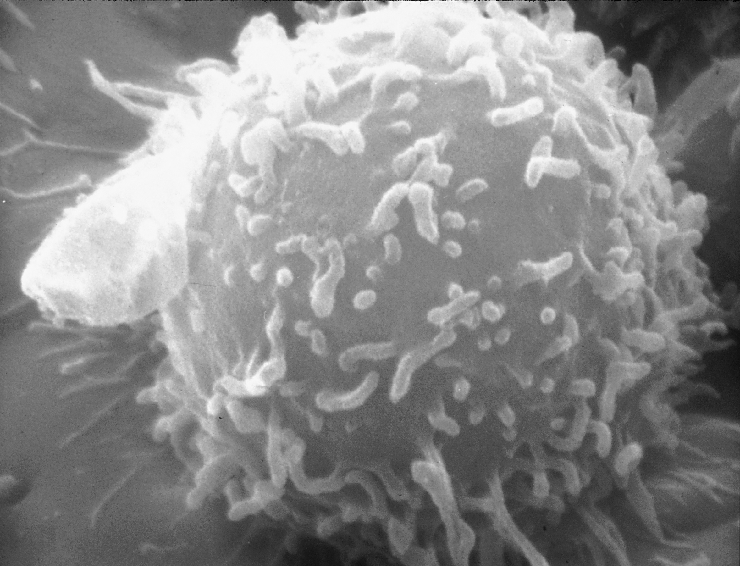
The lymphocyte electron micrograph records a different cell using a different imaging method. Compare the two by asking which boundaries and internal features can be distinguished, then identify what remains uncertain. The lymphocyte’s appearance does not establish whether it is a B or T cell. This comparison teaches appropriate confidence: an image can provide real structural evidence without answering every question about cellular identity or function.
9. Cellular terms cross Latin and modern scientific vocabulary
Cellula, nucleus, nucleolus, and cytoplasma are useful Latin or Latinized expressions. Modern organelle names also belong to an international scientific vocabulary that draws on several linguistic traditions. Do not assume that every name on a cell poster is an official gross-anatomy Latin label. Use the term consistently and pair it with a clearly defined structure or process.
Build a study table with three columns: structure, process, and an image limitation. For the nucleus, the limitation might be that the photograph does not resolve individual chromosomes. For the plasma membrane, a light micrograph may show a boundary without resolving the phospholipid bilayer. This table connects vocabulary with evidence and helps healthcare teams understand why different laboratory methods answer different questions.
| Structure | Selected scientific term | Learning connection |
|---|---|---|
| Cell | Cellula | Specialized living unit |
| Nucleus | Nucleus | Genetic organization |
| Cytoplasm | Cytoplasma | Cytosol plus non-nuclear contents |
| Endoplasmic reticulum | Reticulum endoplasmaticum | Intracellular processing network |
10. Connect cellular structure with healthcare learning
Medical students can use cell organization to explain why tissue specialization matters. Practice assistants and hospital staff encounter terms such as cellular sample, nuclear appearance, and cytoplasm in laboratory communication; the terms describe levels of observation rather than diagnoses by themselves. Physicians can use a simplified diagram to explain what a test examines, while retaining the limits of the actual method.
For eldercare education, relate cells to the tissues and organs involved in an established care discussion rather than using microscopic concepts to infer treatment. Finish revision by explaining one route through the cell and one exception to the typical diagram. If you can describe both, you have learned a model with its limitations, which is more useful than memorizing an idealized colored picture.
Sources and image credits
Sources checked on 8 October 2026. Numerical examples, anatomical classification counts, and research estimates are identified separately in the text.
- OpenStax: cell membrane
- OpenStax: cellular organelles
- OpenStax: nucleus and DNA
- NHGRI: diploid chromosomes
- Molecular Biology of the Cell: chromosome organization
Explanatory prose and simplified SVG graphics are original. Photographs retain their stated Creative Commons licenses; creator and source links appear beside each image. Catalog artwork is educational and decorative; no clinical authorship or peer-review credential is claimed.