1. Endocrine anatomy is a map of communication
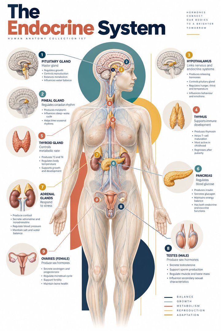
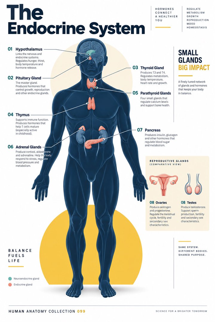
The human endocrine system coordinates functions through hormones and the tissues that respond to them. Its anatomy includes conspicuous glands and less obvious hormone-producing cells within other organs. A whole-body diagram is helpful because the sites are distributed rather than joined into one continuous tube. The connecting relationship is communication through the circulation and interaction with appropriate receptors.
For healthcare learners, separate three questions: where a hormone is produced, how its release is regulated, and which tissues respond. These questions are easy to merge when a poster places one hormone name beside one gland. A richer explanation connects several organs and includes feedback. The same framework helps medical students study physiology and helps clinical teams understand terminology in laboratory requests and reports.
2. Hormone action requires a responsive target
Hormones influence growth, development, metabolism, sexual function, and other processes. A disorder can involve inadequate or excessive secretion, but it can also involve reduced responsiveness to a hormone. The circulating message and the response of the target are therefore different parts of the system. A measurement of concentration alone does not describe every aspect of endocrine function. [NIDDK: endocrine diseases and hormone function]
Use a communication analogy carefully: the gland sends a message, but the recipient must have the relevant machinery to receive and act on it. Unlike a simple telephone call, biological responses depend on receptor expression and intracellular processes. When explaining a test, name what was actually measured. Avoid turning one laboratory value into a complete statement about the gland, target tissues, symptoms, and treatment.
3. The hypothalamus and pituitary form a regulatory junction
The pituitary, or hypophysis, lies in the sella turcica and connects anatomically and functionally with the hypothalamus. The adenohypophysis synthesizes several major hormones. The neurohypophysis stores and releases hormones made by hypothalamic neurons, including antidiuretic hormone and oxytocin. It is inaccurate to describe the posterior pituitary as the site where those hormones are originally synthesized. [OpenStax: pituitary and hypothalamus]
A useful drawing places synthesis, transport, storage, and release in separate locations. Learners can then follow a hormone without assigning every step to the most visible gland. This is also a general lesson in physiology: the location from which a substance enters blood need not be the location in which the substance was produced. Keep both locations explicit when explaining an endocrine pathway.
4. Feedback links glands into an axis
The hypothalamic–pituitary–thyroid axis provides a familiar example: hypothalamic thyrotropin-releasing hormone supports pituitary thyroid-stimulating hormone release, and thyroid hormone feeds back on regulatory levels. This is a connected control arrangement rather than three independent secretions. A pathway illustration should show the direction of stimulation and distinguish feedback from onward hormone delivery. [OpenStax: pituitary and hypothalamus]
Cover the arrows in a diagram and redraw them with two styles: one for the forward regulatory sequence and one for feedback. Then explain what information a gland receives from the wider system. The exercise encourages relational thinking and helps prevent the common mistake of treating the term “master gland” as meaning that the pituitary operates without hypothalamic input or feedback.
5. Thyroid and parathyroid glands are distinct structures
The thyroid has two main lobes joined by an isthmus. Follicles provide a recognizable microscopic organization. The parathyroid glands are separate structures, typically four in number, with individual variation. Parathyroid hormone contributes to calcium regulation. Similar names and close location do not make the thyroid and parathyroids interchangeable. [NCI SEER: thyroid and parathyroids]
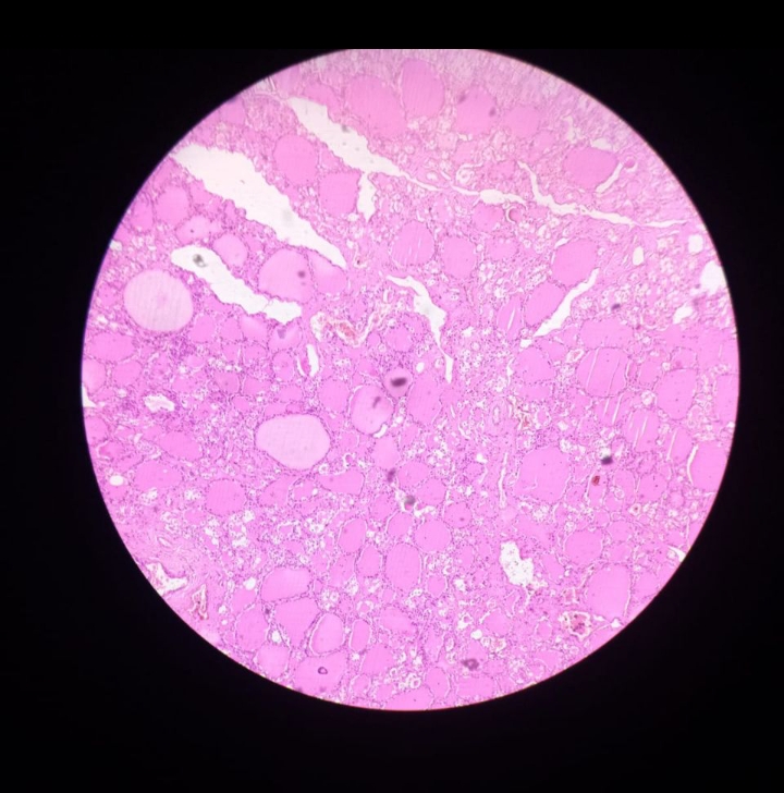
In a diagram exercise, draw the thyroid first and then add the parathyroids as distinct structures rather than dots labeled with thyroid hormones. On a microscopic image, identify the overall follicular pattern before trying to connect it with a whole-body outline. The comparison helps explain why a gland can have a simple external silhouette while containing specialized tissue arrangements that support its endocrine role.
6. Adrenal cortex and medulla have different identities
Two adrenal glands lie near the kidneys. Each has an outer cortex and inner medulla. The cortex is described with three zones and produces steroid hormones, including cortisol and aldosterone. The medulla produces catecholamines such as epinephrine and norepinephrine. A single adrenal label on a body poster therefore conceals functionally different tissue compartments. [NCI SEER: adrenal glands]
Study the organ at two scales: its position relative to the kidney and its internal tissue organization. A location image cannot establish which compartment is producing a particular hormone. Use a color key that remains consistent across views, and state when a drawing is a section. This habit connects anatomical depth with physiology while avoiding the idea that all adrenal hormones arise from one uniform tissue.
7. The pancreas illustrates an organ with two routes
Pancreatic islets contain endocrine cell populations, including insulin-producing beta cells and glucagon-producing alpha cells. The pancreas also has exocrine tissue whose secretions travel through ducts toward the duodenum. Endocrine release into the circulation and exocrine secretion through a duct are different routes within the same organ. [NCI SEER: endocrine pancreas]
Trace both routes on a simple sketch. If a hormone arrow enters a pancreatic duct, reconsider what the diagram is claiming. This exercise explains why classifying an entire organ with one label can be insufficient. For healthcare teams, a clear explanation of the route also helps interpret whether a report concerns glandular anatomy, circulating hormones, or a digestive function.
8. Other organs extend the endocrine map
The endocrine map is broader than the major glands usually highlighted in an introductory poster. The kidneys, for example, have hormone-related roles connected with red-cell production and mineral balance. Gonadal tissues combine hormone production with reproductive functions. These examples show how the endocrine system overlaps with other organ systems rather than occupying a separate, sealed anatomical territory. [NIDDK: how the kidneys work] [NIDDK: endocrine diseases and hormone function]
Create a study card for an organ you usually associate with another system. Add an endocrine connection and specify the responding tissue or physiological relationship. This encourages integrated learning. In a clinical conversation, however, do not infer a person’s endocrine status from the presence or position of the organ alone; function requires the relevant history, assessment, and measurements.
9. Numerical anchors are classification facts
Useful introductory counts include two thyroid lobes, two adrenal glands, and typically four parathyroid glands. The adrenal cortex is conventionally divided into three zones. These counts belong to different anatomical categories, and the parathyroid count varies. They are learning anchors rather than epidemiological rates, measurements of hormone output, or criteria for diagnosing an individual. [NCI SEER: thyroid and parathyroids] [NCI SEER: adrenal glands]
When making a numerical graphic, put the counted structure beside the value. Explain variation where appropriate and avoid presenting these counts as comparable measures of organ size or importance. The graphic below groups selected conventional counts only. It intentionally avoids hormone concentration ranges, because laboratory reference intervals depend on the assay, population, timing, and clinical context of the actual measurement.
10. Latin terms and real tissue support the same map
Selected anatomical names include glandula thyroidea, glandula parathyroidea, glandula suprarenalis, and hypophysis. Adenohypophysis and neurohypophysis retain the pituitary’s important internal distinction. Insulae pancreaticae refers to pancreatic islets. Learn a term with its location and a functional relationship, rather than assuming its familiar appearance tells you all of its physiology. [FIPAT: Terminologia Anatomica, second edition]
The real thyroid micrograph below adds tissue organization to the external gland outline. Its colors come from histological staining, not natural tissue coloration. A single micrograph cannot establish the complete hormonal state of the person from whom tissue originated. Use it to compare visible structure with the simplified poster, then return to the cited sources for the physiological relationships that the image cannot directly show.
| English | Selected Latin |
|---|---|
| Thyroid gland | Glandula thyroidea |
| Adrenal gland | Glandula suprarenalis |
| Pituitary gland | Hypophysis |
| Pancreatic islets | Insulae pancreaticae |
11. A practical framework for study and communication
For medical students, follow one endocrine axis and explain its feedback before memorizing additional hormones. For practice assistants and hospital personnel, retain the name of the requested test, collection context, and reported units. Physicians can use a gland map to introduce a discussion while explaining that hormone effects depend on responsive tissues and the wider regulatory system.
In eldercare teaching, connect the anatomy with the person’s established condition and care plan. A general endocrine illustration supports understanding but does not set medication schedules or interpretation thresholds. Finish a teaching session by asking learners to distinguish a gland’s location, a hormone’s source, and its target. Clear answers to those three questions provide a reusable foundation for later physiology.
Sources and image credits
Sources checked on 8 October 2026. Numerical examples, anatomical classification counts, and research estimates are identified separately in the text.
- FIPAT: Terminologia Anatomica, second edition
- NIDDK: endocrine diseases and hormone function
- OpenStax: pituitary and hypothalamus
- NCI SEER: adrenal glands
- NCI SEER: thyroid and parathyroids
- NCI SEER: endocrine pancreas
- NIDDK: how the kidneys work
Explanatory prose and simplified SVG graphics are original. Photographs retain their stated Creative Commons licenses; creator and source links appear beside each image. Catalog artwork is educational and decorative; no clinical authorship or peer-review credential is claimed.