1. The nervous system is a network of relationships
The human nervous system supports sensation, movement, regulation, and the integration of experience. Its anatomy is easiest to learn when you connect a structure with the information it receives and the response it helps produce. A brain diagram, a nerve chart, and a microscopic neuron image are different views of that network. None should be mistaken for a complete explanation of how a person thinks or acts.
For medical students and healthcare teams, the starting point is a clear description of the task or change being discussed. “Difficulty lifting the left foot” is a different kind of statement from “a nerve problem.” The first describes a function and side; the second suggests an interpretation that needs further assessment. Anatomy becomes clinically useful when it improves that description before it is used to construct an explanation.
2. Distinguish central and peripheral organization
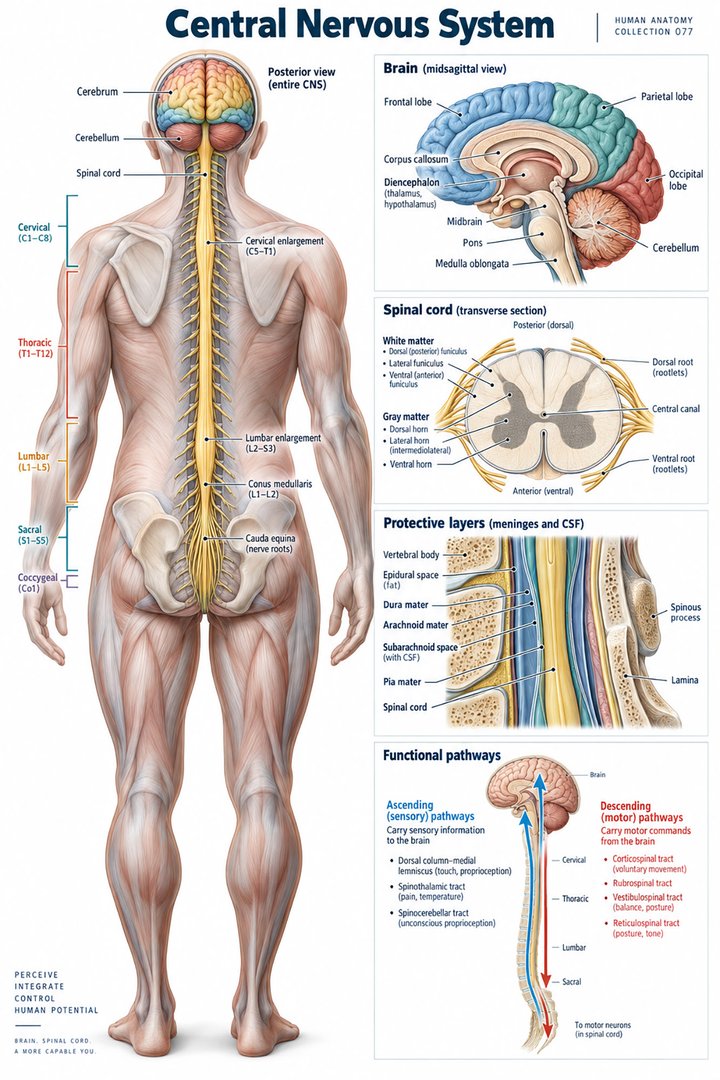
The central nervous system comprises the brain and spinal cord. Peripheral nerves and ganglia connect central structures with much of the rest of the body. Sensory input, integration, and a response are useful functional categories, but they do not map onto three isolated anatomical boxes. The same network can participate in several tasks. [OpenStax: nervous system organization]
Start a study chart with two columns: where a structure is and what relationship it serves. A spinal cord label belongs in the central column, while a named limb nerve belongs in the peripheral column. Add an arrow that crosses the boundary. This prevents a common learning problem in which “central” and “peripheral” are recalled as disconnected lists rather than parts of one communication system.
3. Neurons communicate; glia have active supporting roles
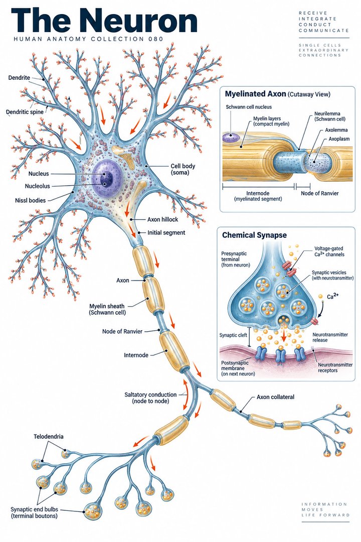
A typical teaching neuron has a cell body, dendrites, and an axon. This pattern is a helpful introduction, but real neurons differ in shape and arrangement. Glial cells perform functions that include support, myelination, and immune surveillance. Oligodendrocytes form myelin in the central nervous system, while Schwann cells perform myelination in the peripheral nervous system. Calling glia merely “glue” loses much of their importance. [OpenStax: neurons and glia]
When reading a cell diagram, distinguish the whole cell from one process and from a bundle containing many processes. A long colored line on a nerve poster is not necessarily one neuron. Ask which scale is being represented. This habit helps connect microscopic organization with regional anatomy without allowing an attractive simplified cell drawing to stand in for every form of nervous tissue.
4. Electrical and chemical signaling are linked
Changes in membrane voltage can generate an action potential when the relevant threshold conditions are met. At a chemical synapse, neurotransmitter release affects receptors on a target cell, changing its activity. A pathway therefore involves more than a signal traveling along an uninterrupted wire: transmission between cells and integration within circuits matter as well. [OpenStax: neural signaling]
Use different marks for an axon and a synapse when sketching a pathway. Say where the signal travels along a cell and where communication crosses to another cell. This does not require drawing every molecular event. Its value lies in preventing the misleading impression that a sensation can be followed through a single continuous line from skin to brain and back to muscle.
5. Sensory and motor describe the direction of information
Sensory input carries information toward central processing; motor output carries commands toward targets. A peripheral nerve can contain both kinds of axons. The dorsal and ventral roots associated with spinal nerves provide an important introductory contrast: sensory fibers enter through dorsal roots, while motor fibers emerge through ventral roots. Once fibers are organized into peripheral routes, a named nerve can carry a mixed set of functions. [OpenStax: peripheral nervous system]
Read the caption before deciding what an arrow means. Some drawings show the direction of information, while others simply point to a structure. A useful revision task is to draw two arrows with different meanings and label both explicitly. This practice is especially valuable when moving between a reflex diagram, a regional nerve chart, and an image that includes both blood vessels and nerves.
6. Somatic, autonomic, and enteric labels describe function
Somatic pathways are associated with body sensation and skeletal muscle responses. Autonomic pathways regulate targets such as smooth muscle, cardiac muscle, and glands. Enteric networks are associated with the digestive tract. These functional labels overlap with central and peripheral anatomy rather than replacing that anatomical division. [OpenStax: nervous system organization]
Avoid reading “autonomic” as meaning unrelated to the brain or unaffected by the wider body. For teaching, compare the labels using the target and route rather than a slogan. A chart showing one organ receiving more than one neural input is an opportunity to discuss coordinated regulation. Its arrows should be read as relationships in a network, not as a pair of switches with identical effects in every situation.
7. Quantitative anchors: cranial and spinal nerve pairs
Introductory anatomy describes twelve cranial nerve pairs and thirty-one spinal nerve pairs. The spinal pairs are grouped as eight cervical, twelve thoracic, five lumbar, five sacral, and one coccygeal. These are anatomical classification counts, not measurements of nerve health or prevalence of disease. In particular, eight cervical nerve pairs should not be confused with the conventional seven cervical vertebrae. [OpenStax: peripheral nervous system]
The regional counts make a useful checking exercise. Add the five spinal groups yourself and explain what the total represents. Then point to the cervical region on a spine chart and state whether the number you are reading refers to nerves, vertebrae, or spinal cord segments. Similar-looking labels can describe different organizational levels, and clear naming prevents a small numerical mistake from becoming a larger spatial misunderstanding.
8. Latin names make the map easier to compare
Selected names include encephalon for the brain, medulla spinalis for the spinal cord, nervus for a nerve, and ganglion for a collection of peripheral neuron cell bodies. The plural nervi appears in grouped nerve terminology. Anatomical Latin can make atlas comparison easier, but the name must still be paired with side, level, and viewpoint. [FIPAT: Terminologia Anatomica, second edition]
Build vocabulary around a small region rather than copying an entire list at once. Identify one central structure, one peripheral connection, and one target. Write each name in English and Latin, then describe their relationship aloud. This exercise checks whether you can use the terminology to explain anatomy. A memorized translation without a route is much less useful in a practical teaching conversation.
| English | Selected Latin | Orientation cue |
|---|---|---|
| Brain | Encephalon | Within the cranial cavity |
| Spinal cord | Medulla spinalis | Within the vertebral canal |
| Nerve | Nervus | Plural: nervi |
| Cerebellum | Cerebellum | Distinct from the cerebral hemispheres |
9. Compare a scan with a specimen photograph
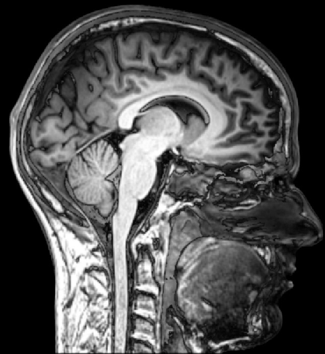
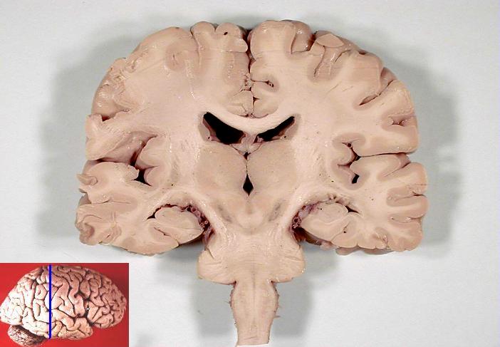
The sagittal MRI below shows a slice through a human head, while the specimen photograph shows a coronal section of a human brain. Their viewpoints differ. The MRI’s face points toward the image’s right, and the coronal photograph includes an original inset indicating the section plane. Establish these orientations before trying to match individual structures.
Notice the folded outer cortex and the different appearance of deeper regions without assuming that image brightness has the same meaning in a scan and a specimen. Preparation, imaging method, and section level all influence what is visible. The images are provided for spatial learning. They do not establish a diagnosis, and neither depicts the entire nervous system or reveals every connection involved in a task.
10. Turn anatomy into clear observation
Practice assistants, hospital personnel, and eldercare teams can use anatomical vocabulary to make observations specific: side, body region, task, and change from the person’s usual abilities. It is often more informative to describe what happened during an activity than to assign an unconfirmed lesion location. Record and communicate through the local care process, keeping the person’s symptoms and observed performance distinct.
For students and physicians, use those descriptions as the beginning of a pathway question. Ask which levels of the network could contribute and what information would distinguish them. A regional chart helps organize the discussion, but it cannot replace examination or the broader clinical context. Explain uncertainty clearly when teaching; the most confident label is not always the most useful description.
11. A study routine that tests connections
Choose one everyday action, such as reaching for a cup. Describe the information needed to notice the cup, plan the action, and control the movement. Mark the central and peripheral portions on a simple chart. Then explain why that task cannot be assigned to a single nerve or one colored brain region. This is a learning exercise, not an attempt to diagnose a person’s difficulty.
Return to the same action with a second representation: a cell diagram, a spinal root chart, or a brain section. State what the new image adds and what it leaves out. The nervous system becomes easier to understand when each picture contributes one layer to a connected explanation. Names and counts then support that explanation instead of becoming an isolated memory test.
Sources and image credits
Sources checked on 8 October 2026. Numerical examples, anatomical classification counts, and research estimates are identified separately in the text.
- OpenStax: nervous system organization
- OpenStax: neurons and glia
- OpenStax: neural signaling
- OpenStax: peripheral nervous system
- FIPAT: Terminologia Anatomica, second edition
Explanatory prose and simplified SVG graphics are original. Photographs retain their stated Creative Commons licenses; creator and source links appear beside each image. Catalog artwork is educational and decorative; no clinical authorship or peer-review credential is claimed.