1. The heart is a layered, three-dimensional organ
A heart illustration often begins with four colored chambers, but the organ is also a muscular wall, a set of valves, a blood supply, and a system that coordinates electrical activity. Learning its structure means connecting those parts rather than memorizing them as separate lists. The same picture can be used to trace flow, compare wall thickness, or establish a valve’s position, provided its viewpoint is understood first.
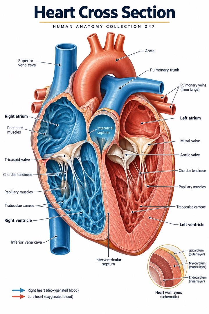
For healthcare learners, a useful starting question is: “What has been removed to make this visible?” A cutaway exposes structures that are enclosed in an intact heart. The apparent spaces and straight routes on the page are teaching choices. Recognizing that choice helps when moving from a poster to a clinical image, a model, or a report describing a particular chamber or valve.
2. Establish position and viewpoint
The heart lies within the mediastinum between the lungs, with much of its anterior surface deep to the sternum. The base and apex describe different parts of its three-dimensional orientation. A frontal cutaway does not reproduce the exact appearance of every surface, and the heart is not situated entirely within the left half of the chest. [OpenStax: heart anatomy]
Say the viewpoint aloud before assigning right and left. In an anterior illustration, the patient’s right is on the viewer’s left. Then find one unmistakable anchor, such as the aorta, and relate the chambers to it. This brief orientation step is particularly useful when comparing diagrams that rotate the heart or use a different plane to expose an internal structure.
3. Four chambers participate in two connected routes
The right and left atria are receiving chambers, while the right and left ventricles eject blood onward. The right ventricle supplies the pulmonary route and the left ventricle supplies the systemic route. Septal tissue separates the right and left sides. The chambers work as parts of a connected system, so the simple description “upper and lower” should be paired with the route rather than learned only as a page position. [NHLBI: heart chambers and tissue]
Trace the receiving vessel, chamber, outgoing connection, and next destination. Then choose another chamber and repeat. This task tests whether the labels form a continuous model. It is also a useful way to explain why two adjacent regions in a cutaway are not automatically connected by a normal opening: the section exposes their relationship, but the intact walls and septa still matter.
4. Valves occupy specific junctions
The tricuspid valve lies between the right atrium and ventricle, and the mitral valve lies between the left atrium and ventricle. The pulmonary valve is at the right ventricular outflow, while the aortic valve is at the left ventricular outflow. These four valve names identify junctions in the route rather than four interchangeable structures scattered through the organ. [NHLBI: valves and blood flow]
For study, replace each valve label with a question: “What lies before this junction, and what lies after it?” Answer with a chamber or vessel, then restore the valve name. This approach is especially effective when a diagram shows an opened leaflet. The leaflet’s appearance is important, but the connection it regulates remains the first relationship to establish.
5. Valve support differs between valve groups
The atrioventricular valves have chordae tendineae connected with papillary muscles. These supporting structures help prevent leaflet prolapse during ventricular contraction; they do not pull the valves open. The aortic and pulmonary semilunar valves have a different arrangement and do not have the same chordal apparatus. Keeping the two groups distinct prevents a common error in simplified valve explanations. [OpenStax: heart anatomy]
When comparing a photograph or model with a diagram, look for the supporting structures as well as the valve opening. State what you can actually identify. A cut surface can make a papillary muscle look like an isolated projection, while a schematic may make the chordae look unnaturally regular. The learning aim is to understand their connection, not to expect a specimen to match a drawing line for line.
6. The wall and surrounding sac are related but distinct
The heart wall is described with three layers: endocardium internally, myocardium in the middle, and epicardium externally. The epicardium is the visceral layer of serous pericardium. The pericardial sac and its fluid-containing space should therefore be distinguished from the muscular wall itself. The myocardium provides the contractile tissue, while the surrounding arrangement permits movement with reduced friction. [OpenStax: heart anatomy]
Draw a nested outline and label the chamber space, wall, and surrounding pericardial relationship separately. This exercise helps when encountering similar-sounding terms. A student who can place endocardium and pericardium correctly has established more than vocabulary: they can explain where a structure lies relative to the blood-filled cavity and to the outside of the organ.
7. Coronary vessels supply the working muscle
The myocardium has its own coronary arterial supply and venous drainage. It should not be imagined as receiving all its requirements directly from blood inside the chambers. Coronary vessels follow routes on and within the heart, connecting the organ’s pumping role with the circulation needed by its own tissue. [OpenStax: circulatory pathways]
This relationship makes a useful teaching contrast: blood passing through the heart is not the same description as blood supplying the heart muscle. Mark these routes with different arrow styles. Explain both without assuming that a colored coronary branch indicates an individual person’s exact pattern. A general chart gives a framework for learning; regional details and clinical findings require their own evidence.
8. Electrical coordination precedes mechanical events
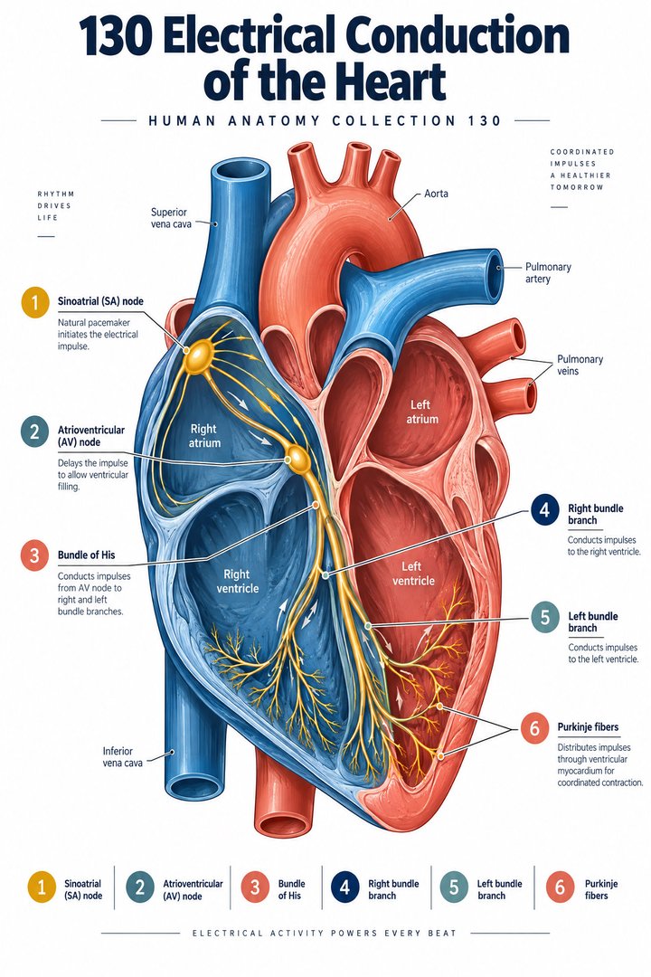
The sinoatrial node normally initiates activity that spreads through the atria, with conduction onward through the atrioventricular system and ventricular conducting tissues. This arrangement supports coordinated activation. An electrocardiogram records electrical activity at the body surface; it is not a direct photograph of valves opening or a measurement of every mechanical aspect of pumping. [NHLBI: how the heart beats] [OpenStax: cardiac electrical activity]
Place an electrical sequence beside a chamber diagram and ask what each adds. The first describes activation order; the second describes structure and route. In a clinical explanation, specify which type of information a test or diagram is providing. This prevents an electrical waveform from being treated as a complete substitute for anatomical or functional imaging.
9. Quantitative anchors: four chambers, four valves, three wall layers
Three useful counts are four chambers, four main valves, and three named heart-wall layers. These counts belong to different categories and should not be compared as measures of size or importance. The graphic groups them as learning anchors, with the number beside the category. It is a classification display rather than a statistical sample of patients. [NHLBI: heart chambers and tissue] [NHLBI: valves and blood flow] [OpenStax: heart anatomy]
A second quantitative exercise is purely mathematical: if a hypothetical rate remained at seventy-five beats per minute for twenty-four hours, the count would be 108,000 beats. That assumption is deliberately simplified; real heart rate changes with circumstances. Keep the assumed rate and duration beside the answer. The calculation teaches how a small rate accumulates over time, without claiming an actual daily total for a particular person.
10. Latin terms support the structural map
Cor is the anatomical Latin term for heart. Selected chamber names include atrium dextrum, atrium sinistrum, ventriculus dexter, and ventriculus sinister. Septum interventriculare identifies the partition between the ventricles. The endings differ because anatomical Latin follows grammatical relationships; learning the complete expression is more reliable than replacing one word mechanically. [FIPAT: Terminologia Anatomica, second edition]
Use the terms to give a short route description rather than reciting a translation list. Name a chamber, identify its side, and add the next connection. Then explain the same route in ordinary English. For physicians and other healthcare staff, the ability to switch between precise terminology and accessible language is especially useful when discussing an investigation with a patient or colleague.
| English | Selected Latin |
|---|---|
| Heart | Cor |
| Right / left atrium | Atrium dextrum / atrium sinistrum |
| Right / left ventricle | Ventriculus dexter / ventriculus sinister |
| Interventricular septum | Septum interventriculare |
11. Real tissue and thoracic images answer different questions
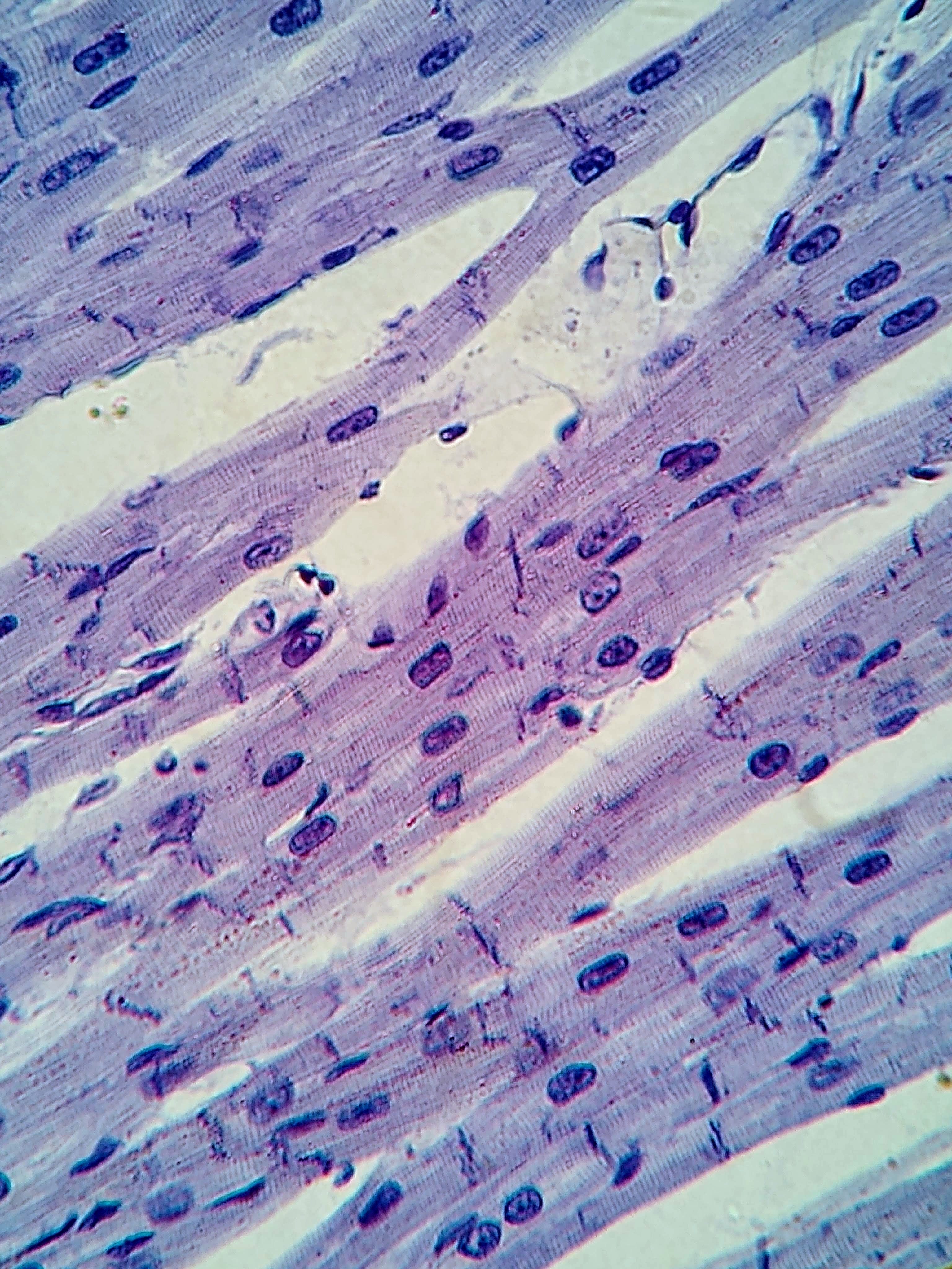
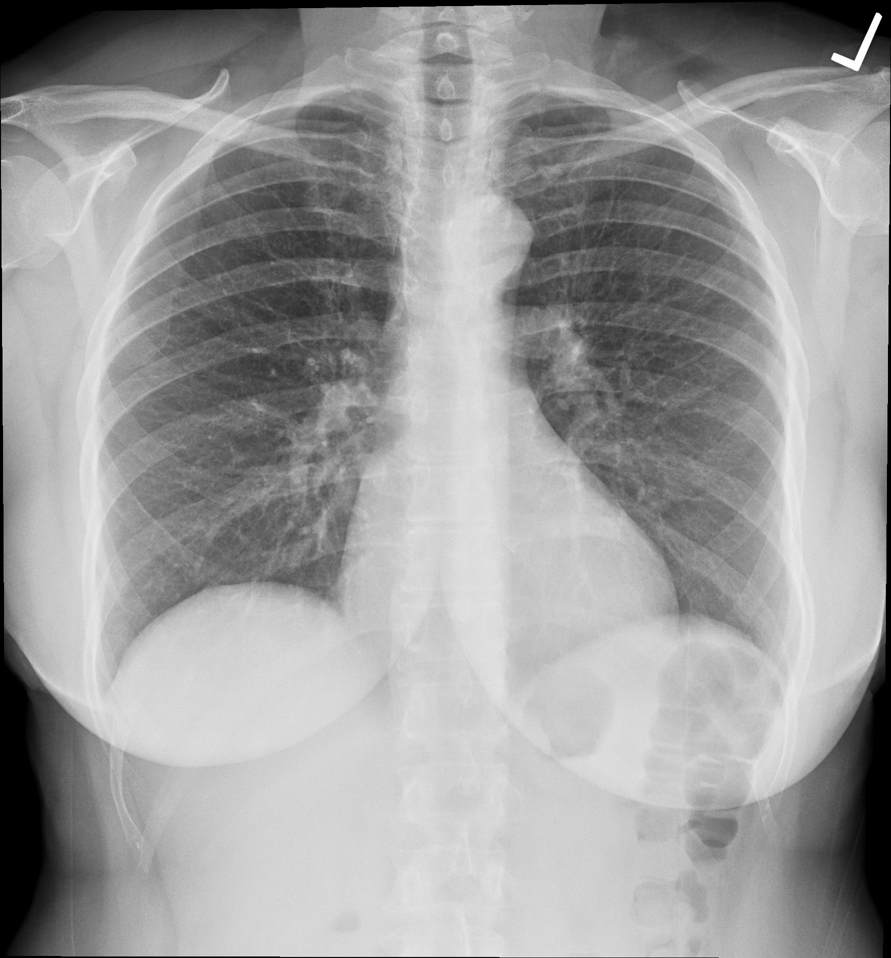
The cardiac muscle micrograph below records a real stained tissue preparation at the source’s stated magnification. Striated tissue profiles and nuclei are visible, while the source does not specify a human specimen. The chest radiograph records a human thorax as a projection. It shows a cardiac silhouette among overlapping structures rather than separate atria, ventricles, and valves.
Compare the images by naming the question each can support. A tissue photograph helps connect the organ with its cellular organization; a thoracic projection helps establish broad location. Neither reproduces the opened-chamber view of the featured poster. This comparison encourages learners to treat the images as complementary representations and to retain their captions, creator credits, and preparation context.
12. A practical study sequence for healthcare teams
Start with orientation, then chambers, junctions, wall layers, blood supply, and electrical coordination. Each step adds a relationship to the preceding one. Medical students can cover the labels and reconstruct that sequence. Practice assistants and hospital personnel can use the same framework to understand the anatomical term named in a report without extending the report beyond what it actually says.
For eldercare teaching, keep explanations tied to the individual’s established care plan and the clinical information provided by the treating team. A general heart guide can clarify vocabulary; it cannot determine a person’s treatment or activity limits. End a learning session by explaining one chamber-to-vessel route and one wall-to-tissue relationship. Those two explanations test both movement through the organ and the structure that makes movement possible.
Sources and image credits
Sources checked on 8 October 2026. Numerical examples, anatomical classification counts, and research estimates are identified separately in the text.
- OpenStax: circulatory pathways
- OpenStax: heart anatomy
- OpenStax: cardiac electrical activity
- NHLBI: heart chambers and tissue
- NHLBI: valves and blood flow
- NHLBI: how the heart beats
- FIPAT: Terminologia Anatomica, second edition
Explanatory prose and simplified SVG graphics are original. Photographs retain their stated Creative Commons licenses; creator and source links appear beside each image. Catalog artwork is educational and decorative; no clinical authorship or peer-review credential is claimed.