1. Breathing and gas exchange are connected processes
The respiratory system brings air into contact with an exchange surface and helps link that surface with the circulation. Its functions extend beyond a pair of lungs expanding in the chest. The upper airway conditions incoming air, the respiratory muscles support ventilation, and the exchange interface connects the air spaces with blood. For healthcare learners, distinguishing these activities is more useful than treating the entire system as one undifferentiated process.
Begin with three questions: Where does air travel? What moves it? Where do gases cross into or out of blood? These questions organize the anatomy around a route, a mechanical action, and an interface. They also make explanations easier for patients and colleagues. A clear description of breathing should state which part of this sequence is being discussed before introducing a measurement or a clinical interpretation.
2. Trace the airway from entry to exchange
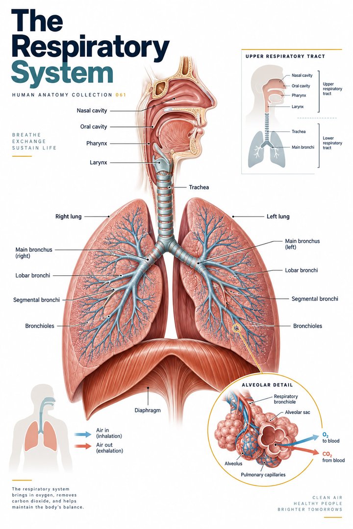
Air can enter through the nasal or oral passages and continue through the pharynx, larynx, and trachea toward the bronchi. The conducting zone provides a route and helps warm, humidify, and filter incoming air. The respiratory zone includes structures where gas exchange occurs. These zones are functional categories, so an illustration should explain their meaning rather than merely coloring two regions differently. [OpenStax: respiratory structures]
Follow the branching pattern from a large airway toward smaller structures. At each step, say whether the drawing shows a whole organ, a tube, or an enlarged detail. An inset of an alveolus may occupy more space on the page than an entire lung. That is a change of scale for teaching, not a representation of their relative size. Always read inset captions before making a spatial comparison.
3. Ventilation depends on changing pressure relationships
During quiet inspiration, diaphragm contraction expands the thoracic space and supports a fall in pressure that draws air inward. Quiet expiration is largely associated with relaxation and elastic recoil. Forced breathing recruits additional muscular activity. This sequence describes a pressure-driven movement of air rather than a lung actively pulling air through its own independent muscular contraction. [OpenStax: breathing mechanics]
Explain the sequence with verbs: the diaphragm contracts, thoracic volume changes, pressure changes, and air moves. A set of verbs is often easier to understand than a static picture of “inhalation.” When teaching, ask a colleague to reverse the explanation for expiration. If the sequence cannot be reversed clearly, revisit the mechanical relationships before adding more anatomical labels.
4. Gas exchange requires an appropriate interface
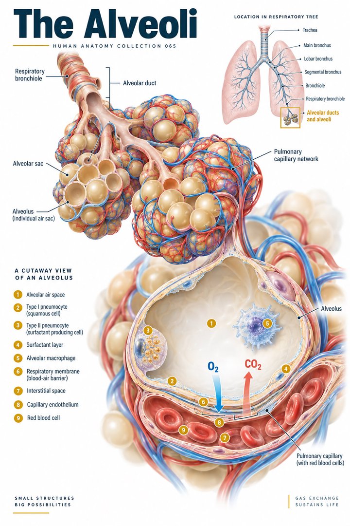
Oxygen and carbon dioxide move across the respiratory interface according to their partial-pressure relationships. Ventilation brings air to the exchange region, while perfusion brings blood through nearby capillaries. These are different processes that must work together. An alveolus drawn beside a capillary is therefore a relationship between two routes, not a single tube carrying both air and blood. [OpenStax: gas exchange]
When sketching the interface, label the air space, the tissue barrier, and the blood compartment separately. Draw oxygen and carbon dioxide arrows in opposite directions at the appropriate interface. Then explain what each arrow means without relying on red or blue. This exercise makes it easier to recognize a diagram that accidentally confuses gas movement with the direction of bulk airflow or blood flow.
5. Alveolar cells and airway defenses have different roles
Type I alveolar cells provide a thin surface for gas exchange, while type II cells produce surfactant. Alveolar macrophages participate in local defense. In conducting airways, mucus and ciliary activity contribute to handling material carried in with the air. These structures support different tasks, and a magnified alveolar picture should not be assumed to describe the epithelial organization of every airway. [OpenStax: respiratory structures]
Use a comparison table during study with the headings “site,” “cell or structure,” and “main relationship.” This approach keeps a protective mechanism in its anatomical setting. It also helps explain why learning a familiar phrase such as “mucociliary clearance” is only a starting point. A term becomes useful when you can identify the region in which the process is being described.
6. Blood carries gases beyond the lungs
Most oxygen in blood is transported in association with hemoglobin, while a smaller amount is dissolved. Carbon dioxide is carried in several forms, including bicarbonate, dissolved gas, and association with proteins. Gas exchange in the lung therefore connects with transport chemistry rather than ending when an oxygen arrow reaches a capillary. [OpenStax: gas transport]
For an introductory explanation, separate entry into the blood from carriage through the circulation and delivery at tissues. Each is a different step in the wider relationship. Do not use one measurement as a complete description of all three. In teaching discussions, naming the step under consideration is often enough to prevent a misleading comparison between quantities that describe different physiological processes.
7. Quantitative facts: tidal volume and a worked example
An introductory value for tidal volume during quiet breathing is about 500 milliliters, or half a liter, although actual volumes vary with the person and conditions. Tidal volume describes air moved in one ordinary breath; it is not the maximum air the lungs can hold. [OpenStax: breathing mechanics]
For a hypothetical example, twelve breaths per minute at half a liter per breath gives six liters per minute of total minute ventilation. The arithmetic is a teaching calculation, not a prescribed target or a prediction for a patient. Total ventilation is also not identical to ventilation reaching the gas-exchanging region, because part of each breath occupies conducting spaces. Keep the definition beside the calculation rather than presenting the number alone.
8. Research statistics: alveolar counts vary substantially
Ochs and colleagues reported a mean alveolar count of 480 million in a study of six adult human lungs, with values ranging from 274 to 790 million. The paper was published in 2004. The small sample and wide range matter: “480 million” should be read as that study’s mean rather than a precise total assigned to every person. [Ochs et al. (2004): alveolar number in six adult human lungs]
The graph presents the reported minimum, mean, and maximum as summary values. It does not show individual observations or a population distribution. That distinction is worth practicing with any anatomical statistic. Ask what was counted, how many samples were studied, and whether the image shows measurements, a classification, or a calculation. A striking number is most useful when it retains the information needed to interpret it.
9. Latin terms and asymmetric lung anatomy
Selected anatomical names include pulmo for lung, pulmones for lungs, trachea for the windpipe, and diaphragma for the diaphragm. The conventional right lung has three lobes and the left has two. These lobe counts are different from the number of bronchi, bronchopulmonary segments, or alveoli, so a numerical label should always identify the level of organization it describes. [FIPAT: Terminologia Anatomica, second edition] [OpenStax: lung anatomy]
On an anterior body diagram, the person’s right lies on the viewer’s left. Confirm that orientation before counting lobes. A practical revision exercise is to write “right lung: three lobes” beside an anterior sketch and then point to the same side on a posterior sketch. The anatomical label remains tied to the person even when the viewpoint changes.
| English | Selected Latin | Naming cue |
|---|---|---|
| Lung / lungs | Pulmo / pulmones | Singular and plural |
| Diaphragm | Diaphragma | Respiratory muscle |
| Right / left lung | Pulmo dexter / pulmo sinister | Patient’s side |
| Bronchus | Bronchus | Airway designation |
10. Real images restore depth and overlap
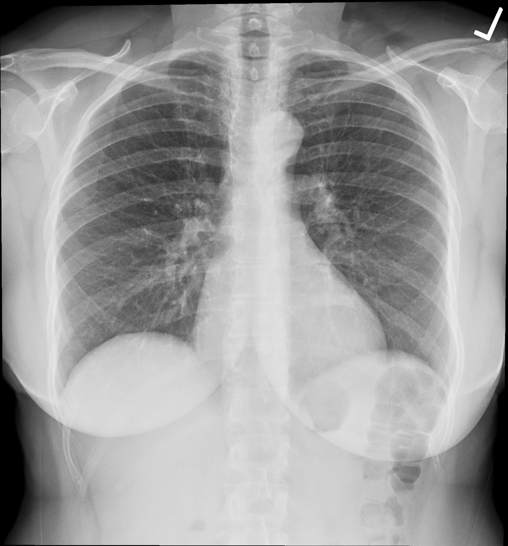
The chest radiograph below shows the thorax as a projection with overlapping structures. The left marker supports orientation, while the cardiac silhouette and diaphragm outlines help establish broad relationships. The source record describes the image as normal; it is not independently interpreted here as a diagnostic study. A projection image should not be read as a direct picture of separated lung lobes.
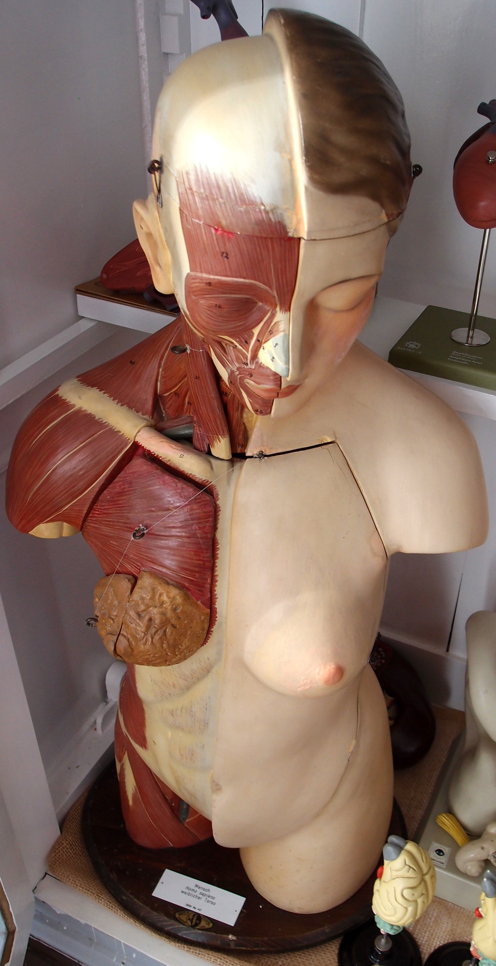
The second image is a real photograph of a torso teaching model. Its exposed muscles and deliberately removed layers help demonstrate how a physical model presents anatomy for education. It is not a photograph of living internal organs. Compare the images by naming what each makes visible and what it conceals. That comparison is often more instructive than asking which one is the “most realistic.”
11. Clinical learning begins with precise description
Practice assistants and hospital teams can strengthen communication by describing the actual observation, its timing, the person’s position or activity, and any change from the usual state. Use locally approved terminology and escalation processes. A respiratory overview supports those descriptions, but it does not supply an individual diagnosis or determine how oxygen, medicines, or equipment should be adjusted.
For students and physicians, organize an explanation around route, mechanics, exchange, and transport. For eldercare teams, use that same framework to understand the purpose of existing care instructions without replacing them with a general diagram. Finish a teaching session by asking the listener to explain the difference between ventilation and gas exchange. A clear answer indicates that the anatomy has become a useful functional model.
Sources and image credits
Sources checked on 8 October 2026. Numerical examples, anatomical classification counts, and research estimates are identified separately in the text.
- OpenStax: respiratory structures
- OpenStax: lung anatomy
- OpenStax: breathing mechanics
- OpenStax: gas exchange
- OpenStax: gas transport
- Ochs et al. (2004): alveolar number in six adult human lungs
- FIPAT: Terminologia Anatomica, second edition
Explanatory prose and simplified SVG graphics are original. Photographs retain their stated Creative Commons licenses; creator and source links appear beside each image. Catalog artwork is educational and decorative; no clinical authorship or peer-review credential is claimed.