Organ location needs three dimensions
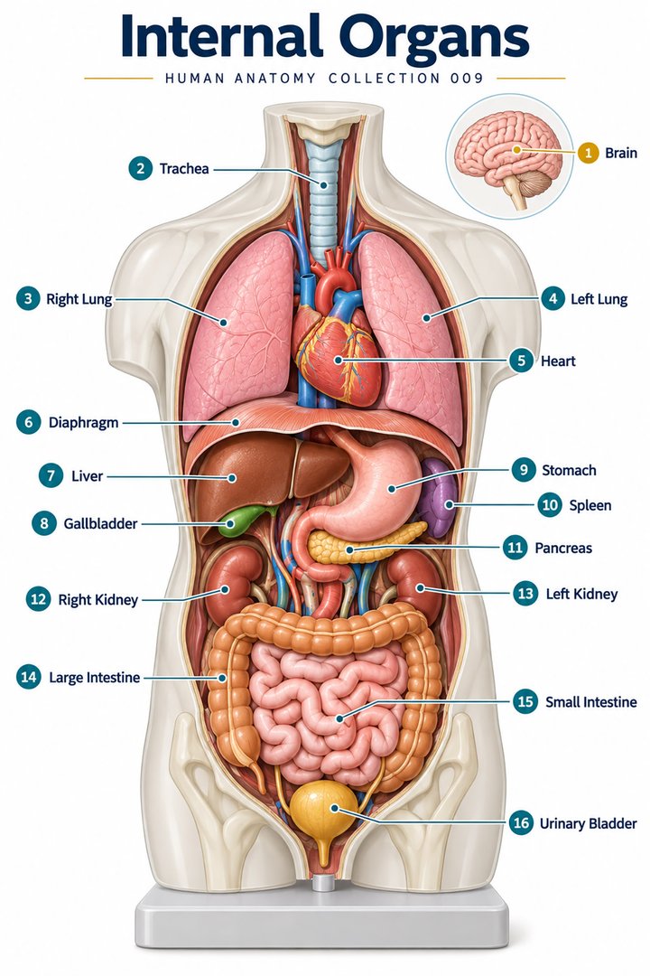
A frontal organ poster is an excellent beginning, but the body does not arrange all organs on one visible sheet. Some are anterior, some posterior, and some span several surface regions. Many overlap in projection. Organ placement therefore requires three kinds of information: the broad region, the depth relationship, and the neighboring structures. A fourth kind of information, physiological state, explains why filling or movement can alter an organ's contour without changing its identity. These distinctions make anatomy useful when moving from a schematic to a real person or a sectional image.
For healthcare learners, “where is it?” should usually be followed by “relative to what?” A liver can be described relative to the diaphragm, rib cage, stomach, and major vessels. A kidney can be described relative to the spine, peritoneum, adrenal gland, and neighboring abdominal structures. Each comparison adds a different coordinate. None requires pretending that every person has exactly the same dimensions. A good location guide teaches representative relationships while preserving room for developmental variation, changes in position, and the different views supplied by imaging.
Four quadrants and nine regions are complementary maps
The abdomen can be divided into four quadrants using intersecting reference lines near the umbilicus. A nine-region map names right and left hypochondriac, lumbar, and iliac regions, with epigastric, umbilical, and hypogastric regions centrally. These are communication maps drawn over a continuous body region, not internal partitions that prevent an organ from crossing a line. The subject's right and left remain the reference sides. NCBI Medical Terminology: Body regions
The numerical facts four and nine describe alternative classifications. A chart comparing them should not suggest that one body has four organs while another has nine. Latin regional labels such as regio epigastrica and regio umbilicalis identify areas rather than organ names. A stomach may project into more than one area, and a bowel loop can occupy a different position within the same broad region. The maps improve description, but they do not identify an organ conclusively from the location of a symptom or a surface mark.
The heart is central with an oblique arrangement
The heart occupies the central thoracic region between the lungs, within the mediastinal and pericardial arrangement. Its position is not adequately described as “entirely in the left chest.” The apex points toward the left, while the chambers and great-vessel connections occupy a three-dimensional, rotated structure. NCBI Bookshelf: Heart anatomy
That geometry matters when reading a diagram. A four-square circulation illustration makes the sequence easy to follow but does not reproduce the heart's complete spatial orientation. A frontal photograph may expose an anterior ventricular surface while hiding posterior atrial structures. A transverse view shows different relations at different levels. The educational task is to translate among those views rather than demand that every representation look identical. One can preserve a correct blood route while acknowledging that the simplified diagram has rearranged structures to improve legibility.
Lung placement follows thoracic surfaces and central roots
The lungs occupy paired pulmonary regions lateral to the mediastinum. Their apices extend superiorly, their bases relate to the diaphragm, and their costal surfaces face the cage. The left lung's cardiac notch and the differing lobar patterns show that paired organs are not necessarily perfect mirror images. Their hila provide regions where roots enter and leave the medial surfaces. NCBI Bookshelf: Lung anatomy
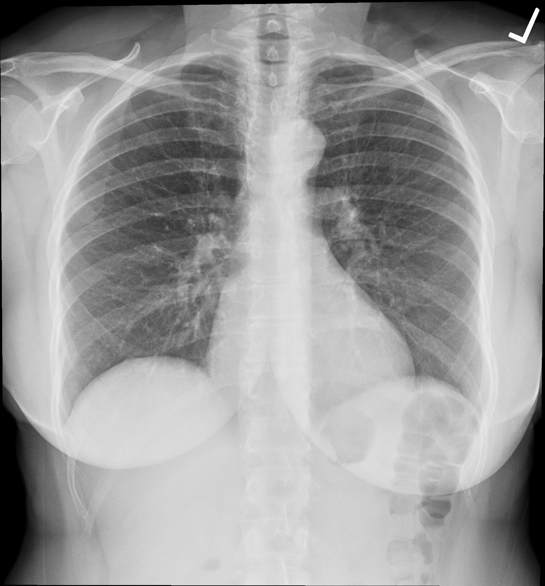
Surface projection should not be confused with the full extent of lung tissue or pleural relationships. A rib counted on an external diagram can help orient a region, but a moving diaphragm changes the inferior contour through breathing. A chest radiograph compresses depth into a projection and can place several structures over the same visible area. These observations support a practical habit: state the method and viewpoint before assigning depth. A shadow on a frontal image is not automatically located at the same anterior surface level as the nearest visible rib.
The liver occupies much of the right upper abdomen
The liver lies predominantly in the right upper abdominal arrangement beneath the right hemidiaphragm, with an extension toward the left. The lower cage provides partial protection, and peritoneal reflections contribute to its position. Its diaphragmatic and visceral relationships differ; a frontal model showing a broad external surface does not show all inferior and posterior associations. Morphological and functional divisions are related but not identical classification schemes. Liver anatomy: Regional anatomical review
Latin hepar identifies the liver. The word “lobe” can refer to an external anatomical division, whereas a functional description follows internal vascular and biliary organization. When teaching placement, begin with the whole organ before introducing those divisions. Otherwise the learner may mistake a line drawn on the liver for a visible wall separating independent organs. The practical spatial lesson is that a large asymmetric organ spans a region and relates to several neighboring structures; one small icon on a quadrant chart cannot convey its entire extent.
Gallbladder placement connects surface and duct anatomy
The gallbladder lies against the visceral liver surface in a defined fossa, commonly between hepatic segments IV and V. Its fundus, body, and neck describe regions within one sac, and its cystic connection joins the broader biliary pathway. The location should be understood together with the duct route rather than as an isolated green object attached decoratively to a liver drawing. NCBI Bookshelf: Gallbladder anatomy
This distinction illustrates a general principle: anatomical proximity does not define functional production. The gallbladder's relationship to the liver is important, but the two do not become synonyms for the same tissue or role. A placement diagram can show the organ on the inferior surface while a second diagram shows how the duct connection relates to neighboring pathways. Separating those purposes improves clarity. A view from below may be more informative for this relationship than the frontal view that is easiest to recognize on a general torso poster.
The stomach changes contour within a stable set of relationships
The stomach occupies the upper abdominal arrangement, largely left of the midline, connecting the esophagus with the duodenum. Its fundus relates to the left diaphragmatic region, and its posterior relationships include a group of neighboring structures often described as the stomach bed. Its size and contour change with filling, making one fixed outline an incomplete description of living anatomy. NCBI Bookshelf: Stomach anatomy
The Latin gaster names the organ, while fundus, corpus, and pylorus specify parts. A learner can understand placement by following the entry, dome, body, and exit instead of memorizing a single shape. An endoscopic image shows the luminal surface rather than the organ's external relation to the pancreas or diaphragm. A gross model shows those relations but cannot establish every internal surface feature. Combining views supplies a fuller map while keeping the evidence from each method distinct.
The spleen is a left upper posterior-lateral landmark
The spleen lies in the left hypochondriac region with relationships to the diaphragm and gastric fundus, typically near the ninth through eleventh ribs. Its visceral and diaphragmatic surfaces differ, and the hilum transmits connected structures. It is a vascular lymphatic organ rather than a segment of the alimentary tube, even though it lies near digestive organs. NCBI Bookshelf: Spleen anatomy
This is a useful correction to an overly flat map. A spleen icon beside a stomach icon may imply that they occupy one plane. Their real relationships include depth, curved surfaces, ligamentous associations, and neighboring kidney or pancreatic arrangements. Naming a side is only the first step. For a teaching discussion, ask learners to identify the diaphragm-facing surface and then the visceral surface. This approach connects the organ's shape with its neighbors and discourages the mistaken assumption that every organ displayed in a frontal atlas can be seen completely from the front.
The pancreas extends across a posterior upper abdominal route
The pancreas has head, neck, body, and tail regions, with an uncinate process associated with the head. The head is related to the duodenal loop on the right; the body crosses the upper posterior abdominal arrangement; the tail extends toward the splenic region. Much of the gland is retroperitoneal, while the tail has distinct peritoneal relationships. Its location behind the stomach makes depth essential to understanding a torso diagram. NCBI Bookshelf: Pancreas anatomy
A primary anatomical and CT study compared 37 autopsy specimens with 32 CT examinations and documented variation in pancreatic tail relationships. This is evidence that a representative picture is not an exact personal template. The study's individual percentages belong to its sample and method rather than an all-purpose rule about every population. Rosen et al.: Pancreatic tail anatomy and CT, 1988
The educational benefit is concrete: a diagram can show a common tail position, but a professional explanation should leave room for variation. Anatomy provides a starting map; an individual's examination or imaging supplies the specific arrangement.
Kidneys occupy a posterior rather than pelvic location
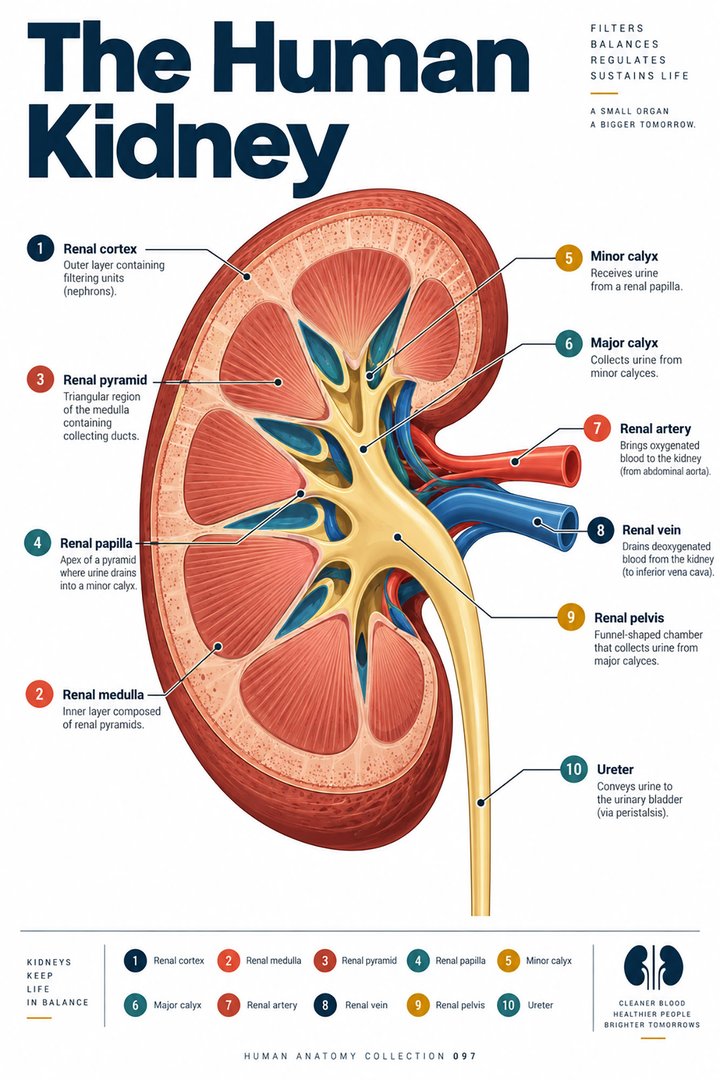
The kidneys lie retroperitoneally on either side of the vertebral column, commonly spanning approximately T12 to L3. The right kidney is usually somewhat lower than the left, related to the liver's position. Each has an upper and lower pole, a medial hilar region, and relations to surrounding fat and fascia. These are representative adult relations, not exact coordinates guaranteed for every body. NCBI Bookshelf: Kidney anatomy
Latin ren names a kidney and hilum renale identifies its entry-exit region. The kidneys should not be confused with the urinary bladder simply because both participate in urinary anatomy. Following urine from kidney through ureter to bladder connects distinct locations and structures. In a frontal model, kidneys may be shifted outward so that they remain visible behind digestive organs. The caption should identify this display choice. Without it, the model may teach names successfully while teaching depth incorrectly.

Suprarenal glands are neighbors with a separate identity
The adrenal or suprarenal glands occupy superior renal relationships within the retroperitoneal arrangement. The right gland is commonly pyramidal and the left more crescentic. Their vascular and regional relationships differ by side, reinforcing that the pair is not simply duplicated mirror anatomy. Each gland has a distinct identity and endocrine organization despite its close relation to a kidney. NCBI Bookshelf: Suprarenal gland anatomy
The Latin glandula suprarenalis makes the positional relationship explicit. A drawing that places the gland like a neat cap directly on a kidney is a useful orientation aid, but it can simplify the actual contour, fascia, and adjacent vessels. Students should learn the broad relationship first and then investigate details in a sectional or specimen view. The same pattern applies to many adjacent organs: a convenient icon communicates vicinity, while a more detailed representation is needed to communicate exact contact surfaces and connected structures.
Bowel routes span regions and differ in mobility
Duodenum, jejunum, and ileum form the small-intestinal sequence. The duodenum has relatively fixed posterior relationships through much of its course, whereas jejunal and ileal loops have mesenteric relationships permitting a different arrangement. Colon regions frame and cross the abdominal map, with the rectal route continuing into the pelvis. A single loop diagram represents the order more reliably than it represents every individual's detailed position. NCBI Bookshelf: Small intestine anatomy
The distinction between order and placement is valuable in clinical communication. An organ can keep its place in the route while occupying a somewhat different position within the abdomen. Likewise, a segment can relate to more than one quadrant without becoming two separate segments. A guide should therefore label continuity clearly and avoid equating every bend of a schematic with an invariant anatomical flexure. Route, mobility, support, and surface projection are connected topics, but they are not interchangeable descriptions.
Pelvic organs require filling state and reproductive context
The urinary bladder lies posterior to the pubic symphysis. When relatively empty it occupies the lesser pelvic arrangement; filling can extend its contour superiorly into the lower abdominal region. A placement drawing should therefore identify the represented filling state rather than present one outline as permanent. NCBI Bookshelf: Bladder anatomy
Where a uterus is present, it lies between the bladder anteriorly and rectum posteriorly, with variations in orientation. Anteversion and anteflexion describe different angles and should not be collapsed into one term meaning simply “forward.” NCBI Bookshelf: Uterus anatomy
The rectum occupies a posterior visceral relationship in the pelvis, while the pelvic floor supplies inferior support and passage-related functions. NCBI Bookshelf: Rectum anatomy
These relationships explain why a sagittal pelvic drawing must identify the anatomy it represents. One generalized diagram cannot display every possible reproductive configuration or physiological state. Clear captions are more helpful than pretending that a single figure is exhaustive. The practical goal is to locate structures respectfully and accurately while acknowledging the context necessary to interpret their arrangement.
Sectional views reveal what a surface map conceals
A transverse section shows depth at a particular level; a sagittal section shows anterior-posterior relationships along a defined plane; a coronal view supplies another perspective. Organ borders can appear and disappear as the level changes because organs are three-dimensional and irregular. A circular or oval profile is a section through a structure, not automatically its whole shape. Before naming something in an image, identify orientation markers and the level, then examine neighboring landmarks and continuity across available views.
A useful learning exercise is to take one organ from the frontal map and predict its appearance in two sections. State which neighbors should become visible and which parts may lie outside the selected plane. Then compare the prediction with a labeled representation. The exercise teaches spatial reasoning without claiming that a general atlas establishes an individual's exact anatomy. Organ placement is most useful when it joins surface description, depth, pathways, and variation into one connected account. A precise location statement creates the foundation for interpretation; it does not replace the evidence needed to assess a particular person.
Anatomical Latin: quick reference
| Latin term | English meaning |
|---|---|
| Hepar | Liver |
| Lien | Spleen |
| Ren | Kidney |
| Vesica urinaria | Urinary bladder |
Sources and image credits
Sources reviewed on 9 October 2026. Counts, estimates, study statistics and hypothetical calculations retain their context in the text.
- NCBI Medical Terminology: Body regions
- NHLBI: Heart anatomy
- OpenStax: Lungs and pleura
- Liver anatomy: Regional anatomical review
- NCBI Bookshelf: Gallbladder anatomy
- NCBI Bookshelf: Stomach anatomy
- NCBI Bookshelf: Spleen anatomy
- NCBI Bookshelf: Pancreas anatomy
- Rosen et al.: Pancreatic tail anatomy and CT, 1988
- NCBI Bookshelf: Kidney anatomy
- NCBI Bookshelf: Suprarenal gland anatomy
- NCBI Bookshelf: Small intestine anatomy
- NCBI Bookshelf: Bladder anatomy
- NCBI Bookshelf: Uterus anatomy
- NCBI Bookshelf: Rectum anatomy
- NCBI Bookshelf: Heart anatomy
- NCBI Bookshelf: Lung anatomy
Explanatory prose and SVG graphics are original. Photograph authors, source records and licenses are credited beside each image. Catalog illustrations provide educational context; anatomical facts are supported by the linked sources.