1. Kidney anatomy links three scales of organization
Human kidney anatomy can be approached through external position, internal compartments, and microscopic units. These scales are connected, but they cannot all be represented accurately by one simple drawing. A whole-body image establishes the organ’s location; a section exposes the cortex, medulla, and collecting spaces; microscopy reveals the structures in which filtration and tubular processing occur. Learning becomes more reliable when the scale is named before the labels are interpreted.
For medical students and healthcare teams, the central task is to connect routes without merging them. Blood enters and leaves through vessels, while processed tubular fluid reaches the collecting system and ureter. The two routes interact closely but occupy different compartments. This guide emphasizes those relationships and the landmarks that make a kidney section understandable.
2. Retroperitoneal position establishes important neighbors
The kidneys lie against the posterior abdominal wall behind the parietal peritoneum. Their usual position is approximately between the twelfth thoracic and third lumbar vertebral levels, with the right kidney commonly lower because of the liver. Lower ribs, surrounding fat, and supporting tissues contribute to their anatomical setting. These descriptions are broad orientation points rather than fixed coordinates in every person. [NCI SEER: kidney anatomy]
Compare an anterior body chart with a posterior view. A kidney can appear to lie behind several other organs because the view has flattened depth. State that relationship instead of assuming a colored outline is on the body’s anterior surface. For clinical communication, retain the side and actual structure described in the report; a general position map does not replace individual imaging.
3. Capsule, fat, and fascia are different coverings
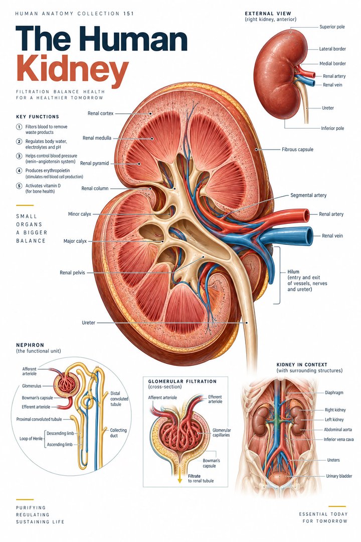
A fibrous capsule closely invests the renal tissue. Perirenal fat and renal fascia form additional surrounding relationships. They are not interchangeable names for the same outer layer. The medial hilum opens toward the renal sinus and provides passage for vessels and the collecting outlet, together with nerves and lymphatics. An external kidney drawing should distinguish the organ surface from the structures attached at this junction. [NCI SEER: kidney anatomy]
A useful study sketch uses separate outlines for the closely applied capsule and the more external supporting arrangement. Then add the hilum as a junction rather than another covering. This makes the drawing helpful when learners encounter a sectional image in which fat and vessels occupy spaces near the collecting system. The different tissues can share a region without becoming one structure.
4. Cortex, medulla, and columns organize the parenchyma
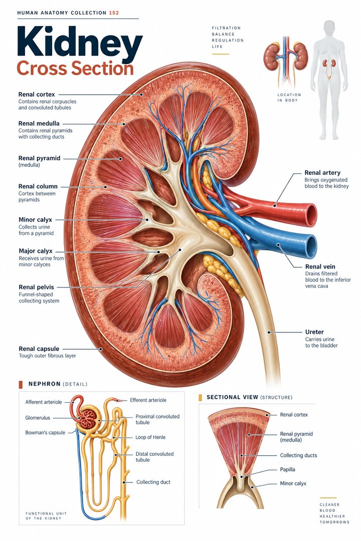
The cortex is the outer renal region, while the medulla contains pyramidal organization. Renal columns are extensions of cortical tissue between pyramids. A pyramid’s papillary end faces toward the collecting spaces. The cortex and medulla together form the renal parenchyma, which should be distinguished from the renal pelvis and other urine-collecting passages. [NCI SEER: kidney anatomy] [OpenStax: gross kidney anatomy]
On an opened-organ poster, first trace the capsule and then identify a pyramid from its broad base to its papillary end. Locate a column beside it. This sequence is more informative than identifying every similarly colored triangular shape as the same structure. The arrangement also explains why the collecting route should be followed from tissue toward larger spaces rather than guessed from the center of the picture outward.
5. Papillae connect processing tissue with collecting spaces
Collecting ducts convey fluid toward papillary outlets. Minor calyces receive it from papillary regions, and several minor calyces connect toward major calyces and the renal pelvis. The pelvis continues toward the ureter. This is a collecting pathway, not the route followed by all incoming blood. Branching and three-dimensional relationships are simplified in a conventional frontal section. [OpenStax: gross kidney anatomy]
Use an arrow to follow one selected outlet rather than drawing every branch at once. Name the change in scale as fluid passes from small ducts to larger collecting spaces. Then follow another outlet and identify where the routes converge. This exercise makes the words minor, major, and pelvis meaningful as relationships rather than a sequence memorized without an image.
6. Renal vessels form a separate branching map
Renal arterial branches lead toward afferent arterioles and glomerular capillaries. Efferent arterioles then connect toward further capillary networks associated with the tubules. Peritubular capillaries and vasa recta participate in this vascular organization. This arrangement differs from a simple artery-to-single-capillary-bed-to-vein illustration used for many other regions. [OpenStax: gross kidney anatomy]
Draw the vascular route beside the tubular route with a clear legend. Where the lines run close together, explain the exchange relationship without connecting the lumens into one pipe. This is particularly useful for students learning why filtration and subsequent tubular processing require closely coordinated but distinct structures. A diagram that preserves the boundary is more valuable than one that merely adds many vessel names.
7. The renal corpuscle contains a filtration interface
A renal corpuscle includes the glomerular capillary tuft and glomerular capsule. The filtration interface involves specialized endothelial, basement-membrane, and podocyte relationships. It should not be represented as a crude sieve with holes that admit every small substance under all conditions. Structural organization and physiological forces together influence the process. [OpenStax: microscopic kidney anatomy]
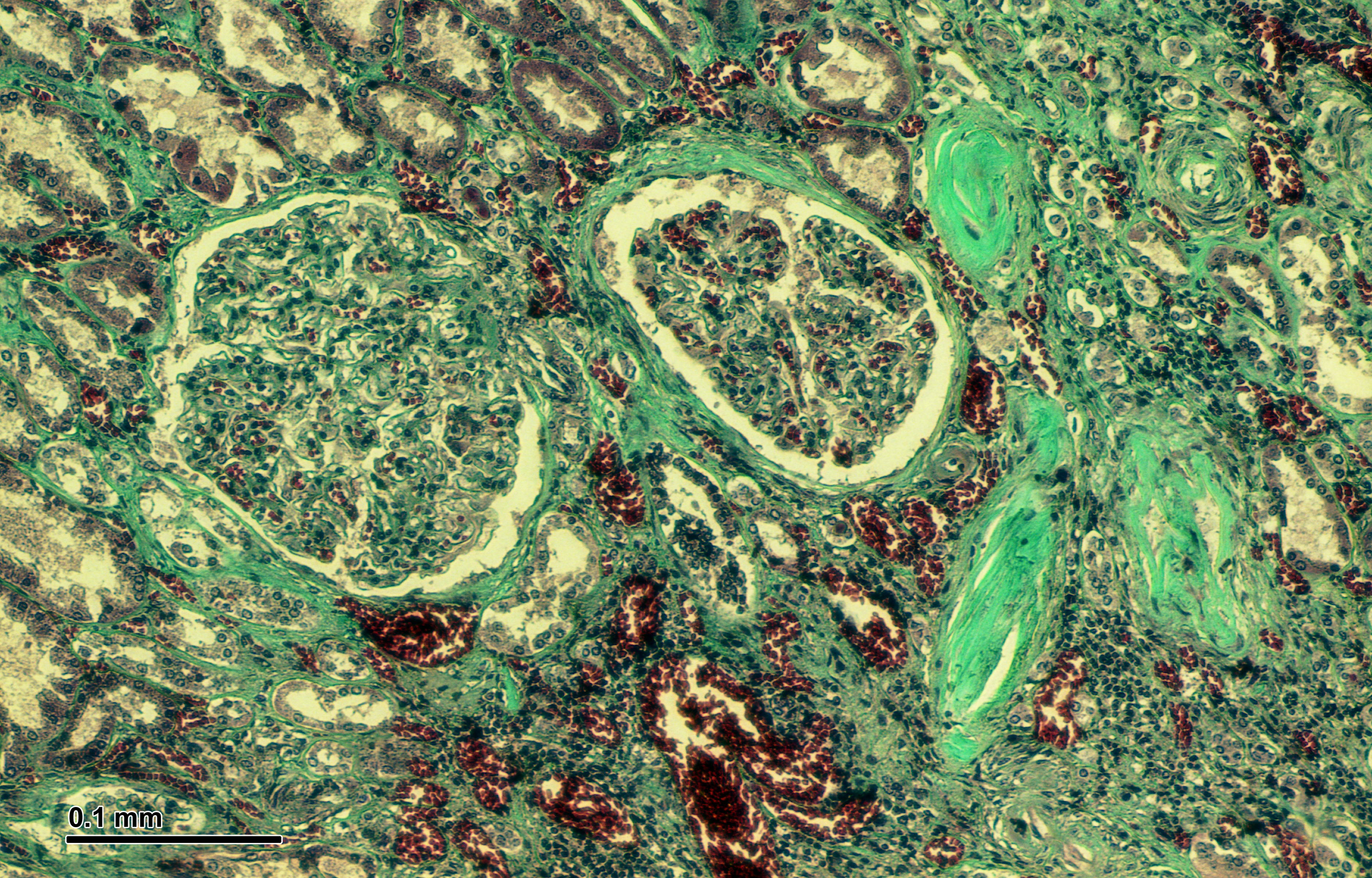
A microscopic field can show a corpuscular profile, but a single light-microscope section does not reveal every part of the filtration interface at the same resolution. Compare a small enlarged schematic with the real tissue image. State what is directly visible and what the explanatory drawing adds. This helps learners use microscopy as evidence without expecting it to display every conceptual layer.
8. Tubular regions differ in location and organization
The proximal convoluted tubule, nephron loop, and distal convoluted tubule are linked portions of the nephron’s tubular route. Collecting ducts receive output from multiple nephrons and are usually distinguished from the nephron proper. Cortical and juxtamedullary arrangements differ, particularly in the extent of their loops. These relationships connect the microscopic route with the cortex–medulla map. [OpenStax: microscopic kidney anatomy]
For study, place one schematic nephron across a cortex–medulla boundary and mark the portions occupying each region. Then compare another nephron arrangement. Avoid assuming that every loop reaches the same depth. The exercise explains why a section contains many tubular profiles even though a colored teaching line presents a continuous route that cannot be followed completely in one microscopic field.
9. Quantitative facts describe scale and variation
NIDDK describes approximately one million nephrons in each kidney. This conveys scale, not an exact count for every person. A commonly taught estimate places about fifteen percent of nephrons in the juxtamedullary group, with the remainder in the cortical group. These proportions are introductory approximations and should not be interpreted as a measurement from the single tissue image below. [NIDDK: how the kidneys work] [OpenStax: gross kidney anatomy]
The graphic displays an approximate eighty-five-to-fifteen split using the same unit, percentage of nephron population. The numbers sum to one hundred by design; they do not report the distribution of kidney volume or the share of urine output. Keeping the denominator explicit makes the numerical comparison useful and prevents one percentage from being borrowed for an unrelated physiological claim.
10. Latin terms retain compartment distinctions
Ren, cortex renalis, medulla renalis, columnae renales, and pelvis renalis provide selected Latin names. Calices renales identifies the collecting calyces, while corpusculum renale names the renal corpuscle. The terms refer to different scales and compartments. Learning them with a route prevents renal pelvis from becoming a synonym for every central space in a kidney section. [FIPAT: Terminologia Anatomica, second edition]
Create a revision table with three categories: parenchyma, vascular structure, and collecting passage. Place each term in the appropriate category, then explain any close relationship between categories. This is useful when interpreting a diagram in which differently functioning structures overlap. For patient-facing discussion, use ordinary language while preserving whether the finding concerns tissue, blood vessels, or the collecting system.
| English | Selected Latin |
|---|---|
| Renal cortex / medulla | Cortex renalis / medulla renalis |
| Renal columns | Columnae renales |
| Renal calyces / pelvis | Calices renales / pelvis renalis |
| Renal corpuscle | Corpusculum renale |
11. Real tissue complements the opened-organ view
The real human kidney micrograph below shows renal tissue in a bright-field preparation, including corpuscular and tubular profiles. The visible colors reflect preparation rather than natural organ coloration. Resizing changes the apparent scale, so a fixed displayed magnification is not claimed. A short scale bar within the source image retains its original context and should not be interpreted without the image’s dimensions.
Medical students can compare recognizable profiles with the organ section and nephron schematic. Practice assistants, hospital staff, physicians, and eldercare teams can use the same structure names to clarify established findings. Finish revision by explaining one tissue compartment, one blood route, and one collecting route. Those three explanations connect renal anatomy across scales without inferring an individual diagnosis from the illustration.
Sources and image credits
Sources checked on 8 October 2026. Numerical examples, anatomical classification counts, and research estimates are identified separately in the text.
- FIPAT: Terminologia Anatomica, second edition
- NIDDK: how the kidneys work
- OpenStax: gross kidney anatomy
- OpenStax: microscopic kidney anatomy
- NCI SEER: kidney anatomy
Explanatory prose and simplified SVG graphics are original. Photographs retain their stated Creative Commons licenses; creator and source links appear beside each image. Catalog artwork is educational and decorative; no clinical authorship or peer-review credential is claimed.