1. Lung anatomy connects a branching tree with a thin interface
The human lungs combine large anatomical regions, a branching airway tree, and microscopic exchange surfaces. These scales are connected, but a single drawing must simplify them to remain readable. A lobe map establishes regional organization; an opened lung shows vessels and airways; a micrograph reveals tissue relationships that are invisible in the external outline. The learning task is to connect these scales without confusing their dimensions.
For medical students and healthcare teams, distinguish the air route from the blood route and the lung surface from the pleural space. A poster can introduce all four, but its arrows, colors, and enlarged details require a clear legend. This guide follows those relationships and uses real radiographic and microscopic images to show what different methods reveal.
2. Paired lungs occupy different sides of the mediastinum
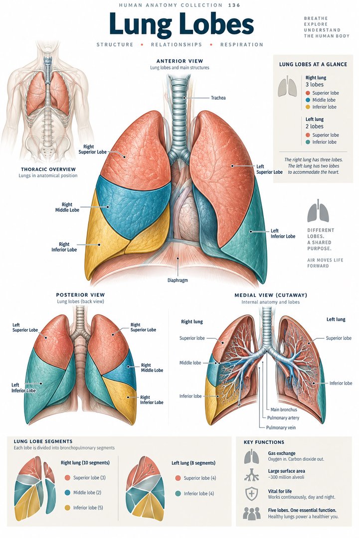
The lungs lie on either side of the mediastinum. Each has an apex, a base related to the diaphragm, and costal and mediastinal surfaces. Their external shapes reflect neighboring structures rather than perfect symmetry. A frontal body chart can establish their broad location while concealing the depth and posterior relationships present in the thorax. [OpenStax: lung anatomy]
Compare a whole-body view with a lung removed from its surroundings in a diagram. Name what the second image omits: chest wall, diaphragm, mediastinal neighbors, and supporting connections. This helps learners avoid treating an isolated organ outline as its complete anatomical setting. The same habit is useful when explaining why a chest projection contains overlapping structures rather than a neatly separated pair of organs.
3. Right and left lobes have a conventional organization
The right lung normally has superior, middle, and inferior lobes separated by horizontal and oblique fissures. The left normally has superior and inferior lobes separated by an oblique fissure. The lingula belongs to the left upper lobe and is not a separate left middle lobe. Variations in fissure completeness and appearance occur. [OpenStax: lung anatomy] [NCBI Bookshelf: lung imaging anatomy]
Trace a fissure before naming the lobe beside it. Then identify which side of the body the diagram shows. This sequence prevents a color patch from being treated as sufficient evidence of a regional boundary. It also makes the lingula easier to understand as a named projection within a lobe rather than an additional unit added to make the two lungs look numerically symmetrical.
4. The hilum and root are related but different terms
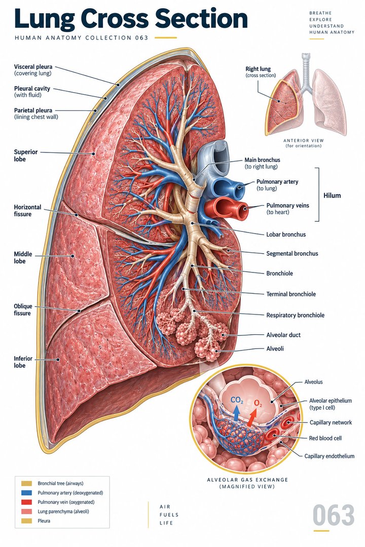
The pulmonary hilum is the entry and exit region on the mediastinal aspect. The root is the bundle of bronchi, vessels, lymphatic structures, and nerves passing between lung and mediastinum. Calling every visible structure in this region “the hilum” can conceal the distinction between a junctional area and the actual structures crossing it. [NCBI Bookshelf: lung anatomy]
Use a label for the region and separate labels for its contents. Then follow one bronchus and one pulmonary vessel away from the junction. This adds depth to a sectional poster and helps students recognize that several distinct routes share an entry area. A location in the same bundle does not make their contents or physiological roles interchangeable.
5. Main, lobar, and segmental bronchi form a hierarchy
Main bronchi supply the lungs, lobar bronchi relate to lobes, and segmental bronchi relate to bronchopulmonary segments. The right main bronchus is generally shorter, wider, and more vertical than the left. This broad relationship is an anatomical description, not a rule that determines the location of every inhaled object in every circumstance. [NCBI Bookshelf: bronchial anatomy] [NCBI Bookshelf: lung imaging anatomy]
Draw a branching tree with the level named beside each branch. A learner who labels every branch “bronchus” has recognized the structure category but not the hierarchy. The levels explain why a lobe and a bronchopulmonary segment are different regional units. Keep the branch level explicit when comparing a whole-organ poster with an imaging report.
6. Segment counts depend on the classification used
A commonly taught segmental arrangement uses ten bronchopulmonary segments on the right. Left-sided descriptions often use approximately eight to ten, depending on whether combined segments are counted separately. A source that uses a different left count may be using a different naming convention rather than describing an entirely different organ. [NCBI Bookshelf: bronchial anatomy]
When preparing a teaching diagram, state the classification and avoid comparing counts without checking how the units were defined. Ask students to identify a segment’s supplying branch and regional relationship rather than only reciting a total. This approach is more useful when a report or surgical discussion uses a specific segment name that cannot be understood from a lobe color alone.
7. Microscopic organization supports exchange and local defense
Conducting airways end at terminal bronchioles, while respiratory bronchioles begin the gas-exchanging zone and connect toward alveolar ducts and alveoli. Type I alveolar cells contribute much of the thin exchange surface; type II cells produce surfactant. Alveolar macrophages add a local phagocytic role. These cell types and routes should be distinguished in an enlarged detail. [OpenStax: respiratory structures]
Compare the enlarged alveolar inset with the size of the whole lung in a poster. State explicitly that the scales differ. A few prominently drawn air spaces cannot be used to count the organ’s full alveolar population. Similarly, a cell icon explains a type and role but does not provide a literal image of every cellular relationship at the interface.
8. Pleural layers enclose a potential space
Visceral pleura covers the lung and extends into fissures. Parietal pleura lines related thoracic surfaces. The pleural cavity is the potential space between these layers, normally with a thin lubricating fluid film. It is not an ordinary air-filled chamber containing the lung. The lung occupies its thoracic region while the pleural relationship follows its surface. [OpenStax: lung anatomy]
Use a close-up layer diagram beside the whole-organ view. Identify the two surfaces and the narrow space between them before discussing movement. This prevents a large gap drawn for visibility from being read as the normal size of a physical cavity. For clinical communication, retain the distinction between a pleural finding and a finding within pulmonary tissue.
9. Research estimates reveal microscopic scale
Ochs and colleagues reported a mean of approximately four hundred and eighty million alveoli, with estimates ranging from two hundred and seventy-four to seven hundred and ninety million, in six adult human lungs studied using stereology. The sample and method matter: these are research estimates from a small study, not a universal reference interval applicable to every living person. [Ochs et al. (2004): alveolar number in six adult human lungs]
When sharing the statistic, retain the unit, sample, and reported range together. Do not turn the study range into a diagnostic target or imply that a chest radiograph measures alveolar number. The fact is useful because it contrasts the simple organ outline with the enormous microscopic interface, while reminding learners that a numerical estimate requires its own method and evidence.
10. Latin terminology links external regions with internal routes
Pulmo dexter and pulmo sinister name the right and left lungs. Apex pulmonis, basis pulmonis, hilum pulmonis, and radix pulmonis identify external orientation and the junctional relationship. Pleura visceralis and pleura parietalis preserve the layer distinction. Bronchus and bronchiolus refer to different parts of the airway hierarchy. Learn each term with its scale and neighboring structures. [FIPAT: Terminologia Anatomica, second edition]
Build a vocabulary table grouped by surface, entry region, airway, and covering. Add one example sentence locating each term. This makes terminology usable across posters, images, and reports. For patient-facing discussion, use ordinary English while retaining whether the finding concerns an airway, pulmonary tissue, or pleural relationship; those locations should not be collapsed into the generic word chest.
| English | Selected Latin |
|---|---|
| Right / left lung | Pulmo dexter / pulmo sinister |
| Apex / base | Apex pulmonis / basis pulmonis |
| Hilum / root | Hilum pulmonis / radix pulmonis |
| Pleural layers | Pleura visceralis / pleura parietalis |
11. Real images show different levels of anatomy
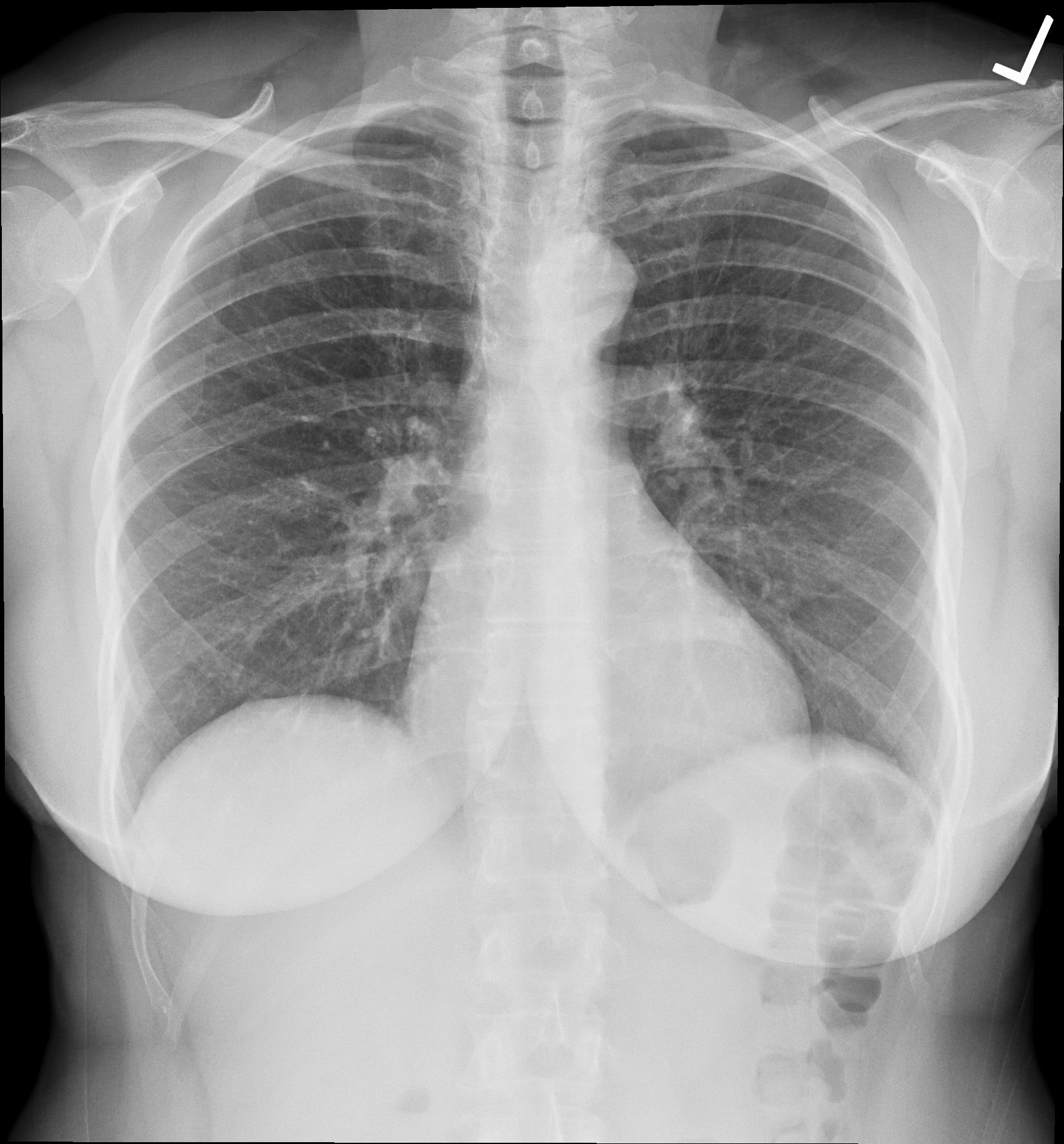
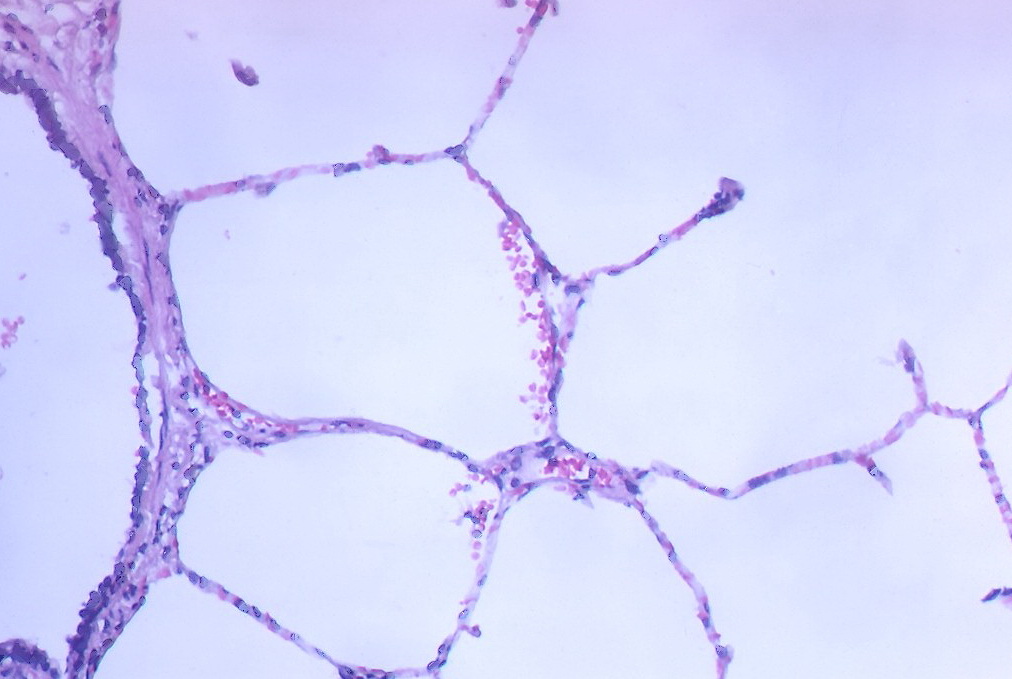
The chest radiograph below records a real human thoracic projection. It shows overlapping structures, not a clean opened-lung view. The lung micrograph shows alveolar tissue around a terminal bronchiole, but the source does not explicitly identify species. Its tissue preparation and microscopic scale differ from the radiograph, and neither image should be expected to display every lobe and segment label.
Medical students can explain what each method reveals before comparing it with the schematic. Practice assistants, hospital staff, physicians, and eldercare teams can use these distinctions when discussing established findings. Finish revision by tracing an airway level, locating a pleural layer, and explaining one microscopic relationship. This connects the lung’s anatomy across scales without making an individual diagnosis from the educational images.
Sources and image credits
Sources checked on 8 October 2026. Numerical examples, anatomical classification counts, and research estimates are identified separately in the text.
- OpenStax: respiratory structures
- OpenStax: lung anatomy
- Ochs et al. (2004): alveolar number in six adult human lungs
- FIPAT: Terminologia Anatomica, second edition
- NCBI Bookshelf: lung imaging anatomy
- NCBI Bookshelf: lung anatomy
- NCBI Bookshelf: bronchial anatomy
Explanatory prose and simplified SVG graphics are original. Photographs retain their stated Creative Commons licenses; creator and source links appear beside each image. Catalog artwork is educational and decorative; no clinical authorship or peer-review credential is claimed.