The gallbladder stores a secretion made by another organ
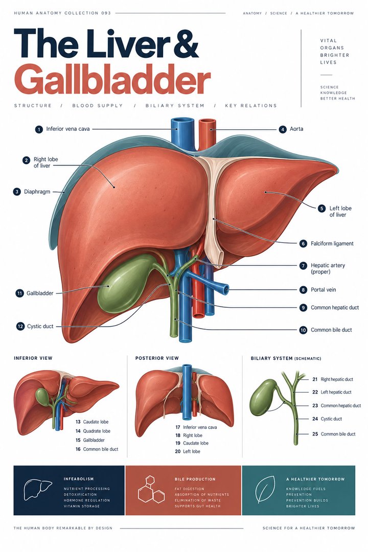
The gallbladder is a small hollow organ associated with the inferior liver region in the right upper abdomen. Its central role is to store and concentrate bile and release it through the biliary route when required for digestion. The liver produces bile; the gallbladder does not manufacture it. This distinction is fundamental because it separates production from temporary storage and coordinated delivery. The organ is also distinct from the urinary bladder, despite the shared English word bladder. The Latin name vesica biliaris, also encountered as vesica fellea, identifies its connection with bile. NIDDK: Gallstones and the biliary tract provides the basic anatomical context.
For medical students, the gallbladder offers a clear example of how a modest-sized structure can influence a connected system of ducts and organs. For healthcare staff, understanding the connections makes terms in reports more precise and helps explain why a gallbladder discussion can involve the liver, duodenum, and pancreas. This guide follows the organ's gross parts, microscopic wall, and duct route before relating them to digestive function and clinical terminology. The aim is to understand the anatomical question being asked rather than to diagnose a person's symptoms from a pear-shaped drawing.
Fundus, body, and neck organize the gross anatomy
The gallbladder's main gross parts are the fundus, body, and neck. The fundus is the rounded distal region, the body occupies the main reservoir, and the neck narrows toward the cystic duct. These are regions of one hollow organ rather than three separate compartments divided by permanent walls. The Latin terms fundus vesicae biliaris, corpus vesicae biliaris, and collum vesicae biliaris connect the names with their anatomical positions. The NCI's liver and gallbladder anatomy describes these gross parts. A diagram should label the duct separately so that the neck is not mistaken for the entire outgoing route.
The familiar pear shape is an introduction, not a promise that every specimen or image will look identical. Filling state, individual variation, and viewing angle change the apparent outline. In a gross anatomical photograph, the liver may obscure part of the organ, while an isolated specimen no longer shows its position in the body. Pairing those views helps students distinguish shape from relationship. A body outline should also identify the patient's right side, especially in an anterior illustration where it appears on the viewer's left. Clear orientation is as important as the names printed beside the organ.
Capacity is a volume, not daily bile production
A commonly described gallbladder storage capacity is approximately 30 to 50 milliliters, with variation according to filling and individual conditions. This is a reservoir-volume description, not the amount of bile the liver produces each day and not a diagnostic threshold for an imaging report. The range is presented in NCBI Bookshelf: Gallbladder anatomy. One milliliter corresponds to one cubic centimeter, so the numbers describe a relatively small hollow space. They do not indicate how many stones may be present or how effectively the organ contracts. Keeping the quantity attached to its definition makes it useful rather than misleading.
An educational graphic can show a simple volume range with a label stating typical approximate storage, avoiding a claim that every healthy organ contains fifty milliliters at all times. The visible size on an image depends on when and how it was obtained, among other factors. A reservoir also changes its contents over time, so a static outline captures only one state. Students can compare the volume label with the functional cycle of storage and release to see why one measurement is not a complete assessment. For staff, preserve the stated imaging description instead of substituting a generic capacity value from a textbook.
The wall is adapted for storage and concentration
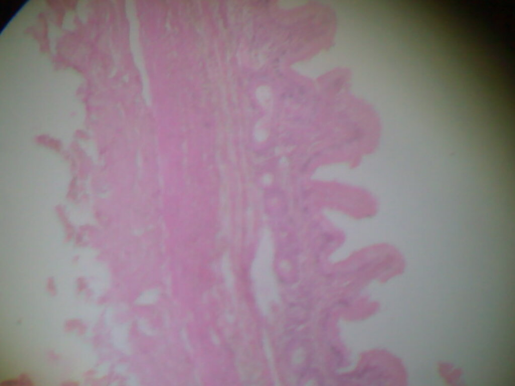
The gallbladder has a folded mucosal lining with an epithelium and underlying connective tissue, together with smooth muscle and an outer covering related to its surface. Unlike much of the gastrointestinal tube, its wall does not have a conventional muscularis mucosae and submucosa arranged in the same way. The muscle fibers are not simply a neatly layered miniature version of the intestinal wall. This makes the gallbladder a useful example of why one generic digestive wall diagram should not be applied to every associated organ. The organization is demonstrated in the University of Leeds Histology Guide.
The lining participates in absorption of water and electrolytes, helping concentrate the stored bile. A folded surface increases the available interface, but a fold visible in one histological section should not be mistaken for a separate gland or a permanent chamber. Sectioning can produce complex profiles from a continuous three-dimensional mucosa. In a real tissue photograph, identify the lumen first, then trace the lining and compare it with the smooth muscle region. The stain gives contrast, not natural organ color. If a source image does not establish species or sampling details, its caption should not invent them.
The cystic duct connects storage with the main biliary route
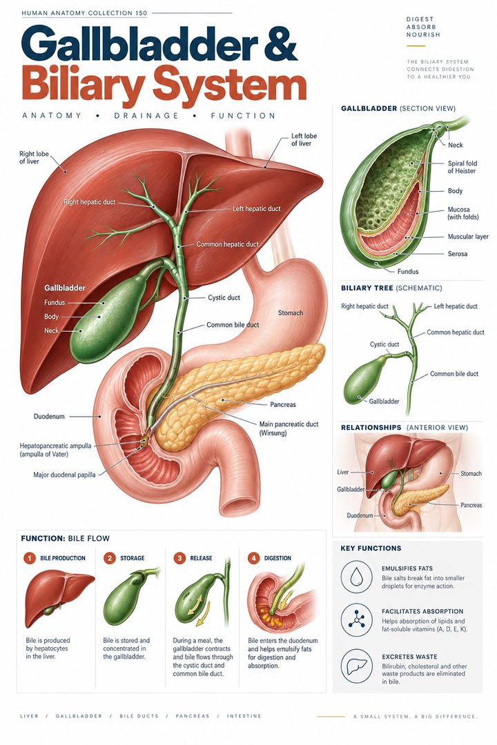
The cystic duct, ductus cysticus, connects the gallbladder neck with the extrahepatic biliary system. Bile can enter the reservoir during storage and leave it during emptying, so a diagram should not portray the cystic duct as a permanently one-way outlet. Mucosal folds in the duct are sometimes called spiral valves, but that name should not be taken to mean a simple mechanical check valve like those drawn in veins. The anatomy and variations of the connection are described in NCBI Bookshelf. Individual routes can differ, making a single idealized joining angle a teaching convention rather than a universal rule.
For learning, draw storage and release as two states of the same network. In the storage panel, indicate movement toward the gallbladder where appropriate; in the release panel, show movement from the reservoir toward the intestinal destination. This is clearer than placing two arrowheads on one line without explaining the timing. The duct also exists in relation to nearby vessels and hepatic structures, so its name should remain distinct from that of the cystic artery. One carries bile and the other supplies blood. Similar labels do not imply that the two structures have the same contents or physiological role.
Hepatic ducts and common bile duct have different connections
The right and left hepatic ducts join to form the common hepatic duct in the standard anatomical arrangement. The common hepatic duct and cystic duct then connect to form the common bile duct, ductus choledochus, which carries bile toward the duodenum. The labels common hepatic duct and common bile duct therefore describe different positions within the route. They should not be treated as interchangeable abbreviations. The duct relationships are summarized in NCI biliary anatomy. A clear figure preserves the junctions, because an unlabeled single tube from liver to intestine hides the storage connection and the change in nomenclature.
This arrangement makes a route-tracing exercise particularly useful. Start in the liver, pass through the hepatic ducts, then compare the pathway into the gallbladder with the pathway onward toward the intestine. Next start within the gallbladder and identify the route that stored bile takes during release. The paths share some segments but begin in different places. Students should name the junction before the next duct, rather than memorizing a disconnected sequence of abbreviations. For healthcare staff, recording the specific duct named in a report is important because a finding in one segment is not automatically a finding in every part of the biliary system.
The duodenal destination links bile with pancreatic secretion
Near the duodenum, the distal bile route lies in close relationship with the pancreatic duct system. In a common arrangement, the ducts meet near their opening into the intestine through the major duodenal papilla, but terminal configurations vary. This relationship helps explain why a problem near the shared destination can involve both biliary and pancreatic processes. It does not mean the gallbladder produces pancreatic enzymes or the pancreas produces bile. The NCI's pancreas and duodenum anatomy shows the neighboring structures, while the two origins should remain distinct in a functional diagram.
The hepatopancreatic ampulla, ampulla hepatopancreatica, and the muscular regulation near the terminal route are terms learners may encounter. These should be placed within the full duct map rather than presented as isolated names at the end of an organ list. A sectional figure can clarify which tube enters which wall and where the luminal destination lies. Avoid drawing an enormous ampulla as though it were a second storage organ equivalent to the gallbladder. The scale is usually exaggerated for readability, and the caption should acknowledge that the figure represents relationships rather than measured dimensions.
Bile supports fat handling without acting as a digestive enzyme
Bile contains substances that support the handling of lipids in the intestinal lumen. Bile salts contribute to emulsification and micellar processes that make lipid digestion and absorption more effective in the coordinated digestive environment. They are not equivalent to pancreatic lipase and should not be described as enzymes that directly hydrolyze all fat. Bile also provides a route for substances such as bilirubin, linking digestion with hepatic processing and excretion. The chemical digestion and absorption context is explained in OpenStax: Digestion and absorption. A useful figure assigns different arrows to physical organization, enzymatic breakdown, and movement toward absorption.
Gallbladder emptying is coordinated with digestive signals and the movement of contents into the small intestine. This timing helps connect reservoir function with the availability of food substrates. It is not a claim that the organ waits until a person consciously notices a meal and then empties all its contents instantaneously. For students, a simple sequence of storage, concentration, and release can communicate the principle without inventing a precise universal schedule. For staff, the lesson helps explain the organ's digestive role while leaving personal dietary advice to the relevant clinician and the individual's assessed needs.
Gallstone terminology should identify the actual location
Cholelithiasis refers to gallstones, commonly within the gallbladder, while choledocholithiasis identifies stones in the common bile duct. Cholecystitis concerns inflammation of the gallbladder, and cholangitis concerns inflammation of bile ducts, often in an infectious clinical context. These terms describe related but distinct conditions and should not be merged into one label because the same anatomical system is involved. NIDDK describes stones and their relationship with bile duct blockage. An anatomical map explains why a stone in one region can have a different consequence from a stone remaining within the reservoir.
The NIDDK educational page, last reviewed in November 2017, cites a United States gallstone prevalence estimate of ten to fifteen percent. This historical educational estimate is not a newly measured statistic for the current year and should not be presented as one. It also does not state that every affected person has symptoms: silent stones and symptomatic obstruction are different contexts. A numerical inset should include the population and source review date and avoid predicting an individual's experience. The more important anatomical learning point is to identify where a finding is located and whether the report describes storage tissue, a duct, inflammation, or a downstream consequence.
Imaging and symptoms provide complementary information
Ultrasound commonly provides structural information about the gallbladder and biliary region. Other investigations may address ducts, flow, inflammation, or associated organs according to the clinical question. A blood test does not produce an image of a stone, and an image alone does not necessarily explain every symptom. NIDDK: Gallstone diagnosis describes how history, examination, laboratory findings, and imaging can contribute together. For students, this is another example of why anatomy matters to test interpretation: the test's target and the anatomical location of a possible problem must remain explicit.
Clinical observations such as pain, vomiting, fever, or jaundice require appropriate assessment in their context and should not be assigned to the gallbladder from location alone. NIDDK: Gallstone symptoms explains relevant symptom patterns and complications. For healthcare staff, clear recording of onset, timing, associated observations, and change from the person's usual state supports communication with the responsible clinician. Avoid the shorthand gallbladder pain unless that clinical conclusion has been established. An anatomical understanding improves the description of the region, but it does not make the region the only possible source of an abdominal complaint.
The reservoir can be removed while hepatic bile production remains
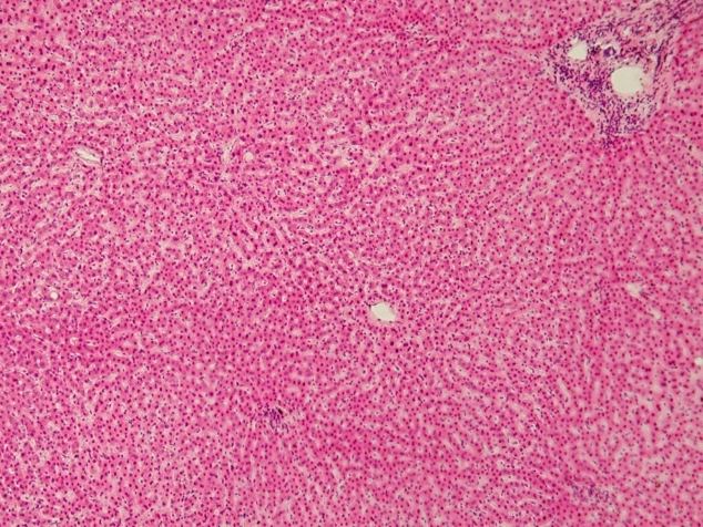
After gallbladder removal, the liver continues producing bile, which can pass through the remaining biliary route toward the intestine without the same storage reservoir. This is why absence of the gallbladder is not equivalent to absence of bile. The anatomical consequence is explained in NIDDK: Gallstone treatment. It is nevertheless inappropriate to promise that every person will have identical digestive experiences after surgery. Individual recovery and ongoing symptoms require the appropriate clinical context. An educational diagram can remove the reservoir while preserving the liver-to-duodenum connection to make the anatomical change clear.
To review the organ, trace three related processes separately: bile production in the liver, storage and concentration in the gallbladder, and delivery through ducts to the intestine. Then place the blood supply and wall tissue alongside that luminal route rather than mixing them into one arrow. This integrated map helps medical students connect gross and microscopic anatomy and helps clinical teams use report terminology precisely. The gallbladder is best understood as a changing reservoir within a connected digestive system. Its anatomy explains the relationships, while actual findings and professional assessment determine what those relationships mean for a particular person.
Three views for one connected organ
A useful learning exercise places a body-position diagram, a duct map, and a tissue photograph beside one another. Begin by marking the liver-facing surface on the position diagram, then find the reservoir connection on the duct map. Finally identify the lumen and lining in the tissue photograph. Each view answers a different question: where the organ lies, where its contents can travel, and how its wall is organized. Ask the learner to explain which relationship cannot be established from each image alone. A close tissue photograph cannot show the complete duct route, while a route schematic cannot establish the appearance of the actual mucosa. This exercise also improves captions by requiring the author to state what the image demonstrates and which details have been simplified.
Anatomical Latin: quick reference
| Latin term | English meaning |
|---|---|
| Vesica biliaris | Gallbladder |
| Ductus cysticus | Cystic duct |
| Ductus hepaticus communis | Common hepatic duct |
| Ductus choledochus | Common bile duct |
Sources and image credits
Sources reviewed on 9 October 2026. Counts, estimates, study statistics and hypothetical calculations retain their context in the text.
- Gallbladder anatomy
- Liver and gallbladder
- Gallbladder histology
- Gallstones and historic estimates
- Gallstone diagnosis
- Gallstone symptoms
- Gallbladder removal anatomy
- Digestive absorption
- Pancreas and duodenum
Explanatory prose and SVG graphics are original. Photograph authors, source records and licenses are credited beside each image. Catalog illustrations provide educational context; anatomical facts are supported by the linked sources.