One organ participates in two different communication routes
The pancreas contributes to digestion and to metabolic regulation through different tissue components. Its exocrine system produces digestive secretions that reach the small intestine through ducts, while its endocrine cells release hormones into the circulation. The two functions share an organ but should not be drawn as a single indistinguishable output. Insulin does not normally travel to the intestine through the pancreatic duct to perform its endocrine role, and pancreatic digestive enzymes are not delivered to the digestive lumen through the systemic blood supply. This route distinction is the most useful starting point for understanding pancreatic function. NIDDK: Exocrine pancreatic insufficiency introduces the organ's dual roles.
For medical students, the pancreas connects abdominal anatomy, histology, digestive chemistry, and endocrine physiology. For healthcare staff, it explains why a discussion of pancreatic health may include nutrient handling as well as glucose regulation. These are related concerns, but a normal result in one domain does not automatically establish normal function in the other. This guide follows both routes from the cells that produce a secretion to the tissues where it acts. The aim is an integrated model that retains the distinction between a duct leading into the digestive tract and a blood vessel carrying a hormonal signal.
Regional anatomy places the pancreas among several neighbors
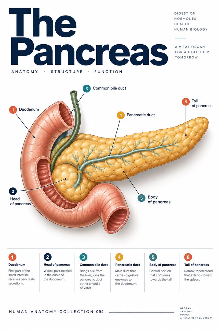
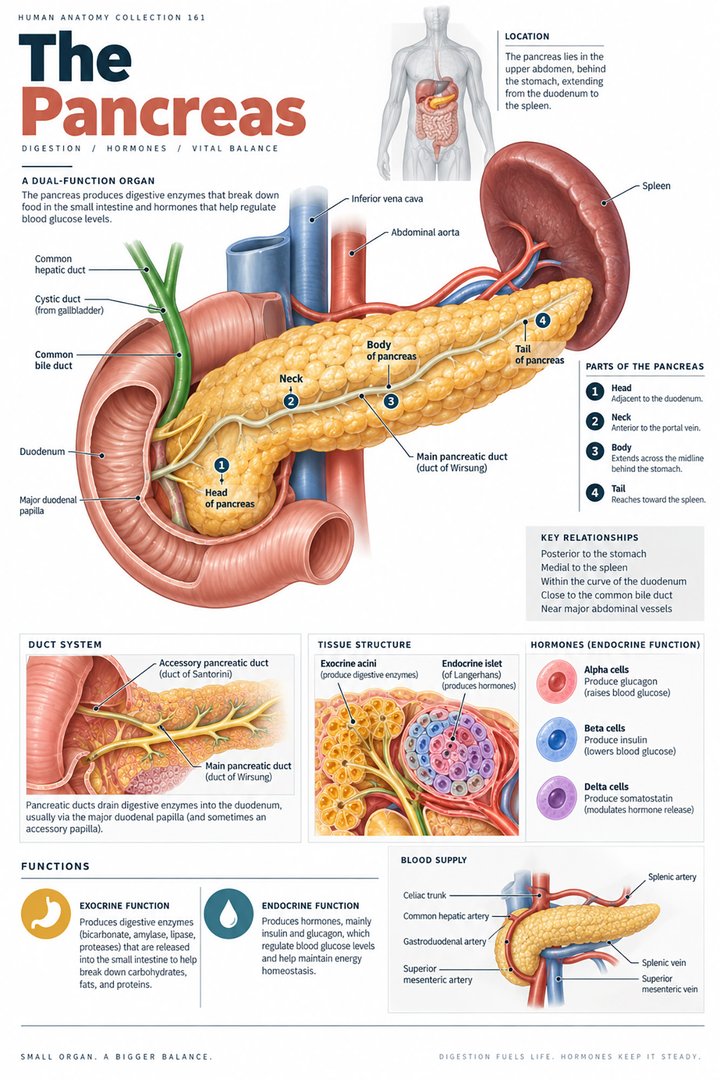
The pancreas lies in the upper abdomen in close relationship to the stomach, duodenum, spleen, major vessels, and biliary tract. The head is associated with the curve of the duodenum, while the body and tail extend toward the left side and the splenic region. Anatomical descriptions commonly distinguish head, neck, body, and tail, and also name the uncinate process as a projection associated with the head. Depending on how components are grouped, a list can contain four main regions or separately include the uncinate process. This is a nomenclature difference rather than a disagreement about an extra organ. See NCBI Bookshelf: Pancreas anatomy.
The Latin terms caput pancreatis, collum pancreatis, corpus pancreatis, cauda pancreatis, and processus uncinatus help label the regional map. Most of the pancreas is related to the posterior abdominal wall, so a simple surface outline cannot show all its depth relationships. A teaching diagram should indicate whether the stomach has been removed or made transparent to reveal it. This matters because an apparently unobstructed view can otherwise suggest that the organ sits directly against the anterior body surface. Position in an atlas is a reference; clinical imaging establishes the relationships in the individual.
Acinar tissue organizes digestive production around small lumens
Exocrine pancreatic tissue is organized into acini, groups of secretory cells associated with small lumens that connect to a duct system. The acinar cells produce enzyme-related secretions, while the ductal system contributes to the fluid environment that carries them toward the intestine. This organization resembles a branching delivery network rather than a reservoir holding all products in one large cavity. The structure is explained in NCBI Bookshelf: Exocrine pancreas anatomy. The word acinus describes a tissue unit, whereas a duct describes a route; using both labels makes the connection between production and delivery explicit.
On a histological image, first distinguish secretory tissue from a duct profile and from any islet region present in the field. A single slice may not show the continuity of the branching ducts, even though the tissue belongs to a connected system. A microscopic photograph therefore benefits from a nearby schematic showing the larger route. The schematic should not imply that an acinus has the same size as an islet or a main duct. Scale and staining affect what becomes visible. Students should ask which structure is demonstrated directly and which connection has been added to explain the physiology.
Digestive enzymes act on different kinds of food molecules
Pancreatic enzymes contribute to the digestion of carbohydrates, fats, and proteins in the small intestine. Amylase acts on appropriate carbohydrate substrates, lipase participates in fat digestion, and several proteases contribute to protein breakdown. These products have different molecular targets rather than being interchangeable forms of one general digestive fluid. Bile helps create conditions for handling fat, but it is not itself a pancreatic enzyme and it is produced by the liver. The roles of accessory organs are introduced in OpenStax: Digestive accessory organs. A figure can show three substrate categories with separate arrows toward smaller absorbable products.
Do not interpret three illustrated categories as the claim that the pancreas produces only three enzymes. The chart groups functions for learning; each category can contain several molecular processes. Mechanical mixing, chemical breakdown, and absorption are also separate steps. Food can be mixed well yet still require chemical digestion, and digestive products must cross an intestinal barrier before entering the body's internal environment. Following this sequence helps learners understand why a functioning pancreas participates in a wider system. Its secretions act in a lumen whose acidity, contents, motility, and epithelial processes also influence the overall result.
Bicarbonate supports the intestinal chemical environment
Pancreatic secretion includes bicarbonate-rich fluid that helps neutralize the acidic contents reaching the duodenum from the stomach. This creates conditions in which intestinal and pancreatic digestive processes can function. Bicarbonate is not an enzyme and should not be shown breaking a food molecule into fragments. It changes the chemical environment, while enzymes catalyze particular reactions. Keeping those roles separate makes the pancreatic model much clearer. The relationship between pancreatic fluid, duodenal contents, and digestive activity is described in Pancreapedia: Human exocrine secretion. An explanatory diagram should show neutralization beside enzyme action rather than using one arrow for both.
The pH environment is dynamic, not a fixed number that is identical everywhere in the small intestine at all times. A teaching scale can indicate movement away from the highly acidic gastric environment without inventing a patient-specific reading. For students, this is a useful example of how physiology depends on conditions as well as on the presence of a product. Producing an enzyme is not the complete story if the enzyme does not reach an appropriate environment or mix effectively with its substrate. For healthcare staff, it helps explain why digestive performance can depend on several connected organs rather than one laboratory label.
Duct connections link the pancreas with the duodenum and bile route
The main pancreatic duct, ductus pancreaticus, collects secretions along the gland and delivers them toward the duodenum. Its terminal relationship with the common bile duct and duodenal opening is important, although individual duct arrangements vary. An accessory pancreatic duct can contribute an additional route. A schematic should identify its chosen typical arrangement instead of implying that every person has exactly the same joining point and openings. The NCI's pancreas and duodenum anatomy illustrates the regional relationships. Pancreatic and biliary secretions may meet near their intestinal destination, but that does not mean they originate from the same tissue.
When learning the route, trace a secretion from an acinar region through progressively larger ducts before naming the final opening. Then trace bile from the hepatic route toward the intestine and compare where the two pathways become neighbors. This approach makes the anatomical relationship easier to understand than memorizing a duct name without its connections. It also explains why a problem near a shared terminal region can have consequences involving more than one organ. The diagram is a model of anatomy, however, not a tool for determining the cause of a patient's abdominal symptoms from location alone.
Enzyme production includes mechanisms that limit premature activation
Some pancreatic proteolytic enzymes are produced and secreted in inactive precursor forms. Their activation is coordinated with the intestinal environment, helping separate production in the gland from digestion of food in the lumen. Trypsinogen is a familiar example of a precursor that becomes trypsin. It is inaccurate to state that every pancreatic enzyme is inactive until reaching the intestine: amylase and lipase do not fit that blanket description. The important principle is selective control of enzyme activity, not a universal rule applied to all secretory proteins. Digestive enzyme organization is discussed in OpenStax.
For a teaching diagram, use separate labels for precursor and active enzyme and specify the example being shown. A large switch icon labeled all enzymes activate here would hide the distinctions. The same care is helpful when discussing inflammation of the pancreas, because disease mechanisms cannot be reduced to one cartoon in which a gland simply digests itself. NIDDK: Pancreatitis introduces pancreatic inflammation and its different contexts. An anatomy guide can explain why controlled delivery and activity matter while leaving diagnosis, severity assessment, and treatment to the appropriate clinical process.
Islets release hormones through a vascular route
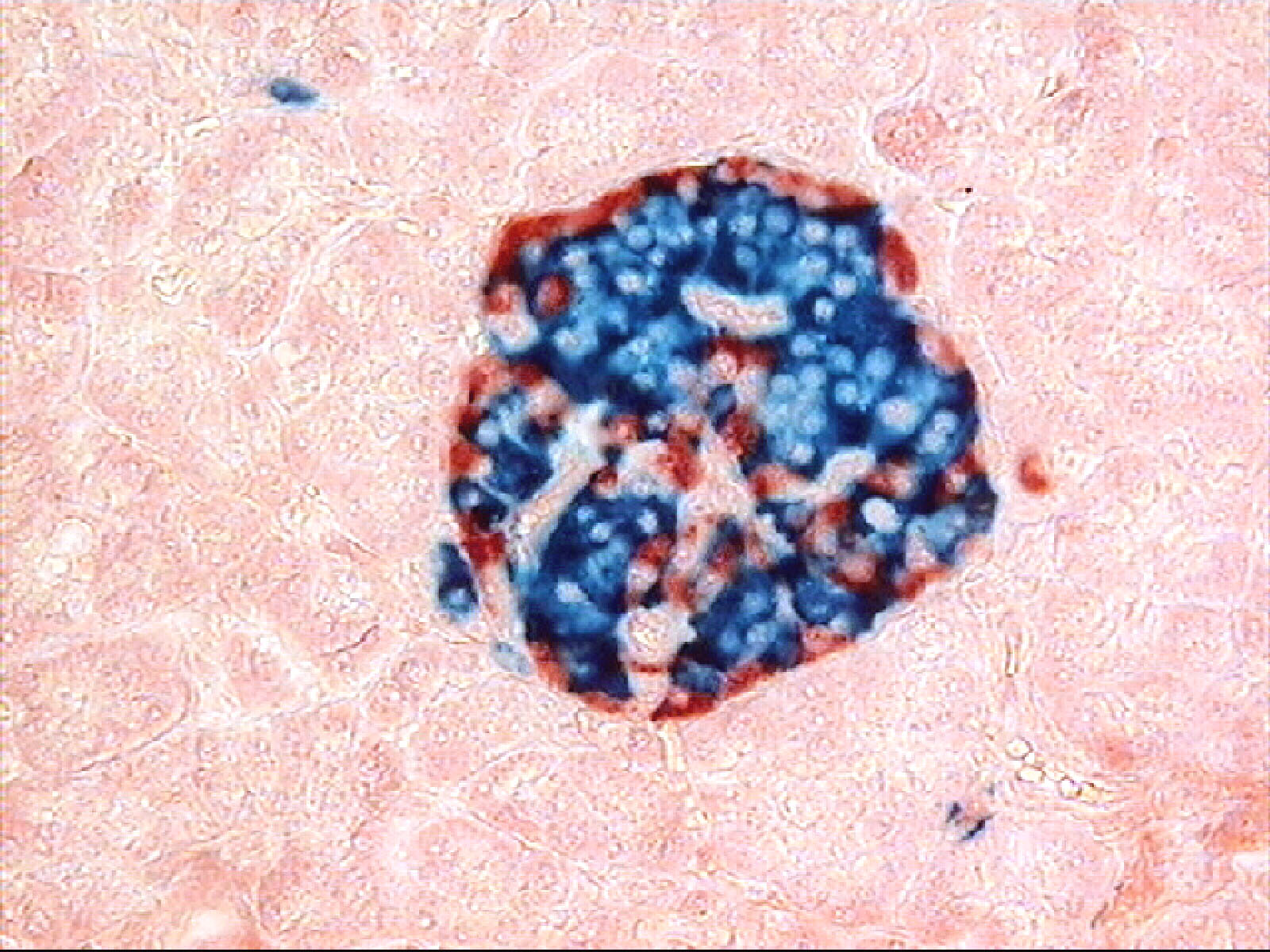
Pancreatic islets, insulae pancreaticae, contain endocrine cell populations embedded among the exocrine tissue. Beta cells produce insulin, alpha cells produce glucagon, and other populations contribute signals including somatostatin and pancreatic polypeptide. These products are associated with local interactions and endocrine communication through blood. A routine section can show an islet region, while immunostaining can identify selected proteins or cell populations. A colored immunostained photograph should be captioned according to the actual method; the colors are not a natural view of the living pancreas. The cell populations are introduced in OpenStax: Endocrine pancreas.
The arrangement of endocrine cells can vary, and human islets should not automatically be described using a single idealized rodent pattern. A schematic can show several cell types without claiming that their exact positions and proportions apply to every human islet. For students, the critical distinction is between the hormone-producing tissue, its blood supply, and the responding tissues elsewhere. The same organ can contain a duct-directed route and a circulation-directed route separated by only a small physical distance. This makes the pancreas a strong example of why tissue organization and secretion destination are more informative than the organ name alone.
Insulin and glucagon coordinate metabolism in different ways
Insulin contributes to nutrient use and storage through effects in responsive tissues, while glucagon supports metabolic responses that include increasing hepatic glucose availability. Their roles are often introduced through opposing influences on blood glucose, but the physiology is broader than two perfectly symmetrical switches. The liver, muscle, adipose tissue, and other tissues have different responses and transport mechanisms. It is therefore too simplistic to say that insulin is required for glucose to enter every cell in the body. The relationship between insulin action and diabetes is introduced in NIDDK: What is diabetes?.
A useful metabolic diagram names the target tissue before showing the response. An arrow from insulin to muscle does not mean the same cellular action as an arrow to the liver, even when both contribute to overall nutrient handling. Likewise, the concentration of one hormone does not provide a complete account of a person's metabolic condition. For healthcare staff, glucose observations and the timing of meals or other relevant events should be documented accurately within the care plan. The anatomical explanation supports understanding of endocrine routes, while individual therapeutic decisions depend on the person's clinical assessment rather than a general two-arrow graphic.
Digestive and endocrine dysfunction are different assessment questions
Exocrine pancreatic insufficiency concerns inadequate effective enzyme support for digestion. This can involve reduced enzyme production, insufficient delivery, or conditions that prevent enzymes from mixing or functioning properly. It is not identical to every form of pancreatic inflammation or to diabetes. Endocrine dysfunction concerns hormone production or action and can exist with a different relationship to digestive function. NIDDK explains this distinction in the digestive context. Recognizing the two domains helps a team avoid assuming that one result has answered every question about the organ.
NIDDK's reviewed educational information reports a broad estimate that enzyme-related problems occur in roughly 30 to 90 percent of people with chronic pancreatitis across differing studies. This is a condition-specific range, not prevalence in the general population, and the wide interval reflects variation in study results and clinical context. It should not be converted into one precise probability for a patient. The figure is useful mainly because it demonstrates why definitions and populations must accompany a statistic. An educational graphic can show the published range with its condition label and source review date, while the explanatory text makes clear that the range does not replace individual assessment.
A functional map combines production, delivery, and response
To review pancreatic function, follow three questions for each pathway: what tissue produces the secretion, how does it reach its destination, and what process occurs there? For the digestive route, the sequence involves exocrine cells, ducts, the intestinal environment, and chemical breakdown before absorption. For the endocrine route, it involves islet cells, blood vessels, responsive tissues, and metabolic coordination. The two routes interact within a person but remain distinct enough to require separate anatomical arrows and separate assessment questions. A good article illustration should make that distinction visible without splitting the pancreas into two imaginary separate organs.
For clinical learning, connect the map with carefully described observations rather than prescribing a conclusion. Changes in digestion, nutrition, abdominal symptoms, or glucose handling can have multiple causes, and the team needs context to interpret them. Record the specific observation, timing, and relevant history according to the setting. For students, compare a gross specimen, a tissue image, and a functional schematic, noting which question each answers. This produces a coherent explanation of a mixed gland: the pancreas helps prepare nutrients for absorption and communicates with tissues about their use, through two differently organized routes.
Testing the route rather than memorizing one organ label
In a teaching session, ask whether a proposed pancreatic observation concerns structure, secretion, delivery, or a target tissue response. A duct image concerns an anatomical route, while a tissue stain can identify selected cells and a metabolic test measures a physiological quantity. These observations can be combined, but none should silently replace the others. Draw the appropriate destination beside each result and explain which connection remains uncertain. This exercise makes pancreatic anatomy practical because it reveals the missing step in a statement such as the pancreas is normal. A mixed organ can only be assessed meaningfully when the question identifies which function or structure is being discussed.
Anatomical Latin: quick reference
| Latin term | English meaning |
|---|---|
| Pancreas | Pancreas |
| Ductus pancreaticus | Pancreatic duct |
| Insulae pancreaticae | Pancreatic islets |
| Duodenum | First segment of the small intestine |
Sources and image credits
Sources reviewed on 9 October 2026. Counts, estimates, study statistics and hypothetical calculations retain their context in the text.
- Pancreas anatomy
- Exocrine anatomy
- Accessory digestive organs
- Exocrine insufficiency
- Diabetes overview
- Pancreatitis overview
- Pancreas and duodenum
- Pancreatic islets
- Pancreapedia: Human exocrine secretion
Explanatory prose and SVG graphics are original. Photograph authors, source records and licenses are credited beside each image. Catalog illustrations provide educational context; anatomical facts are supported by the linked sources.