A small neck gland with effects throughout the body
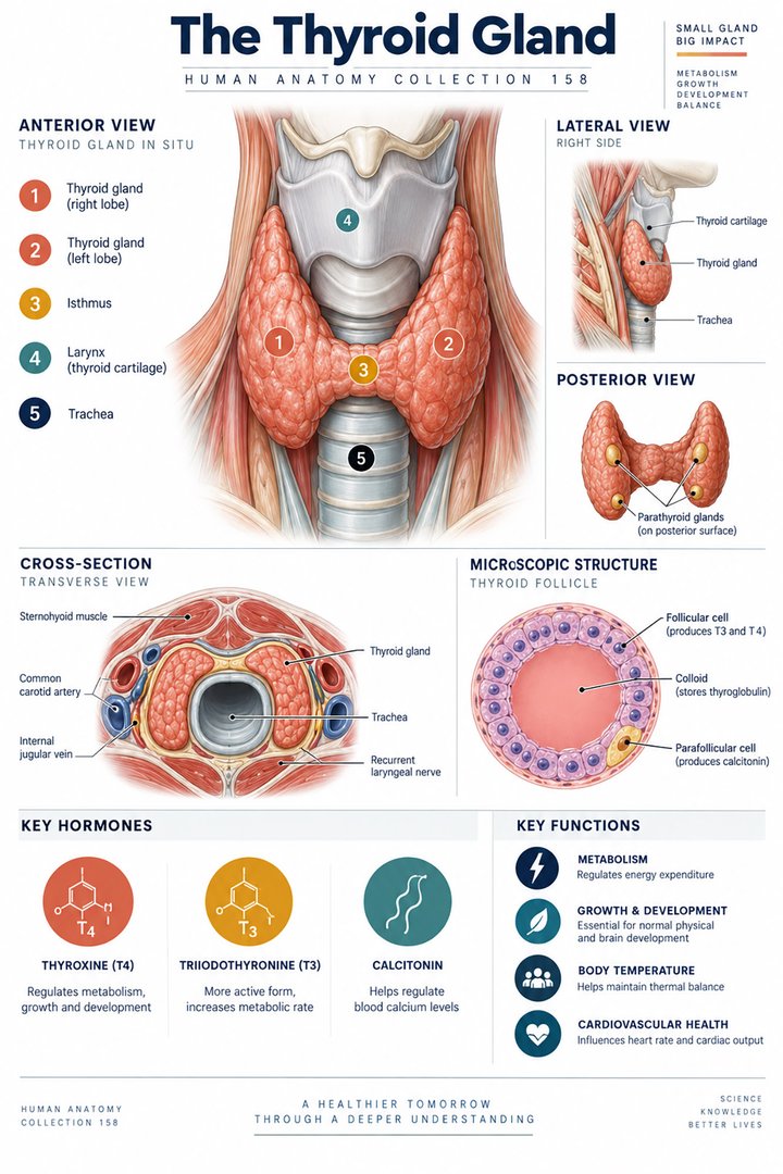
The thyroid gland, glandula thyroidea, is an endocrine organ in the anterior neck. Its familiar butterfly silhouette is useful for finding it on a body map, but the silhouette alone does not explain its function. Thyroid hormones influence energy use and many other processes throughout the body. The gland therefore connects a local anatomical region with effects in distant tissues. Its assessment brings together several kinds of evidence: a person's history, examination, hormone measurements, and sometimes imaging or tissue sampling. Each method answers a different question. NIDDK: Thyroid tests introduces these complementary approaches.
For medical students, the thyroid offers an unusually clear connection between gross anatomy, microscopic units, synthesis, and feedback. For practice assistants and hospital staff, that connection helps explain why a neck image and a blood result should not be treated as duplicates. For care teams supporting older adults, the same knowledge encourages careful documentation of new changes without assuming that they are simply part of aging. This guide follows the thyroid from its neck relationships to its follicles and hormones, then explains how to distinguish a structural finding from a statement about hormone activity.
Two lobes and an isthmus occupy a three-dimensional region
The typical thyroid has right and left lobes connected by an isthmus across the anterior tracheal region. The lobes lie beside the airway and are related to the larynx, surrounding muscles, vessels, and nerves. The gland does not form a complete ring around the trachea. An anterior drawing often hides the posterior relationships, so it is helpful to compare it with an axial view through the neck. The Latin labels lobus dexter, lobus sinister, and isthmus glandulae thyroideae identify the main parts. Regional anatomy is described in NCBI Bookshelf: Thyroid anatomy.
Two lobes is a typical anatomical count, not a statement that every gland has exactly the same outline. A pyramidal extension or other variation can alter the familiar shape. Position and size also need to be interpreted in the appropriate individual context. In a teaching image, specify which view is shown and identify the patient's side rather than the viewer's side. The right lobe in an anterior illustration appears on the viewer's left. This simple orientation point is essential when comparing a labeled diagram with an ultrasound or clinical report, where the image display convention may be different.
Neighboring parathyroids, vessels, and laryngeal nerves matter
The parathyroid glands are usually associated with the posterior thyroid region, but they are distinct endocrine organs with their own function and variable position. The thyroid's vascular supply and nearby recurrent laryngeal nerves make the region anatomically important in surgery. The nerve-vessel relationship can vary; an atlas should not imply that every patient has one guaranteed arrangement. Knowing these neighbors explains why a thyroid discussion can include calcium regulation and voice-related observations without suggesting that thyroid hormones directly perform every function of the region. The anatomical relationships are reviewed in Clinical Methods: Neck and thyroid examination.
A useful diagram separates proximity from membership. The parathyroids should have independent labels, the airway should remain recognizable, and the nerve should be shown as a nearby neural route rather than as a thyroid secretion duct. An operation in one tissue region can involve careful protection of neighboring structures, but a general anatomy guide should not provide procedural instructions. Students can instead learn to name the structures that must be considered and understand why a posterior view adds information missing from the frontal silhouette. For staff, accurate reporting of a new voice or swallowing change is more useful than an assumption about which specific structure caused it.
Development explains variations in adult position
The thyroid develops through a migration process from an early midline pharyngeal region toward its usual neck location. Developmental remnants can help explain some variations and midline structures encountered later. This embryological history is not visible in the standard adult butterfly drawing, yet it is relevant to understanding why thyroid tissue or related remnants may appear outside the most familiar outline. The early anatomical route is described in NCBI Bookshelf: Thyroid embryology. The developmental explanation provides context rather than a method for identifying a neck swelling from its location alone.
For a learning exercise, show the developmental route as a separate schematic beside adult anatomy rather than adding a confusing extra gland to the adult figure. Label it as a developmental concept, and avoid implying that every remnant persists in everyone. This makes embryology useful without overloading a basic map. The broader lesson is that an adult structure has a history: migration, growth, and tissue differentiation can produce variations that a single idealized diagram omits. Medical students should use the typical arrangement as a reference while remaining aware that actual clinical anatomy requires individual evidence.
The follicle is the central histological organization
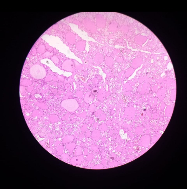
Thyroid tissue is organized predominantly into follicles. Each follicle has an epithelial boundary surrounding a lumen containing colloid, with surrounding connective tissue and vessels. The colloid contains material associated with hormone synthesis and storage, making the follicle more than an empty circular cavity. The cells have an orientation toward the lumen on one side and toward the interstitial and vascular environment on the other. This polarity is important because materials do not simply move through the follicle in one undirected step. The arrangement is introduced in OpenStax: Thyroid gland.
In a real histological image, follicles have different apparent sizes and shapes because a section cuts three-dimensional structures at different positions. A small profile may represent a small follicle or a section passing near the edge of a larger one. The stain gives contrast rather than reproducing the natural color of living tissue. Begin by locating the epithelial border, central colloid, and spaces between follicles, then consider what the image can and cannot establish. Routine appearance does not provide a measured hormone concentration. A careful caption names the specimen and stain and avoids claiming that every visible cell has been individually identified.
Iodine connects chemistry with gland function
Iodine is an essential component of thyroxine and triiodothyronine. T4 contains four iodine atoms and T3 contains three, so the subscripts are molecular information rather than a numerical severity scale. Hormone synthesis involves specialized handling of iodine and thyroglobulin within the follicular system. The anatomical arrangement supports a coordinated sequence of uptake, synthesis, storage, and release rather than immediate passage from food to every target tissue. NIH ODS: Iodine explains the nutrient's role. The simple three-versus-four count is a useful factual inset that should remain clearly separate from laboratory units and clinical interpretation.
Iodine information is often presented as though more must always mean better. That is not an appropriate inference from the fact that the gland requires it. Nutrient physiology and the management of a particular thyroid condition are different questions, and both insufficient and excessive exposure can be relevant in different circumstances. An anatomy article should explain the material requirement without suggesting a personal supplement dose. For students, a useful task is to trace iodine from the blood-facing side of a follicular cell toward the synthesis compartment and then trace released hormone toward the circulation. This reinforces why the follicle's tissue organization matters to its chemistry.
T4, T3, binding proteins, and target tissues are connected
Circulating thyroid hormones exist in protein-bound and free forms, and the measured quantities differ according to what an assay evaluates. Free hormone and total hormone are therefore not interchangeable terms. Target tissues can also convert T4 to T3, connecting gland secretion with regulation outside the gland. The consequence is that thyroid physiology cannot be understood as one organ releasing a fixed number of identical signals directly into every cell. NIDDK explains why binding protein changes can affect total hormone measurements. The distinction is particularly important when a report contains abbreviations that look similar but represent different measurements.
For a laboratory-reading exercise, place TSH, total T4, free T4, and T3 on separate lines and write beside each what has been measured. Do not add a universal reference interval from memory: laboratory methods and clinical contexts matter. The purpose of the exercise is to identify the quantity, not to diagnose a fictional person. A result's unit, reference information, and timing belong with the value. This habit also supports professional communication because it prevents a general phrase such as thyroid level from hiding which analytical result the team is discussing.
Feedback links the thyroid with hypothalamus and pituitary
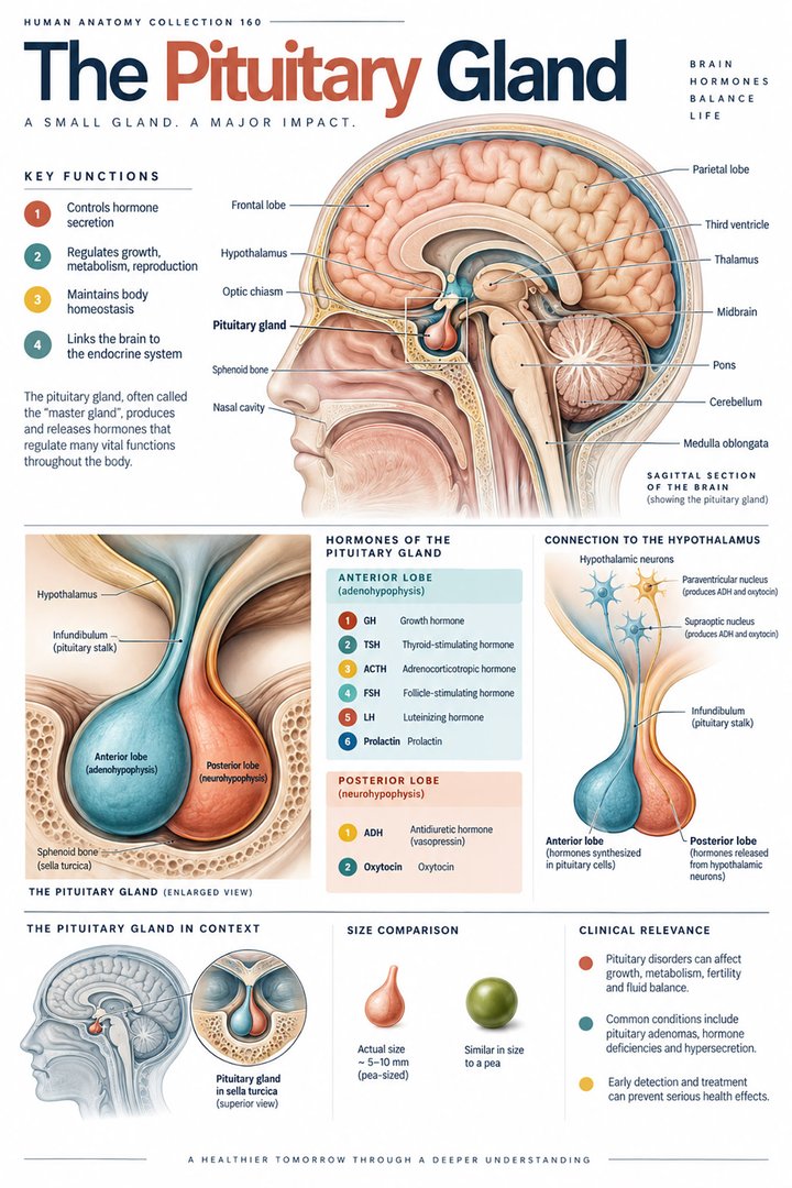
The thyroid participates in an endocrine feedback axis involving hypothalamic thyrotropin-releasing hormone, pituitary thyroid-stimulating hormone, and thyroid hormones. In a simplified negative-feedback diagram, thyroid hormone influences the upstream drive, while TSH stimulates thyroid activity. The abbreviations TRH and TSH describe different signals from different tissues. A figure showing only TSH inside the thyroid would therefore place the source incorrectly. The general relationship is explained in OpenStax: Pituitary and hypothalamus. A useful diagram includes explicit labels for the source and target of each arrow and uses a separate symbol for inhibitory feedback.
This organization explains why an upstream signal can be informative about a downstream gland, but it does not make the signal an infallible stand-alone diagnosis. Primary gland changes, central conditions, physiological states, and analytical issues can require different interpretation. Students should learn the usual relationship while preserving those qualifications. In professional settings, quoting the measured TSH and the documented accompanying result is more precise than asserting that the thyroid has definitely switched on or off. Negative feedback regulates a continuous biological system; it is not a binary light switch with two states.
Thyroid hormone disorders are not identified by one symptom
Hypothyroidism refers to inadequate thyroid hormone activity for the body's needs, while hyperthyroidism concerns excessive production by the gland. Symptoms can involve energy, temperature tolerance, bowel function, skin, and cardiovascular observations, but such findings are not unique to thyroid disease. NIDDK reports that nearly five in every hundred Americans aged twelve or older have hypothyroidism, with many cases mild or not obvious. This is a population estimate for the specified setting, not the probability that any person with fatigue has the condition. The estimate and symptom context are described in NIDDK: Hypothyroidism.
Thyrotoxicosis is a broader term for excess circulating thyroid hormone effects and is not identical to increased synthesis in every case. Inflammation can release stored hormone, while other mechanisms involve active overproduction. NIDDK: Hyperthyroidism explains several causes of excess hormone. These distinctions show why anatomy, synthesis, storage, and release all matter. For healthcare staff, document the person's reported experience and observed change without deciding that one symptom identifies the mechanism. For students, connect each mechanism to the relevant part of the follicular or feedback model rather than placing every abnormal state in one undifferentiated category.
A nodule is a structural finding, not a hormone diagnosis
A thyroid nodule is a localized structural finding within the gland. Its presence does not by itself establish whether overall hormone function is high, low, or normal, and it does not by itself establish malignancy. Ultrasound and other appropriate assessments characterize structure, while blood tests provide information about function. Tissue sampling may answer additional questions in selected circumstances. The American Thyroid Association's thyroid nodule information explains why these methods are complementary. A normal-looking neck or a normal hormone result cannot automatically answer every question about a focal structural change.
An educational comparison can show three columns: what an examination observes, what an image describes, and what a blood assay measures. This makes it easier to understand why a clinician may request more than one method. The chart should not invent a clinical pathway or imply that every nodule requires the same intervention. For patients, a clear explanation of the purpose of the ordered test is often more helpful than a broad reassurance based on one unrelated result. For staff preparing documentation, preserve the report's stated location and description rather than replacing it with a generic thyroid disease label.
Connecting thyroid anatomy with respectful clinical communication
The thyroid offers a practical model for integrated anatomical learning. Start with the neck location, add neighboring structures, identify the follicle, trace a material into synthesis, and follow hormone release toward responsive tissues. Then place the pituitary signal and feedback alongside that route. The model should distinguish molecules, cells, tissue compartments, and whole organs rather than giving them all the same scale. A real photograph, a histological image, and an original diagram can each contribute when their limitations are clearly stated. The numerical facts of two lobes and three or four iodine atoms refer to different levels of organization and should never be combined into a false statistic.
In practice, good communication records what is known: the specific test, the named structural finding, the person's current observations, and the clinical context. Avoid attributing fatigue or a functional change automatically to age, weight, or one previous thyroid diagnosis. Treatment and test interpretation require the responsible healthcare professional's assessment, while anatomical understanding helps the entire team communicate accurately. The enduring lesson is that a small local organ participates in a distributed physiological system. Learning its tissue architecture and connections makes thyroid terminology useful rather than leaving it as a collection of familiar abbreviations.
A focused comparison for a teaching session
Compare a thyroid micrograph with a neck ultrasound image during a teaching session. Ask first what scale each image represents, then identify whether the visible feature is a whole lobe, a focal structural finding, or one microscopic follicle. Next place the available blood assay beside the images and explain why its quantity cannot be read directly from either picture. This comparison trains the learner to connect evidence without confusing it. It also prevents a common communication error in which a reassuring statement about one kind of test is applied to a question that the test did not address. Keeping the anatomical scale and the measurement question explicit makes the thyroid model easier to use in real clinical discussions.
Anatomical Latin: quick reference
| Latin term | English meaning |
|---|---|
| Glandula thyroidea | Thyroid gland |
| Isthmus glandulae thyroideae | Thyroid isthmus |
| Folliculus | Follicle |
| Glandula parathyroidea | Parathyroid gland |
Sources and image credits
Sources reviewed on 9 October 2026. Counts, estimates, study statistics and hypothetical calculations retain their context in the text.
- Thyroid tests
- Hypothyroidism
- Hyperthyroidism
- Thyroid nodules
- Iodine
- Thyroid anatomy
- Thyroid examination
- Thyroid development
- Thyroid follicle
- Pituitary axis
Explanatory prose and SVG graphics are original. Photograph authors, source records and licenses are credited beside each image. Catalog illustrations provide educational context; anatomical facts are supported by the linked sources.