The spinal cord is a central organ within a protective column
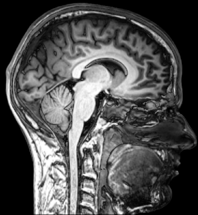
The spinal cord, medulla spinalis, is part of the central nervous system. It connects with the brainstem and occupies the vertebral canal, but it is not the vertebral column itself. Bone, ligaments, meninges, fluid spaces, roots, vessels, and neural tissue share the region while retaining distinct structures and functions. This distinction is essential when interpreting an image or a clinical description. A vertebral injury and a spinal cord injury are related possibilities, not equivalent names for the same tissue. The cord is also more than a passive cable: it contains circuits that participate in processing information and coordinating responses. NINDS: Spinal cord injury introduces its communication role.
For medical students, the most useful learning approach moves between longitudinal and cross-sectional views. The long view explains regional levels and root routes, while the cross section explains the arrangement of neurons and axons. For healthcare teams, these views support precise language about an observed change and the location discussed in a report. A phrase such as lumbar level can be incomplete unless it identifies whether it refers to a vertebra, a spinal segment, or a nerve root. This guide builds those distinctions before linking them with sensation, movement, autonomic function, and the protected environment around the cord.
The lower end of the cord is not the lower end of the spine
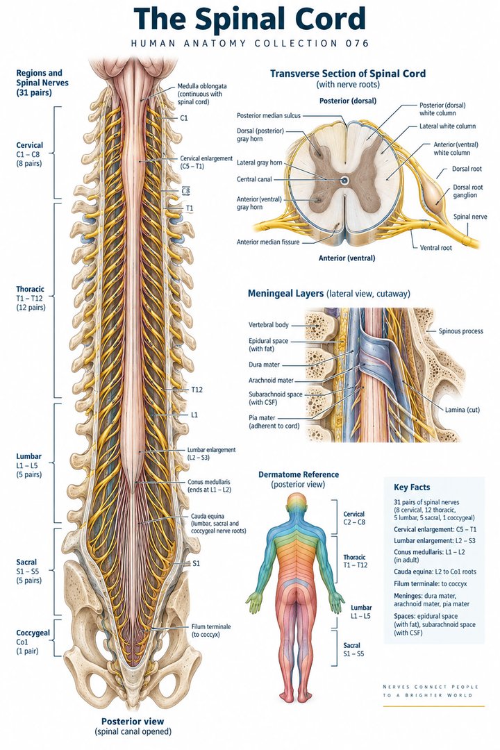
In adults, the tapering lower end of the spinal cord is usually around the first or second lumbar vertebral level, with individual variation. This end is called the conus medullaris. Lower lumbar, sacral, and coccygeal roots then descend within the canal toward their exits, forming the cauda equina. These roots are peripheral neural structures, whereas the cord itself is central tissue. A diagram showing the spinal cord extending as a continuous central tube all the way to the coccyx is therefore misleading. The regional relationship is explained in NCBI Bookshelf: Conus medullaris.
The filum terminale is a slender anchoring structure associated with the lower cord region; it should not be mistaken for an extra bundle carrying the same functions as the cauda equina. On a long anatomical image, roots can visually resemble a continuation of the cord, especially when the scale is small. A useful label set distinguishes conus, descending roots, and filum rather than naming the entire lower region spinal cord. This also explains why some procedures use lumbar spaces below the usual adult cord ending. The anatomical explanation should remain descriptive, since the safety and technique of a procedure depend on professional assessment and training.
Thirty-one segmental pairs organize the peripheral connections
The conventional spinal nerve arrangement comprises 31 pairs: eight cervical, twelve thoracic, five lumbar, five sacral, and one coccygeal. These pairs connect with corresponding spinal cord segments. A segment is defined by neural relationships, not by a visible joint in the spinal cord. The cord itself does not consist of stacked vertebra-like blocks. The numerical map is useful for organizing routes, but a drawing with thirty-one neat rectangles is only a schematic. The external organization is described in Neuroscience: External anatomy of the spinal cord and the root arrangement in OpenStax: Peripheral nervous system.
As the vertebral column and cord develop, their longitudinal relationships change. Consequently, lower cord segments do not simply sit opposite the vertebra with the same number. A learning diagram can place vertebral labels down one side and segmental labels beside the cord, joining the roots to their exit points. This allows students to see why a lumbar nerve can have a long descending route even though the adult cord ends in the upper lumbar vertebral region. When reviewing a report, keep the stated level and the structure it describes together. Substituting a segment name for a vertebral level can change the anatomical meaning considerably.
Enlargements reflect the demands of limb connections
The cord has cervical and lumbosacral enlargement regions associated with the neural supply of the upper and lower limbs. These regions contain substantial circuitry related to limb sensation and movement. The enlargement is not an additional organ inserted into the cord; it is a change in the organization and size of the same continuous central structure. Its shape varies along the length, so one cross section cannot stand for every level. OpenStax: Central nervous system places these regions within the cord's overall anatomy. The relationship between region and function is more informative than memorizing a single diameter as though it were universal.
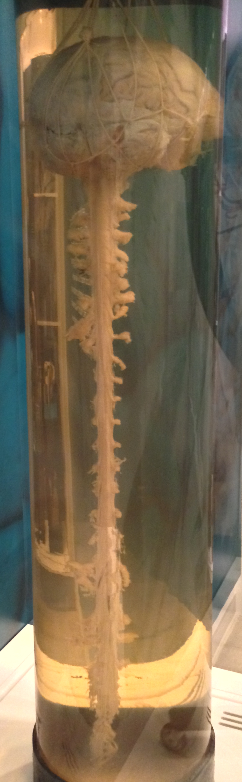
For a teaching activity, compare a thoracic section with a section through an enlargement and look at the relative amount and arrangement of gray and white matter. The differences reflect both local circuitry and axons traveling through the cord. A photographic specimen may show shape without resolving these internal components, whereas a stained section can reveal them in detail. Pairing the two views demonstrates why scale matters. A surface photograph can establish that a region is broader, but it cannot identify the function of every microscopic element. Use each image for the anatomical question it can actually answer.
Gray matter contains organized local neural circuits
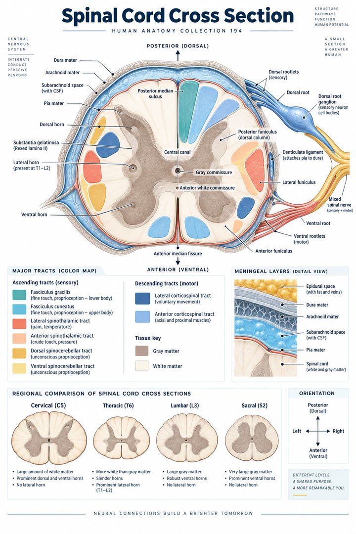
In cross section, spinal gray matter has a central arrangement often compared with a butterfly or an H. Posterior horns are associated with sensory processing, while anterior horns include somatic motor neurons whose axons leave toward skeletal muscles. Intermediate and lateral regions contain circuitry associated with autonomic functions at appropriate levels. These are broad organizational principles, not a promise that every neuron fits into one simplistic category. The Latin labels cornu posterius, cornu anterius, and substantia grisea make the anatomical vocabulary precise. The organization is summarized in Neuroscience: Internal anatomy of the spinal cord.
The term horn describes the appearance of gray matter in a section, while column can describe its longitudinal organization. This is a helpful reminder that the same tissue can be named differently depending on the viewing plane. A section is not a flattened whole cord; it is one cut through a longer three-dimensional structure. Students should add anterior and posterior orientation before labeling horns, especially when viewing a specimen that lacks the familiar textbook colors. The deep anterior median fissure and other landmarks help establish the plane. Correct orientation is necessary before drawing conclusions about which side or pathway a highlighted region represents.
White matter carries multiple ascending and descending routes
Spinal white matter, substantia alba, contains axon pathways arranged into posterior, lateral, and anterior regions. Ascending routes carry information toward higher central structures, while descending routes influence spinal circuits and outputs. A tract is a central pathway, whereas a peripheral nerve is an anatomical bundle outside the central system. The two should not be treated as interchangeable simply because both contain axons. A cross-sectional tract diagram is a map of functional pathways through white matter, not a view of separate plastic wires that can always be pulled apart. OpenStax: Central processing explains the organization of major sensory pathways.
Pathways also differ in where connections and crossings occur. Some sensory information ascends on the same side of the cord before crossing in the brainstem, while other routes cross relatively near their entry into the cord. This is why the side of a symptom cannot be inferred simply from a left-or-right colored tract diagram. The full route matters. For learning, follow one pathway from receptor to root, through the cord, and toward its later connections. Mark the crossing as a separate event. This makes the anatomical reasoning clearer while avoiding the unsupported assumption that one simple pattern predicts every real lesion.
Roots connect central tissue with peripheral pathways
Sensory axons enter through posterior roots, while motor axons leave through anterior roots. The sensory neuron cell bodies for spinal pathways lie in dorsal root ganglia outside the spinal cord. The roots join to form a mixed spinal nerve, which then branches. This arrangement makes the central-peripheral boundary visible: a motor neuron's cell body may be inside the cord while its axon extends outward into a peripheral route. A sensory neuron has its cell body outside the cord while a central process enters it. The nerve connection therefore does not require every part of a neuron to lie in the same anatomical compartment.
When reading a diagram, separate root, spinal nerve, and ramus. A posterior root is sensory, but a posterior ramus is a mixed branch supplying structures in its territory. The similar directional names can hide this distinction if the junction is omitted. OpenStax provides the relevant peripheral connections. Students can practice by drawing an empty outline with only the cord and then adding the roots, ganglion, mixed nerve, and branches in order. This small exercise is more reliable than memorizing that everything behind the cord is sensory. It preserves both the direction of information and the actual route taken by the axons.
Spinal circuits participate in reflexes and movement control
A spinal reflex includes sensory input, central connections, and motor output. Some circuits are relatively direct, while others involve several interneurons and coordination across muscles. The cord can therefore contribute to a response without waiting for a conscious plan to initiate it. Descending influences from the brain still shape spinal activity, and sensory feedback continues to modify the system. This is not a choice between a brain-controlled body and an independently operating cord. They are parts of a connected organization. OpenStax: Motor responses introduces reflex and motor relationships that make this distinction clear.
The stretch reflex is a useful teaching example because it links a receptor in muscle with a spinal circuit and a muscle response. Its diagram should include the sensory and motor limbs rather than showing a tendon directly activating a muscle. For clinical learners, an observed reflex reflects the condition of several components and the examination context. It cannot locate every possible problem on its own. Staff documenting an examination should preserve the stated finding, side, and relevant circumstances rather than infer a wider diagnosis. The anatomy lesson explains why a local circuit can be affected by both peripheral routes and central influences.
Three meninges and fluid spaces protect the cord
The spinal cord is associated with three meningeal layers: dura mater, arachnoid mater, and pia mater. Cerebrospinal fluid occupies the subarachnoid space around it. In the spinal region, the epidural space contains tissues outside the dura, including fat and venous structures; it is not the same space as the fluid around the neural tissue. A diagram should show the layers in order and distinguish a membrane from a space. Counting three meninges does not mean there are only three structures between skin and cord. The protective environment is described in OpenStax: CNS circulation and coverings.
This layered anatomy is especially useful when interpreting terms used in imaging or procedures. Epidural and intrathecal refer to different relationships to the coverings, so they should not be used as casual synonyms. A cross-sectional educational figure can exaggerate the spaces for clarity, but the caption should explain that the widths are schematic. The same caution applies to the small central canal within the cord, which is not the main surrounding fluid space and may vary in patency in adults. The most informative illustration separates vertebral canal, meningeal coverings, fluid environment, neural tissue, and nerve roots with clear labels.
The cord depends on a regional vascular network
Longitudinal arterial routes include an anterior spinal artery and posterior spinal arterial pathways, reinforced by regional vessels. Radiculomedullary contributions connect segmental sources with the cord's longitudinal circulation. The artery of Adamkiewicz is an important example, but its origin and course vary among individuals. A diagram should not present one origin as a universal guarantee. The vascular network supplies different tissue regions through connected routes, and the blood supply cannot be reduced to a single line running the cord's full length. The basic arrangement is outlined in NCBI Bookshelf: Spinal cord arteries.
This organization adds another level to clinical reasoning. A neurological finding can arise from neural tissue affected by a vascular process as well as from direct mechanical injury or another mechanism. An anatomical account of blood supply helps explain why preserving perfusion is important, but a teaching diagram cannot determine the cause of a patient's symptoms. For students, compare a white-matter tract map with a vascular territory illustration and identify that they are different classifications of the same region. One follows information routes; the other follows arterial supply. Their overlap is clinically relevant, yet they should not be merged into one set of labels.
Describe levels, functions, and changes precisely
Spinal cord injury terminology requires care. Complete and incomplete injuries have formal neurological classification meanings; complete does not simply mean that a scan shows a severe-looking lesion or that every pathway below the injury is physically severed. Standards consider preserved function, including sacral sparing, through a structured examination. ASIA: ISNCSCI educational resources describes the examination framework. An educational guide should not assign an impairment grade from a symptom description. The distinction between neurological level, vertebral location, and functional outcome remains essential when discussing findings across a team.
For medical students, the most useful review is to trace an observation across possible anatomical levels: peripheral receptor and nerve, root, local spinal circuit, long tract, and higher central connection. For hospital and care staff, record new changes in movement, sensation, or relevant bodily function promptly according to the clinical setting, with their timing and distribution. Avoid attributing them automatically to age or one pre-existing condition. Spinal cord anatomy becomes practical when it supports accurate descriptions and clear escalation of observations. It helps people understand a central organ within a complex protected region without confusing the cord, the spine, and the roots that connect them.
Anatomical Latin: quick reference
| Latin term | English meaning |
|---|---|
| Medulla spinalis | Spinal cord |
| Substantia grisea | Gray matter |
| Substantia alba | White matter |
| Cauda equina | Descending lumbar, sacral and coccygeal nerve roots |
Sources and image credits
Sources reviewed on 9 October 2026. Counts, estimates, study statistics and hypothetical calculations retain their context in the text.
- External cord anatomy
- Internal cord anatomy
- Central nervous system
- CNS coverings
- Conus medullaris
- Spinal cord arteries
- Spinal cord injury
- ISNCSCI education
- Central processing
- Peripheral roots
- Motor responses
Explanatory prose and SVG graphics are original. Photograph authors, source records and licenses are credited beside each image. Catalog illustrations provide educational context; anatomical facts are supported by the linked sources.