Circulation is continuous transport through a connected network
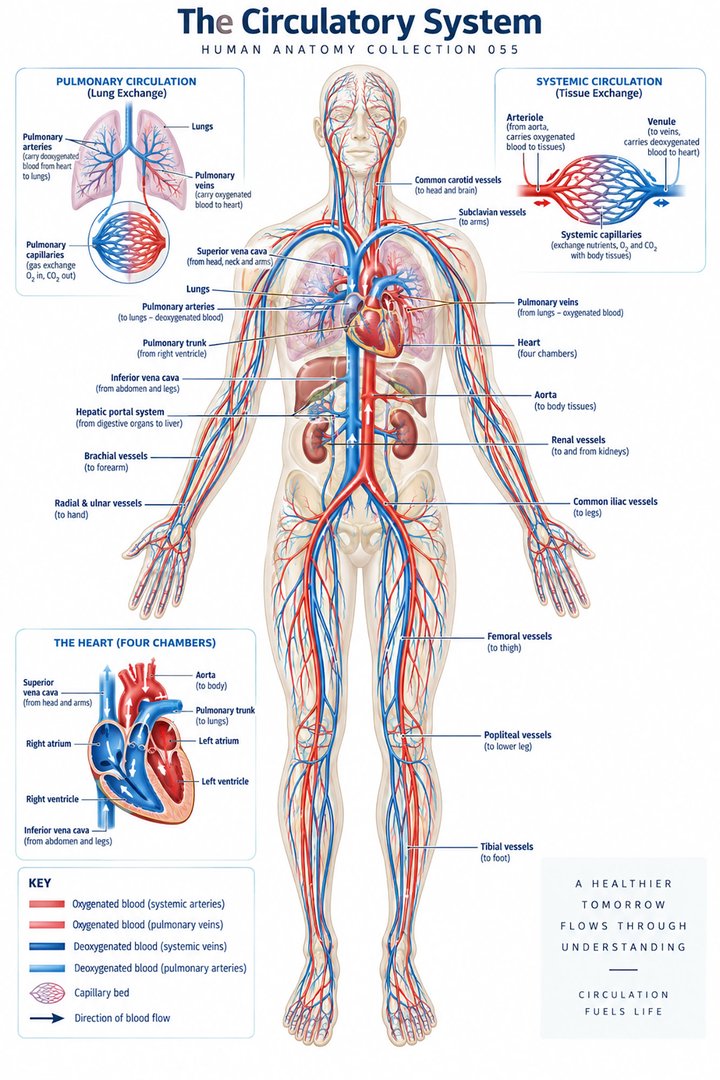
Human blood circulation links the heart, lungs, blood vessels, and tissues in a continuous transport process. It carries oxygen and nutrients toward cells, moves carbon dioxide toward the lungs, and supports the distribution of hormones, immune cells, and heat. The familiar red-and-blue diagram is helpful, but circulation is more than a loop around a body outline. Every point in the loop has a direction of flow, a pressure environment, and a relationship with surrounding tissue. Understanding those differences makes the diagram useful for interpreting physiology rather than just remembering where colored lines are drawn. NHLBI: How the heart works introduces this transport role.
For healthcare teams, circulation knowledge helps separate several observations that are often bundled together. A heart rate is not a direct measure of cardiac output. A blood pressure is not a complete measure of organ perfusion. An oxygen saturation is not the same as the amount of oxygen delivered to a tissue each minute. These distinctions do not make the measurements unhelpful; they explain why clinicians combine them with history, examination, and other findings. This guide builds the anatomical routes and then connects them with the quantities needed to describe transport accurately, using explicitly hypothetical calculations where they clarify a principle.
The pulmonary and systemic circuits are arranged in series
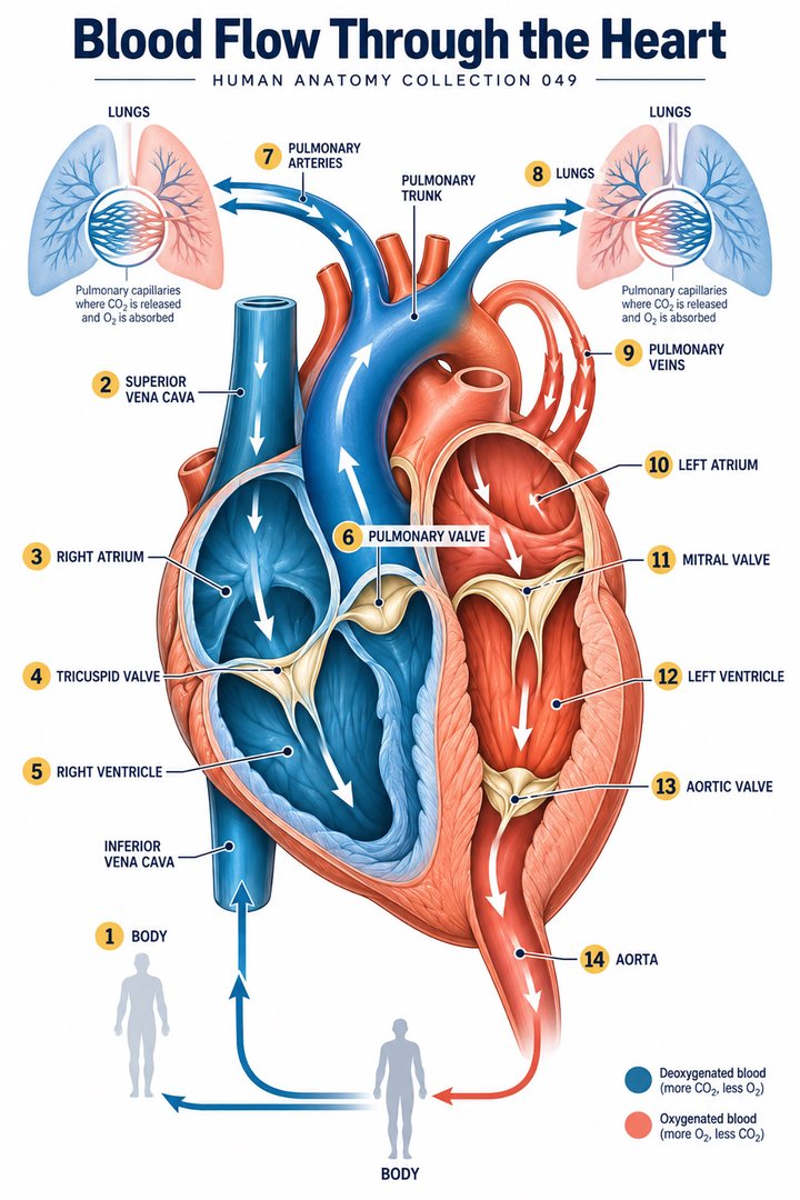
Blood returning from the body enters the right side of the heart and is sent toward the lungs. After gas exchange, it returns to the left side and is propelled through the systemic circulation. The two circuits are connected in series, meaning that blood passes through one and then the other rather than choosing between them as two unrelated destinations. Over time, the right and left ventricles move matching volumes in a stable circulation, although instantaneous flows vary through the cardiac cycle. A diagram should show one connected route and avoid adding the outputs of both ventricles to claim twice the body's cardiac output. The basic route is described in NHLBI: Blood flow through the heart.
The pulmonary circuit operates in a different pressure environment from the systemic circuit. This helps explain why the heart's two sides have different structural demands even though they move the same sustained volume. On a schematic, the two loops can be drawn at equal size for readability, but that does not mean the vessel lengths, pressures, or resistances are equal. A good teaching exercise asks learners to follow one imagined red cell through both loops and name every transition. Following the route continuously makes it easier to recognize a missing connection or an arrow pointing the wrong way.
Artery and vein describe direction, not oxygen color
An artery carries blood away from the heart, while a vein carries blood toward it. In the systemic circulation, arterial blood is generally relatively oxygen-rich and venous blood relatively oxygen-poor. The pulmonary route reverses that oxygen association: pulmonary arteries carry blood toward the lungs before gas exchange, and pulmonary veins return blood afterward. Vessel names therefore cannot be defined solely by the amount of oxygen in their blood. The Latin terms arteria, vena, aorta, and vena cava identify different parts of this directional network. Their basic organization is explained in OpenStax: Blood vessels.
Blue on a chart is a convention, not the actual color of oxygen-poor blood. Human blood remains red, with its appearance influenced by oxygenation and viewing conditions. The colors seen through skin or in an illustration should not be substituted for a physiological measurement. In an educational graphic, include a legend saying relatively oxygen-rich and relatively oxygen-poor and use arrowheads to encode direction independently. This makes the illustration clearer for readers who cannot distinguish the selected colors and prevents the color itself from becoming the entire explanation. The path remains understandable even when the figure is printed in grayscale.
Vessel walls and branching change the flow environment
Large vessels distribute blood, smaller arteries and arterioles influence resistance and local delivery, and capillary networks support exchange. Veins provide the returning route and substantial capacity for blood volume. These roles emerge from both wall structure and network organization. The names tunica intima, tunica media, and tunica adventitia describe the principal wall layers of many vessels. Capillaries are structurally much simpler than a large artery and should not be drawn as miniature thick-walled versions of it. The arrangement links tissue design with transport function, helping learners understand why different regions of circulation behave differently.
Blood velocity in one vessel and total flow through a whole vascular region are different quantities. A large combined cross-sectional area across many parallel capillaries permits relatively slow movement through individual exchange vessels while the circulation continues to transport a substantial volume overall. A single capillary can be narrow, yet the capillary network contains many such channels. This is a useful example of why looking at one vessel in isolation can be misleading. For a teaching diagram, show branching into multiple parallel pathways rather than enlarging one capillary to occupy the entire route. The graphical choice then reflects the network concept without claiming an exact anatomical count.
Pressure, resistance, and flow answer different questions
A pressure difference drives flow through a resistance. The relationship is often summarized as flow equals pressure difference divided by resistance. The pressure difference is important: one pressure value without a downstream comparison does not fully describe the driving condition. Resistance is affected by vessel radius, length, fluid properties, and the organization of the network. This relationship is introduced in OpenStax: Flow, pressure, and resistance. It is a physiological model rather than a promise that one blood pressure reading uniquely determines every organ's flow. Active vessels and changing cardiac conditions make the living circulation more complex than a fixed laboratory tube.
In a simplified laminar-flow tube model, resistance varies inversely with the fourth power of radius. Halving the radius would therefore raise resistance sixteenfold if other assumptions remained unchanged. That calculation demonstrates sensitivity to size; it does not predict the precise effect of a narrowing in a real patient, where branching, compliance, flow disturbance, and compensatory responses also matter. A small mathematical inset can compare hypothetical relative resistances of one and sixteen with an explicit model label. For students, the important learning outcome is recognizing which assumptions support the equation and which living features lie outside it.
Cardiac output combines rate with volume per beat
Cardiac output is the volume ejected by a ventricle per unit time, commonly expressed in liters per minute. It can be calculated as heart rate multiplied by stroke volume. Stroke volume is the volume ejected in one beat, not the whole volume contained in a ventricle. Both quantities can change, so a faster rate does not automatically produce a proportional rise in output. Filling, contractile conditions, and the resistance against which the ventricle ejects also matter. OpenStax: Cardiac physiology explains these relationships. Keeping rate and volume separate prevents a common shortcut in which pulse frequency is treated as a complete measure of pump performance.
Suppose a fictional teaching example uses 60 beats per minute and a stroke volume of 80 milliliters per beat. Multiplying gives 4,800 milliliters per minute, or 4.8 liters per minute. If the same rate were paired with 50 milliliters per beat, the output would be 3.0 liters per minute. Neither example is a patient reference range or a proposed clinical target. The pair simply shows that the same rate can accompany different flows. A diagram should include units on both inputs and the resulting output. Without units, learners can perform the arithmetic while missing what quantity has actually been calculated.
Oxygen delivery also depends on what the blood carries
Flow matters because tissues need transported substances, but volume alone does not state how much oxygen reaches them. Oxygen delivery depends on blood flow and arterial oxygen content. The latter is strongly influenced by hemoglobin and its oxygenation, with a smaller contribution from oxygen dissolved in plasma. Saturation describes the proportion of available hemoglobin binding sites occupied by oxygen; it does not directly count the amount of hemoglobin present. This distinction is explained in the respiratory transport context by OpenStax: Transport of gases. Circulatory and respiratory concepts therefore meet at the level of tissue supply.
Using fictional values only, a flow of 4.8 liters per minute multiplied by an oxygen content of 200 milliliters of oxygen per liter of blood yields a delivery of 960 milliliters of oxygen per minute. Delivery is not identical to consumption: tissues do not extract every available oxygen molecule, and the returning blood retains oxygen. This calculation is a teaching demonstration rather than a clinical estimate for an actual person. It helps readers see why a seemingly reassuring saturation cannot, by itself, describe all oxygen transport. A complete assessment considers the quantity being carried, the flow carrying it, and the tissues receiving it.
Capillary exchange connects circulating blood with cells
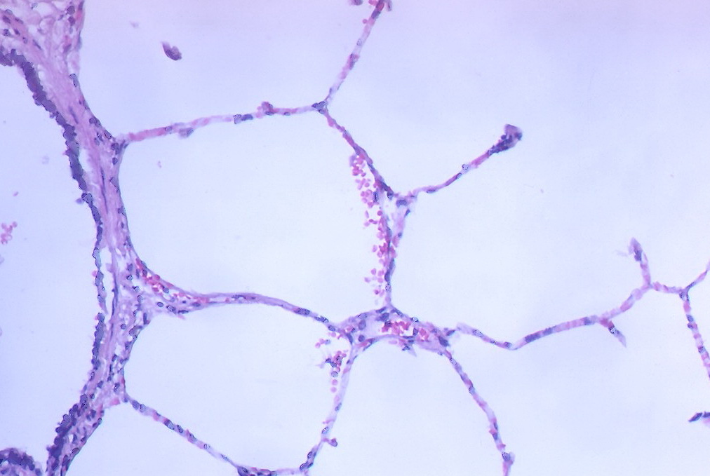
Capillary exchange involves movement between blood and the surrounding interstitial environment. Diffusion is important for gases and other substances, while fluid movement depends on pressure relationships, permeability, and the properties of the endothelial barrier. Capillary beds differ among organs; an exchange surface suitable for a lung is not structurally identical to one in every other tissue. OpenStax: Capillary exchange introduces these processes. The endothelial layer is a living, regulated interface rather than a passive perforated pipe. A simplified picture should show that blood remains inside the vascular lumen while selected substances cross the barrier.
Avoid presenting all fluid filtered from capillaries as automatically reabsorbed at the venous end. Sustained fluid balance also depends on lymphatic return and varies with tissue conditions. A useful diagram shows arterial delivery, a capillary exchange region, venous return, and a separate lymphatic route for interstitial fluid. It should not imply that red cells normally leave the capillary to deliver oxygen directly to neighboring cells. Oxygen moves across boundaries while the red cells circulate. This simple distinction helps prevent errors when students interpret a magnified tissue image or explain why a circulation diagram must include an exchange step between the large vessels and the cells.
Venous return links movement, valves, and breathing
Blood returning to the heart moves through a lower-pressure system with large capacity. Many limb veins contain valves that support directional flow, while muscle contractions can compress nearby veins and help move blood through them. Changes in thoracic pressure associated with breathing also influence venous return. These are parts of a connected process rather than three independent pumps moving separate supplies of blood. OpenStax explains the contribution of venous mechanisms. A diagram showing a vein beside a muscle should include valve direction and should distinguish a contraction phase from relaxation instead of depicting a permanently compressed vessel.
This anatomy is relevant to mobility, but it should not be converted into a universal activity instruction for every patient. Surgical conditions, injuries, illness, and a person's care plan affect what is appropriate. For staff, the useful contribution is understanding why changes in movement or position may be clinically relevant and recording them accurately. For students, venous return also links back to cardiac filling: the amount returning and the heart's ability to accommodate and eject it are interdependent. A circulatory explanation that ends at the capillary leaves out the return route needed to complete the loop.
Coronary and portal pathways add important special routes
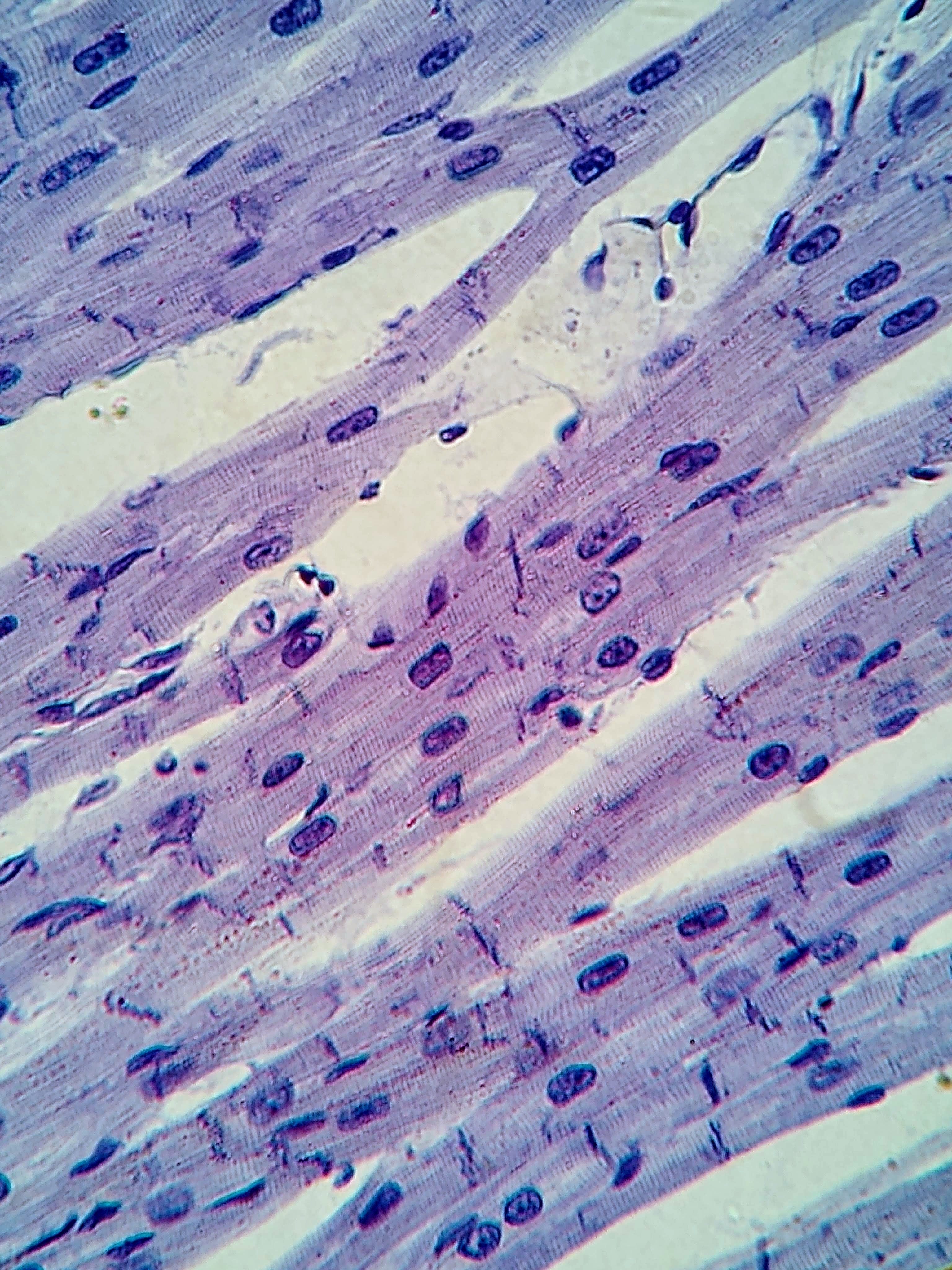
The myocardium requires its own blood supply through the coronary circulation. Blood within a heart chamber does not directly supply all the oxygen needed by the thick muscle wall. Coronary arteries arise from the aortic region and branch across and within the heart, while venous pathways return blood from the myocardium. Separately, the hepatic portal circulation carries blood from much of the digestive region toward the liver before it returns through hepatic veins to the systemic venous route. The portal arrangement connects two capillary regions in sequence rather than returning directly from the first region to the heart. These pathways are described in OpenStax: Circulatory pathways.
The Latin labels arteriae coronariae, vena portae hepatis, and venae hepaticae help distinguish the routes. The portal vein and hepatic veins should not be treated as synonyms simply because both relate to the liver. For a teaching activity, trace an absorbed substance from an intestinal capillary toward the liver, then compare that route with a red cell traveling through a coronary vessel. Each pathway has a different immediate destination and purpose, yet both remain part of the same overall circulation. This comparison illustrates why a body diagram with only one artery and one vein cannot fully represent organ-specific transport.
Reading observations without reducing circulation to one number
Clinical observations describe different aspects of circulation at different moments. Heart rate concerns frequency, blood pressure concerns a pressure signal, skin appearance concerns a visible tissue surface, and an oxygen measurement concerns a particular property of oxygenation. Their relationship depends on the clinical setting. No single observation should be treated as an all-purpose statement that circulation is adequate or inadequate everywhere. The anatomical lesson is to connect the observation with the process it can reasonably describe. For example, a pulse at one site is not a direct measurement of blood flow through every organ, and a diagram cannot establish an individual patient's perfusion.
Useful professional communication states the observation, its timing, and the relevant context. Record a new change relative to the person's usual condition rather than assuming that it is explained by age or one known diagnosis. For students, revisit the complete transport chain: cardiac pumping, arterial distribution, local resistance, capillary exchange, venous return, and lymphatic fluid balance. Then add oxygen content to show what the moving blood carries. This integrated approach gives blood circulation its full meaning. It turns a colored loop into an explanation of delivery and return, while maintaining the distinction between educational physiology and the clinical assessment of a person.
Anatomical Latin: quick reference
| Latin term | English meaning |
|---|---|
| Circulatio sanguinis | Blood circulation |
| Aorta | Aorta |
| Vena cava inferior | Inferior vena cava |
| Vena portae hepatis | Hepatic portal vein |
Sources and image credits
Sources reviewed on 9 October 2026. Counts, estimates, study statistics and hypothetical calculations retain their context in the text.
- Heart overview
- Blood flow through heart
- Vessel structure
- Pressure and resistance
- Capillary exchange
- Circulatory pathways
- Cardiac physiology
- Gas transport
Explanatory prose and SVG graphics are original. Photograph authors, source records and licenses are credited beside each image. Catalog illustrations provide educational context; anatomical facts are supported by the linked sources.