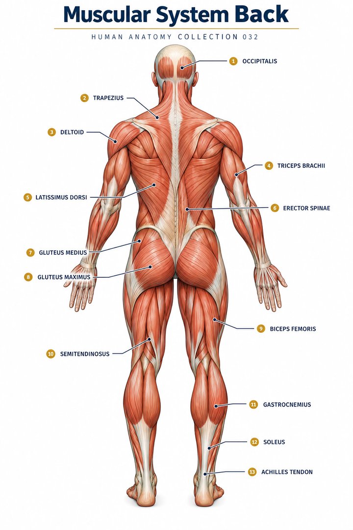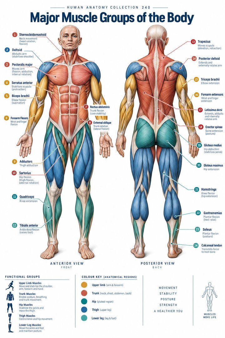
Muscle groups are ways of organizing coordinated tissue
Human muscle groups can be organized by body region, compartment, joint action, or shared function. These systems overlap but do not answer exactly the same question. The anterior thigh is a region, the quadriceps is a named muscle group, and knee extension is an action. A muscle can belong to more than one useful classification, and a familiar task can involve several regions at once. Understanding the relationship between these classifications helps learners avoid treating a list of muscles as a complete explanation of movement. OpenStax: Muscle interactions introduces the cooperation between muscles and the skeleton.
Most clinical movement is not an isolated action performed by one muscle while every other muscle rests. Standing up, adjusting clothing, supporting posture, and bringing a cup to the mouth involve movement, stabilization, and sensory feedback. A muscle group chart becomes more useful when it connects names with a task and identifies which joints are involved. This guide follows the body regionally, while emphasizing the difference between visible movement and the activity that helps control it. The focus is anatomical understanding for medical students and healthcare staff, rather than prescribing exercises or inferring a diagnosis from one difficulty with movement.
Names encode location, shape, and attachments
The Latin term musculus, abbreviated m., means muscle. Names often describe location, shape, fiber direction, relative size, action, or number of heads. Musculus rectus abdominis identifies a straight abdominal muscle, while musculus biceps brachii refers to a two-headed muscle of the arm. A head is an anatomical origin or component, not a separately counted body muscle. Likewise, the three heads of triceps do not mean that the posterior arm has only three muscles. Reading the name as a compact description is more reliable than learning it as an arbitrary sound. See OpenStax: Naming skeletal muscles.
Some terms have a different scope in anatomy from their everyday use. The anatomical arm is the region between shoulder and elbow, while the forearm extends from elbow to wrist. The anatomical leg is between knee and ankle, and the thigh lies above the knee. This vocabulary makes regional muscle descriptions more precise. A handover saying lower limb is broader than one saying anterior leg, and the distinction can matter when recording an observation. Students should place muscle names on a skeleton with the relevant regional terms, then identify what each name tells them and what must still be learned from its attachments and course.
Attachments and joint position explain action
A muscle develops tension and transmits it through attachments, often by tendons, to influence the body. Which movement results depends on the position of the muscle's line of pull relative to the joint axis. Origin and insertion are conventional descriptions of attachments, but the more fixed and more mobile ends can change with the task. For example, moving a limb in space and moving the trunk over a planted limb can use related anatomy under different mechanical conditions. A diagram should therefore identify the joint and the direction of movement rather than relying on an attachment label alone.
Agonist, antagonist, synergist, and stabilizer describe roles within an action, not permanent identities. A muscle that helps produce one movement can oppose a different movement, and an antagonist may contribute controlled resistance rather than remaining silent. Fascicle arrangement also matters: parallel and pennate designs connect fibers with tendons differently. These principles are described in OpenStax. For learning, compare two tasks using the same joint and ask how the roles change. This creates a functional explanation instead of the misleading rule that every joint always operates through a single pair of muscles taking turns.
Axial groups support the head, trunk, and breathing
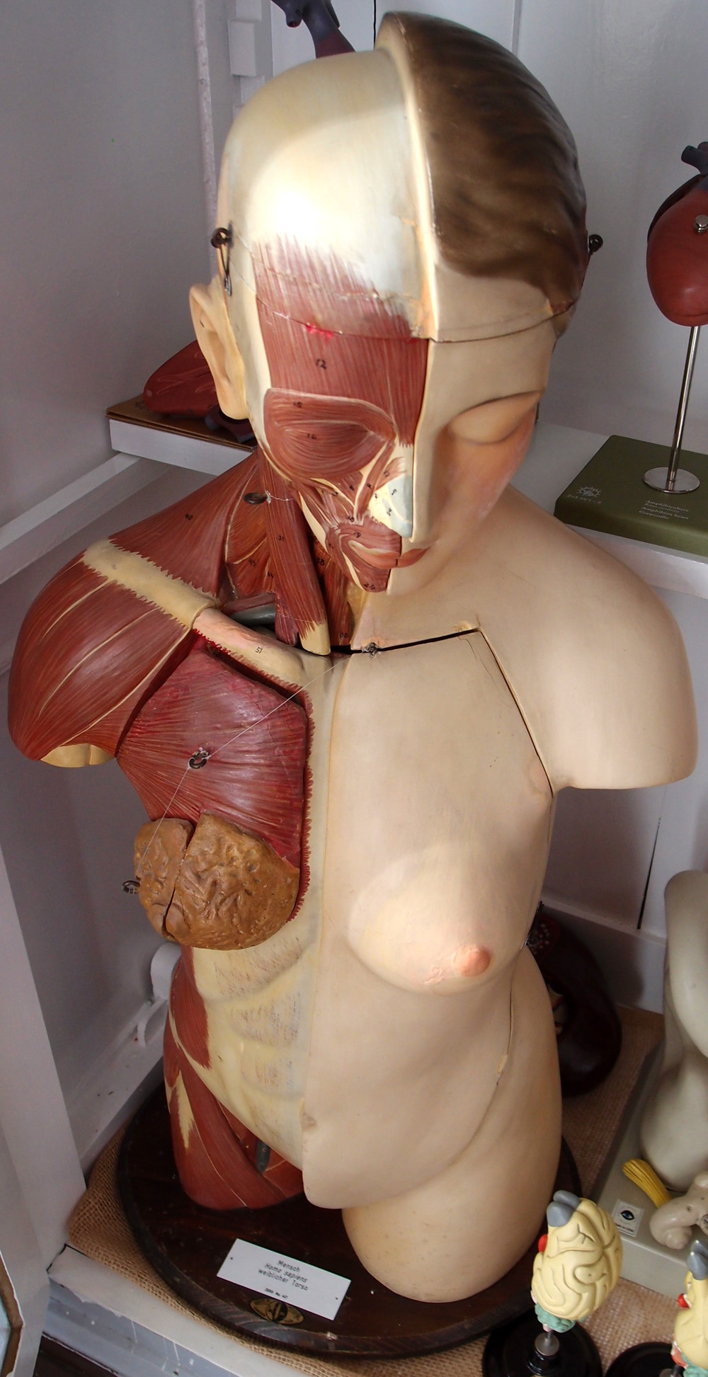
Axial muscles include groups related to the head, neck, vertebral column, and trunk. The sternocleidomastoid, musculus sternocleidomastoideus, and posterior neck muscles contribute to head position, while deeper and superficial back groups participate in maintaining and changing spinal posture. The erector spinae is a group rather than one uniform strip of tissue. Its organization is better understood along the vertebral column than from a single surface outline. OpenStax: Axial muscles of head, neck, and back provides a regional overview. Some axial muscles act on the trunk, while others link the axial skeleton with the shoulder region.
The diaphragm, diaphragma, and intercostal muscles connect muscle anatomy with ventilation. Their actions change thoracic relationships and contribute to pressure changes that move air, rather than directly pulling air as a solid object. For students, this is an important reminder that skeletal muscle is involved in more than conspicuous limb movement. For staff, posture and breathing observations can coexist without implying a single cause. A person who changes their head or trunk position during an activity may be compensating for pain, fatigue, balance, or other needs. Describe the visible behavior and the context before attributing it to one named muscle.
Abdominal wall and pelvic support have layered organization
The abdominal wall contains rectus abdominis and the external oblique, internal oblique, and transversus abdominis, with different fiber directions and broad connective tissue relationships. These muscles contribute to trunk control and abdominal pressure in coordinated ways. A superficial illustration showing only the visible rectus outline omits much of the wall's architecture. The term core is useful in everyday language but has no single universally fixed anatomical membership. When accuracy matters, name the muscles or region being discussed. Their arrangement and relationship to the thorax and pelvis are introduced in OpenStax: Abdominal wall and thorax.
The pelvic floor is another organized region with muscular and connective tissue components supporting pelvic structures and contributing to continence and other functions. It should not be represented as one simple band that tightens independently of the rest of the body. A labeled pelvic view can show depth and orientation more clearly than a full-body surface poster. In professional education, distinguishing support function from a specific exercise instruction is important. An anatomical explanation helps learners understand why trunk pressure and pelvic structures interact, while individual assessment determines what clinical guidance is appropriate. Respectful language and clear orientation are particularly useful when discussing this region with patients or colleagues.
Shoulder movement requires the girdle as well as the arm
The shoulder region combines movement of the humerus with movement and stabilization of the scapula. Deltoid, pectoralis major, latissimus dorsi, trapezius, and serratus anterior contribute through different attachments and actions. The four rotator cuff muscles are supraspinatus, infraspinatus, teres minor, and subscapularis. The count of four refers to these named muscles, not the total number acting at the shoulder. Teres major lies nearby but is not a rotator cuff member. This distinction is a useful numerical fact for a teaching figure because spatial proximity can otherwise make the group boundary unclear. See OpenStax: Shoulder girdle and upper limb.
The cuff's role includes dynamic control of the humeral head while other muscles contribute to movement. A shoulder diagram showing only an arrow beside deltoid cannot explain the full coordination required to raise an arm. A better sequence shows scapular movement and the humeral relationship together. When someone has difficulty reaching a shelf, the observed task includes shoulder, elbow, wrist, hand, posture, and potentially balance. An anatomical group map can suggest what to consider, but it cannot identify the cause from the task alone. Students should describe the movement and joint positions before reducing the explanation to one prominent surface muscle.
Elbow, forearm, and hand groups create position and precision
Biceps brachii, brachialis, and brachioradialis contribute to elbow flexion under different mechanical conditions, while triceps brachii is a principal elbow extensor. Biceps also participates in forearm supination. The two-head and three-head names are useful reminders of structure, but visible arm size is not a direct measure of functional performance. In the forearm, flexor and extensor groups send many tendons toward the wrist and fingers. The NCI's upper extremity muscle overview summarizes the regional relationship. A tendon crossing a joint can transmit tension from a muscle belly located some distance away.
Hand function also depends on intrinsic muscles whose bellies are within the hand. Precision movements combine these muscles with extrinsic tendons from the forearm, sensory information, and joint stability. This explains why holding a key and supporting a heavy bag are not interchangeable tests of the same simple grip. For teaching, compare the position of the muscle belly and the route of its tendon, then identify the joint or joints influenced. For staff recording a difficulty, describe whether the person struggles to grasp, maintain a hold, manipulate a small object, or release it. Those task descriptions carry more information than an unsupported statement that the hand muscles are weak.
Hip groups transfer force between pelvis and lower limb
Hip muscles connect the pelvis and femur through several regional groups. Gluteus maximus contributes strongly to hip extension, while gluteus medius and minimus contribute to abduction and control of the pelvis during appropriate tasks. Iliopsoas contributes to hip flexion, and the adductor group occupies the medial thigh. A muscle's action depends on position and task, so a short label such as abductor is a useful introduction rather than its complete mechanical biography. OpenStax: Pelvic girdle and lower limb describes these relationships. The gluteal group should not be described collectively as simply adducting the thigh.
When a foot is on the ground, controlling the pelvis is part of whole-body balance. This closed-chain situation differs from moving the same limb freely in the air. An illustration can show the stance side and the opposite side of the pelvis to explain the control problem without assigning a diagnostic meaning to a particular gait. For older-person care, a change in transfers or walking deserves a clear record of support needs and observed performance. It may involve muscle capacity, joint pain, neurological change, cardiovascular tolerance, or several interacting factors. Anatomical knowledge helps describe the task precisely while keeping these possible contributors distinct.
Quadriceps and hamstrings show why group membership matters
Quadriceps femoris comprises four named components: rectus femoris, vastus lateralis, vastus medialis, and vastus intermedius. All contribute to knee extension, while rectus femoris also crosses the hip and can contribute to hip flexion. The hamstring group conventionally refers to biceps femoris, semitendinosus, and semimembranosus in the posterior thigh. The short head of biceps femoris does not cross the hip and differs from the classic biarticular hamstring arrangement. These distinctions matter because a group name can hide meaningful differences among its members. OpenStax introduces the regional muscles; the count should always specify whether named muscles, heads, or components are being counted.
A two-joint muscle has its length and mechanical situation affected by both joints. It cannot be fully described by one isolated joint arrow. For a learning exercise, draw the hip and knee in two different positions and trace rectus femoris or a biarticular hamstring between them. This shows why position changes an anatomical task even before force or speed is considered. It also prevents the simplified idea that knee extension and flexion are always produced by two identical teams working separately. Control during a movement can involve simultaneous activity, changing loads, and stabilization at neighboring joints.
Leg and foot groups support contact, propulsion, and balance
The anterior leg includes muscles such as tibialis anterior associated with dorsiflexion, while the posterior region includes gastrocnemius and soleus associated with plantarflexion. Gastrocnemius crosses the knee as well as the ankle, while soleus does not cross the knee. Lateral compartment muscles and other groups contribute to positioning and control of the foot. The NCI's lower extremity overview identifies the major compartmental arrangement. A labeled chart should use leg for the knee-to-ankle region and lower limb when including the thigh, avoiding a common mismatch between everyday and anatomical terminology.
Walking places different demands on the foot during contact, support, and progression. Muscle groups act within a changing sequence rather than repeating one static posture. Intrinsic foot muscles, tendons, joints, and sensory input all contribute to the contact relationship with the ground. A person catching the forefoot during walking has displayed a functional observation, but the observation alone does not establish which muscle or nerve is responsible. Students can use a phase diagram to explain the task, while staff should report when and how the difficulty occurs. Linking anatomy with movement phases makes the regional map more clinically meaningful than a picture of a calf alone.
Muscle activity is not synonymous with visible shortening
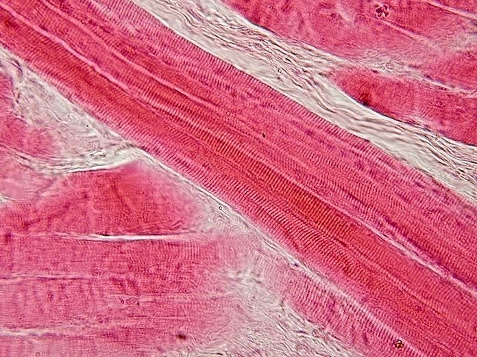
A muscle can generate tension while shortening, while lengthening under load, or with little change in overall length. These situations are commonly described as concentric, eccentric, and isometric actions. They help explain how a group can control lowering or maintain posture even when it is not producing an obvious lifting movement. At the cellular level, contraction involves interactions within the sarcomere and regulation of activation. OpenStax: Muscle contraction provides that microscopic framework. Whole-muscle behavior and fiber-level events should be connected carefully because tendon compliance and architecture also affect the relationship between them.
Motor units and recruitment add another layer of control. A motor unit includes a motor neuron and the muscle fibers it supplies, and changes in recruitment help vary muscle tension. OpenStax: Control of muscle tension explains this organization. For professional learning, begin with the task, identify the relevant regional groups and joints, and then consider the control needed to perform it. This sequence supports clear documentation without prescribing an exercise or assigning a cause. A muscle group guide is most useful when it shows how tissue, attachments, neural activity, and movement interact to produce the everyday functions healthcare teams observe.
Anatomical Latin: quick reference
| Latin term | English meaning |
|---|---|
| Musculus | Muscle |
| Tendo | Tendon |
| Musculus deltoideus | Deltoid muscle |
| Musculus quadriceps femoris | Quadriceps femoris muscle |
Sources and image credits
Sources reviewed on 9 October 2026. Counts, estimates, study statistics and hypothetical calculations retain their context in the text.
- Muscle interactions
- Naming muscles
- Head neck and back
- Abdominal wall and thorax
- Upper limb
- Lower limb
- NCI upper muscles
- NCI lower muscles
- Muscle contraction
- Motor tension control
Explanatory prose and SVG graphics are original. Photograph authors, source records and licenses are credited beside each image. Catalog illustrations provide educational context; anatomical facts are supported by the linked sources.