The communication network beyond the central nervous system
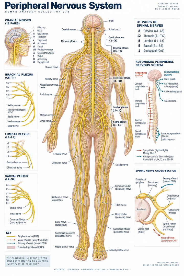
The peripheral nervous system connects the brain and spinal cord with tissues throughout the body. Its importance becomes clear during an ordinary clinical encounter: a person feels the chair beneath them, adjusts their posture, grasps a cup, and regulates circulation without consciously planning every response. These activities require several kinds of information to travel along different routes. Understanding the routes makes peripheral anatomy more useful than memorizing a collection of nerve names. It helps students distinguish where information begins, where it enters the central nervous system, and how an output reaches a muscle, gland, or organ. NINDS: Peripheral neuropathy provides an authoritative introduction to this communication network.
The term peripheral describes location rather than importance. A peripheral lesion can have major consequences for movement, balance, protective sensation, and everyday independence. Conversely, an abnormal movement does not automatically indicate a peripheral problem; the brain, spinal cord, muscle, and neuromuscular junction also participate. An anatomical description should therefore state what has been observed before assigning a cause. For healthcare staff, an accurate account of the affected region, the timing of change, and the tasks that have become difficult can be more useful than an unsupported diagnostic label. The goal of this guide is to build that spatial and functional vocabulary.
A nerve contains more than axons
A peripheral nerve is an organized living structure containing axons, supporting cells, connective tissue, and a blood supply. In a cross section, individual fibers sit within small connective tissue spaces, groups of fibers form fascicles, and the fascicles are assembled into the whole nerve. The names endoneurium, perineurium, and epineurium describe these nested coverings. They should not be confused with myelin, which is a specialized wrapping closely associated with certain axons. A teaching diagram that draws one thick cable without its internal arrangement may be convenient, but it hides the distinction between an individual nerve fiber and the complete nerve. See OpenStax: Peripheral nervous system.
The blood vessels supplying a nerve are often called vasa nervorum. Their presence reminds us that nerve tissue needs oxygen and nutrients just as other tissues do. Pressure on a nerve may affect its mechanical environment and its local perfusion; these processes are related but not identical. A nerve seen during dissection also follows connective tissue planes, passes through openings, and changes its relationship to muscles and bones along its course. Learning those relationships explains why the same nerve can be vulnerable at one location yet relatively protected at another. The useful question is not simply which nerve is present, but what surrounds it at that point.
Schwann cells, myelin, and the speed of signaling
Schwann cells support peripheral axons. Some form a myelin sheath around a segment of one axon, while others support axons without making the same multilayered wrapping. Gaps between adjacent myelinated segments are called nodes of Ranvier. This arrangement supports rapid propagation because the membrane events underlying an action potential are concentrated at successive nodes. The impulse does not travel as a little object inside a tube: it is a changing electrical state of the axon membrane. That distinction helps students understand why axon damage and damage to myelin can disturb signaling in different ways. The basic cellular organization is described in OpenStax: Nervous tissue.
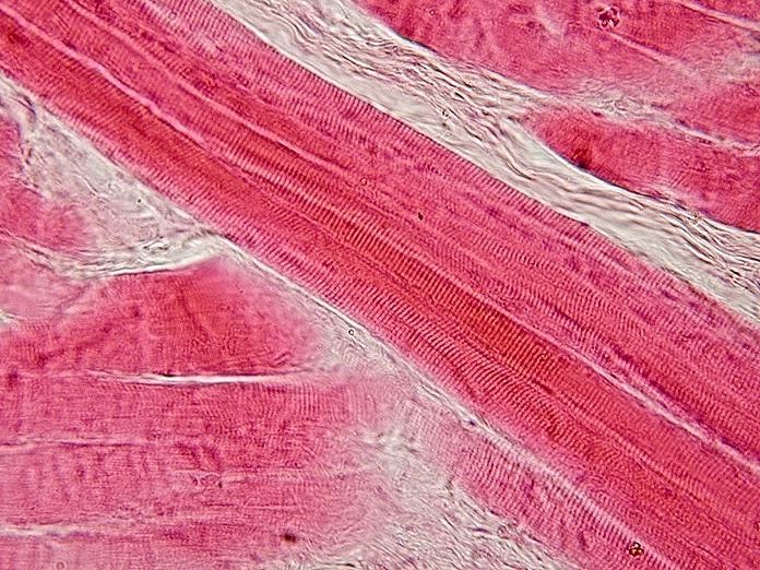
Myelin is one factor influencing conduction; axon diameter and physiological conditions also matter. A single nerve can contain a mixture of fiber types with different diameters, degrees of myelination, and functions. It is therefore misleading to assign one universal conduction speed to every peripheral nerve. A colorful diagram usually shows myelin as a regular series of identical sleeves, but real tissue has considerable microscopic complexity. When reviewing a histological image, first identify whether it is a longitudinal or transverse section, then consider the stain and scale. A pattern that appears discontinuous in one section may represent an intact structure viewed from a different angle.
Sensory input and motor output are directions, not destinations
Afferent fibers carry information toward the central nervous system; efferent fibers carry signals away from it. These directional terms remain useful whether the target is skin, skeletal muscle, or a visceral organ. Many named peripheral nerves are mixed, containing both sensory and motor axons. Calling a whole mixed nerve sensory because a patient reports numbness overlooks its other components. Likewise, motor describes the role of an output pathway rather than the visible size of the nerve. A small nerve branch may control a muscle that is essential for a precise movement, and a large nerve may carry several distinct functions through the same anatomical corridor.
Somatic sensation includes touch, temperature, pain, vibration, and information about body position. These experiences depend on different receptors and pathways rather than one undifferentiated sensory channel. Proprioception allows the nervous system to use information from muscles and joints when coordinating posture and movement. In an observation, loss of one kind of sensation should not be treated as evidence that all sensation is absent. For example, a person may describe an unusual response to light touch while still detecting pressure. The appropriate account records the particular observation and its distribution. NINDS distinguishes the motor, sensory, and autonomic manifestations of peripheral nerve disorders.
Spinal roots, ganglia, and the numerical map
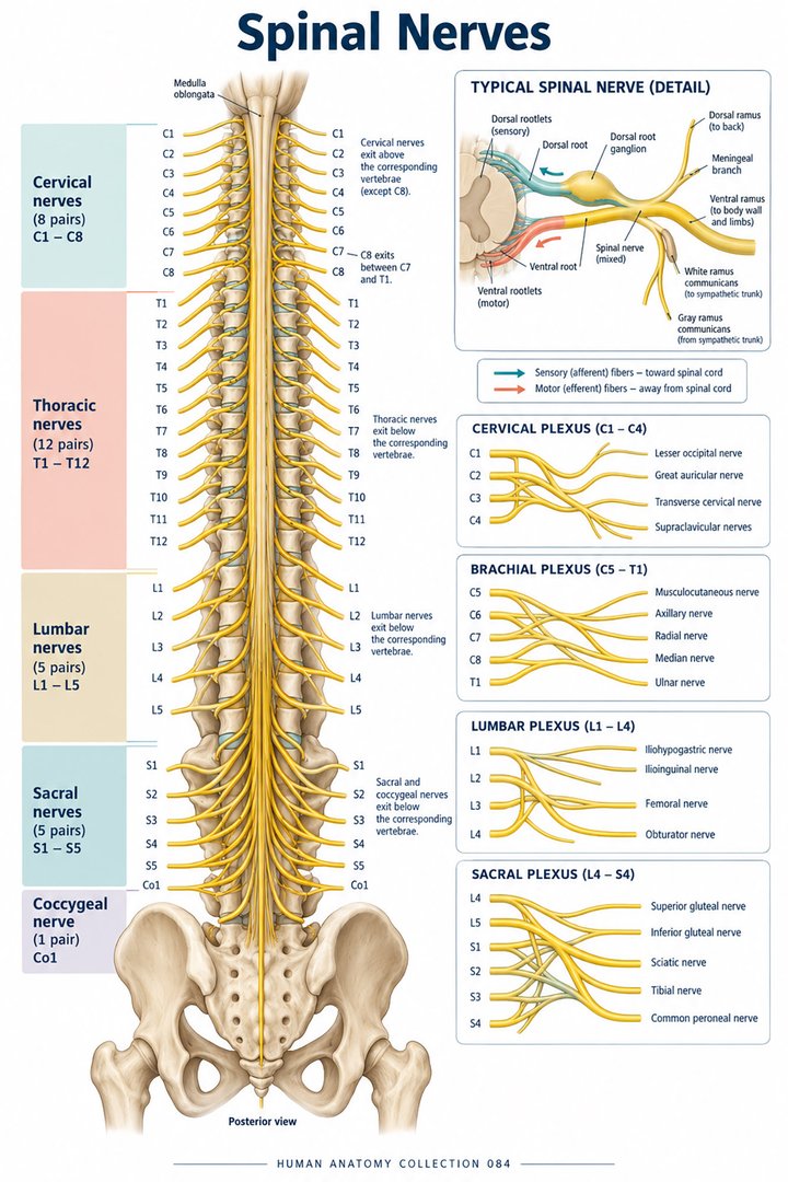
There are 31 pairs of spinal nerves in the conventional adult anatomical map: eight cervical, twelve thoracic, five lumbar, five sacral, and one coccygeal pair. A pair means one nerve on each side, so 31 pairs correspond to 62 individual spinal nerves before their many branches are counted. The eight cervical pairs are especially worth noticing because the adult cervical vertebral column has seven vertebrae. A nerve label is therefore not interchangeable with the name of a vertebra. The count provides a framework for learning, not a claim that every individual has an identical branching pattern. The regional distribution is summarized in OpenStax.
Posterior, or dorsal, roots convey sensory axons toward the spinal cord, while anterior, or ventral, roots carry motor axons away. The sensory neuron cell bodies are grouped in dorsal root ganglia, ganglia spinalia. The roots join to form a mixed spinal nerve before further branching. Both the posterior and anterior rami are mixed branches; the word posterior does not make a ramus purely sensory. This is a common source of confusion because roots and rami occupy neighboring positions on a diagram. Trace the connections in order: spinal cord, roots, mixed nerve, and then rami. Keeping these structures separate makes later clinical localization considerably clearer.
Plexuses rearrange segmental contributions
A nerve plexus is a network in which axons from several spinal levels are redistributed into peripheral nerves. The cervical, brachial, lumbar, and sacral plexuses are the major regional examples. A named limb nerve therefore usually represents contributions from more than one spinal segment. The plexus brachialis is a useful model: its usual main roots are C5 to T1, and its branches supply the shoulder and upper limb. Variations exist, so a simplified diagram should identify itself as a typical arrangement. A plexus does not create a new sensory or motor signal; it rearranges the route taken by existing axons.
This arrangement explains why a root lesion and a peripheral nerve lesion can produce different patterns even when some affected movements overlap. The median nerve, nervus medianus, and ulnar nerve, nervus ulnaris, carry fibers along different routes into the forearm and hand. A root contributing to both can influence more than one named nerve territory. In the lower limb, the femoral nerve and sciatic nerve arise through different plexus arrangements and occupy different anatomical relationships. For learning, draw the spine at one side and the named nerve at the other, then connect several segmental contributions between them. This emphasizes redistribution rather than presenting each peripheral nerve as the continuation of one isolated root.
Cranial nerves and the important exception of the optic nerve
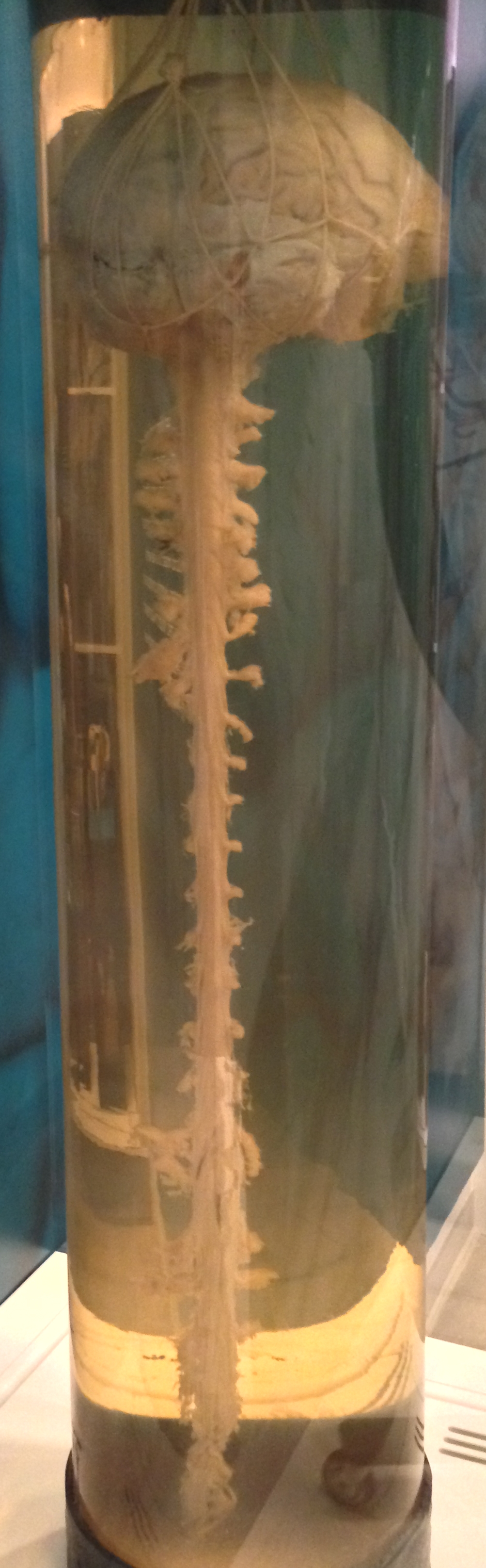
The traditional cranial nerve list contains twelve pairs, numbered I to XII. The list includes routes involved in smell, vision, eye movement, facial sensation, facial expression, hearing, balance, swallowing, and other functions. Some carry predominantly sensory information, some predominantly motor output, and others combine several components. The vagus nerve, nervus vagus, extends beyond the head and neck to influence thoracic and abdominal organs. Its wide distribution demonstrates why cranial does not mean confined to the skull. A learner should connect each number to its name, course, and major functions while recognizing that a short introductory list cannot describe every branch.
Classification also has exceptions. The optic nerve is conventionally numbered cranial nerve II, but developmentally and structurally it is a central nervous system pathway, with central myelin and meningeal coverings. A statement that every cranial nerve is an ordinary peripheral nerve is consequently too broad. The olfactory system also has special relationships at the central boundary. These details are useful because they show that nomenclature, embryology, and tissue structure answer different questions. In practice, choose the classification that matches the issue being discussed, and explain the exception when it matters. A diagram can preserve the familiar twelve-pair list while noting that anatomical membership is more nuanced than a single color coding scheme.
Autonomic and enteric pathways reach internal tissues
Autonomic peripheral pathways regulate smooth muscle, cardiac muscle, and glands. Their sympathetic and parasympathetic components use ganglia outside the central nervous system, where one neuron can communicate with another before the signal reaches its target. In a typical autonomic motor route, a preganglionic neuron begins in the central nervous system and a postganglionic neuron projects from a peripheral ganglion. This contrasts with a somatic motor neuron reaching skeletal muscle directly from its central cell body. The contrast is useful for a schematic, although not every physiological detail fits neatly into two identical chains. OpenStax: Autonomic reflexes and homeostasis explains the organization of visceral regulation.
The enteric nervous system includes networks in the gastrointestinal wall. The myenteric plexus is associated with control of gut movement, while the submucosal plexus contributes to local secretory and vascular regulation. These networks interact with autonomic input but also contain local circuits. They illustrate how peripheral neural tissue can be embedded within another organ rather than appearing as a prominent cable in a dissection. For healthcare staff, autonomic anatomy offers a framework for recording changes such as altered sweating or symptoms occurring on standing. Such observations require clinical interpretation; they should not be assigned to one nerve merely because the word autonomic appears in an educational chart.
Reflexes show that the periphery participates in circuits
A reflex arc includes a receptor, an afferent route, central integration, an efferent route, and an effector. The familiar tendon reflex does not simply make a tendon contract. Tapping the tendon changes the associated muscle's length, activating muscle spindle afferents and a spinal circuit that influences muscle contraction. A diagram should therefore label the muscle receptor and neural connections rather than showing a direct arrow from tendon to movement. Reflexes help demonstrate that input and output are distinct limbs of a connected process. They also remind learners that a movement can begin through spinal circuitry without a conscious decision to initiate it. OpenStax: Motor responses covers this organization.
An observed reflex depends on multiple parts of the circuit and on the examination context. Its presence or absence cannot, by itself, identify every possible site of disease. Muscle condition, peripheral axons, spinal segments, descending influences, and the method of observation all matter. For students, comparing a reflex circuit with a voluntary movement pathway is especially productive: both can reach the same muscle, but their initiation and organization differ. For staff documenting a clinical examination, report the specified finding and side accurately without inventing a broader conclusion. Anatomical literacy strengthens communication by making the circuit visible, while the diagnosis still depends on a complete professional assessment.
Using peripheral anatomy in patient-centered documentation
Terms such as mononeuropathy, polyneuropathy, and radiculopathy describe different patterns or levels of nerve involvement. Mononeuropathy refers to one nerve, whereas polyneuropathy concerns multiple peripheral nerves; radiculopathy concerns a nerve root. These labels are not interchangeable. A person describing symptoms in both feet has not necessarily identified which of these categories applies. Duration, symmetry, associated weakness, and clinical findings affect interpretation. NINDS notes that more than 100 types of peripheral neuropathy have been identified, illustrating the breadth of possible mechanisms. This is a classification fact, not an estimate of how likely any one person is to have a particular condition.
A useful handover distinguishes sensation from performance. Describe whether the person reports tingling, has difficulty holding an object, catches the front of a foot during walking, or needs additional support for a familiar task. State when the change was noticed and whether it is new relative to the person's usual function. Respect the limits of the observation: inability to complete a task may reflect pain, joint restriction, fatigue, or central as well as peripheral changes. The best anatomical explanation links tissue, pathway, and function without assuming that every complaint has one cause. This approach makes peripheral nervous system knowledge relevant to outpatient care, hospital teamwork, rehabilitation, and support for older adults.
Comparing two anatomical maps
A dermatome map and a peripheral nerve map describe different ways of organizing cutaneous sensation. The first follows a spinal segmental relationship; the second follows the distribution of a named nerve after peripheral routing. Their borders overlap and vary, so neither should be treated as a set of sharply bounded colored stickers on a patient. In a teaching exercise, place both maps beside the same hand outline and identify which questions each can answer. Then describe an observation using a concrete surface landmark rather than saying only that the picture matches. This approach supports localization while preserving the uncertainty that exists in real clinical observations. It also makes the distinction between a nerve root contribution and a terminal branch easier to remember.
Anatomical Latin: quick reference
| Latin term | English meaning |
|---|---|
| Nervus | Nerve |
| Ganglion spinale | Spinal sensory ganglion |
| Plexus brachialis | Brachial plexus |
| Nervus ischiadicus | Sciatic nerve |
Sources and image credits
Sources reviewed on 9 October 2026. Counts, estimates, study statistics and hypothetical calculations retain their context in the text.
Explanatory prose and SVG graphics are original. Photograph authors, source records and licenses are credited beside each image. Catalog illustrations provide educational context; anatomical facts are supported by the linked sources.