1. Circulation connects the entire body
The human circulatory system is a moving connection between organs that otherwise seem separate on a poster. A meal supplies nutrients, the lungs support gas exchange, and the kidneys process blood continuously. Circulation links these activities with tissues throughout the body. For healthcare learners, the central question is therefore not simply where a vessel lies, but what route it belongs to and what relationship its contents have with the surrounding tissue.
Begin with a practical distinction: a diagram describes structure, whereas a clinical measurement describes one aspect of function at a particular time. A colorful artery on a chart cannot tell you a patient’s blood pressure, and a pressure reading alone cannot describe every part of the vascular tree. Keeping those two kinds of information connected, but distinct, makes teaching and documentation more precise.
2. Follow the pulmonary and systemic circuits
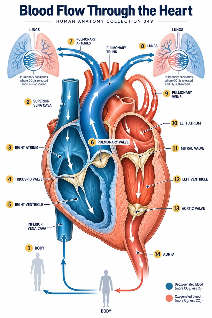
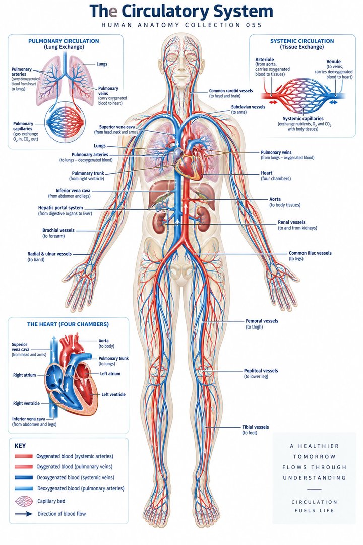
Pulmonary circulation links the right side of the heart with the lungs; systemic circulation links the left side with the body’s tissues. Blood travels through the right atrium and ventricle toward pulmonary vessels, returns through pulmonary veins to the left atrium, and leaves the left ventricle through the aorta. The two routes operate as a connected sequence. [OpenStax: circulatory pathways]
For study, trace one complete circuit without lifting your finger from the chart. Say the chamber, vessel, and destination aloud. Then restart at a different point. If you can only recite the route beginning with the right atrium, you may have learned a sentence rather than a connected pathway. A useful second exercise is to explain which part of the route passes through the lung and which passes through another organ.
3. Artery and vein describe direction
Arteries carry blood away from the heart, and veins carry it back. These names do not specify oxygen content. Pulmonary arteries carry relatively oxygen-poor blood toward the lungs, while pulmonary veins return oxygen-rich blood. Capillaries connect small arterial and venous vessels and provide an extensive interface for exchange. [OpenStax: vessel structure]
Check the legend before reading color as information. Red and blue are teaching conventions, and the blood inside a blue-coded vessel is not literally blue. Ask whether a graphic uses its colors for vessel type, relative oxygenation, or an artistic grouping. The answer matters when comparing a whole-body poster with a diagram focused only on one regional circulation.
4. Vessel walls reflect different mechanical roles
Most arteries and veins have three named wall layers: an inner tunica intima, a middle tunica media, and an outer tunica externa. Their proportions differ among vessels. The middle layer contains smooth muscle, and large elastic arteries have substantial elastic tissue. Capillary walls are much thinner, supporting exchange rather than resembling a miniature version of the full arterial wall. [OpenStax: vessel structure]
Try comparing a cross-section with a longitudinal view. The cross-section shows the lumen and wall; the longitudinal view shows a route through the region. Neither alone gives all the information. In a teaching session, ask one person to identify the layers and another to explain the direction of flow. That combination turns a static ring into a functioning part of the circulation.
5. Pressure, flow, and resistance are different quantities
Flow depends on a pressure difference and the resistance along the route. Vessel radius is particularly influential in simplified fluid models: resistance varies inversely with the fourth power of radius when the other modeled conditions remain constant. Halving radius therefore corresponds to a sixteenfold resistance increase in that model. Real vessels are living, branching, compliant structures, so this mathematical relationship should not be used as a direct bedside prediction. [OpenStax: flow, pressure and resistance]
This distinction helps when explaining why “high pressure” and “high flow” cannot be treated as synonyms. Always name the quantity and its units. When reviewing a record, notice whether the number represents a pressure, a rate, a volume, or a calculated index. A number written without its measurement context can appear more informative than it actually is.
6. Capillary exchange connects blood with tissue fluid
Substances can cross the capillary interface by processes that include diffusion and fluid movement. Hydrostatic and osmotic influences help describe fluid exchange, and lymphatic vessels provide a return route for fluid that enters the interstitial compartment. Capillary exchange is therefore part of a larger fluid relationship, rather than a sealed delivery tube ending at a cell. [OpenStax: capillary exchange]
A useful diagram-reading habit is to separate three spaces: the vessel lumen, the interstitial fluid, and the inside of a cell. An arrow leaving a capillary does not automatically mean that the substance has entered a cell. Draw these spaces separately during revision and label each boundary. This simple exercise clarifies why an organ’s tissue environment matters alongside its blood supply.
7. Numerical facts become useful when units are retained
Cardiac output is heart rate multiplied by stroke volume. Using an introductory example of 75 beats per minute and 70 milliliters per beat gives 5,250 milliliters per minute, or 5.25 liters per minute. This describes output from one ventricle, not a sum of both ventricles. The assumed values are teaching examples, not a universal measurement for every resting adult. [OpenStax: cardiac physiology]
Repeat the calculation with a hypothetical stroke volume of 50 milliliters at the same rate: the result is 3.75 liters per minute. The arithmetic illustrates that rate alone does not determine output. It does not predict what will happen to a particular patient, because the values and their regulation must be measured or assessed in context. Always keep the input assumptions beside the result.
8. Latin terminology supports accurate naming
Selected anatomical Latin includes arteria for artery, vena for vein, cor for heart, and atrium dextrum for the right atrium. The plural venae cavae refers to the major caval veins. Learning these terms alongside a route is more useful than memorizing translations in isolation. Standardized terminology gives learners a shared naming framework across languages. [FIPAT: Terminologia Anatomica, second edition]
Write an English label beside a Latin label and add a destination: “pulmonary trunk, toward the lungs.” Then remove the destination and try to recall it. This makes vocabulary part of a spatial explanation. In patient-facing communication, use the plain-language description that best supports understanding, while retaining precise anatomical terms in professional records when appropriate.
| English | Selected Latin | Learning relationship |
|---|---|---|
| Heart | Cor | Pump within the thorax |
| Right atrium | Atrium dextrum | Receives systemic venous return |
| Left ventricle | Ventriculus sinister | Ejects toward the aorta |
| Artery / vein | Arteria / vena | Away from / toward the heart |
9. Real images show different scales
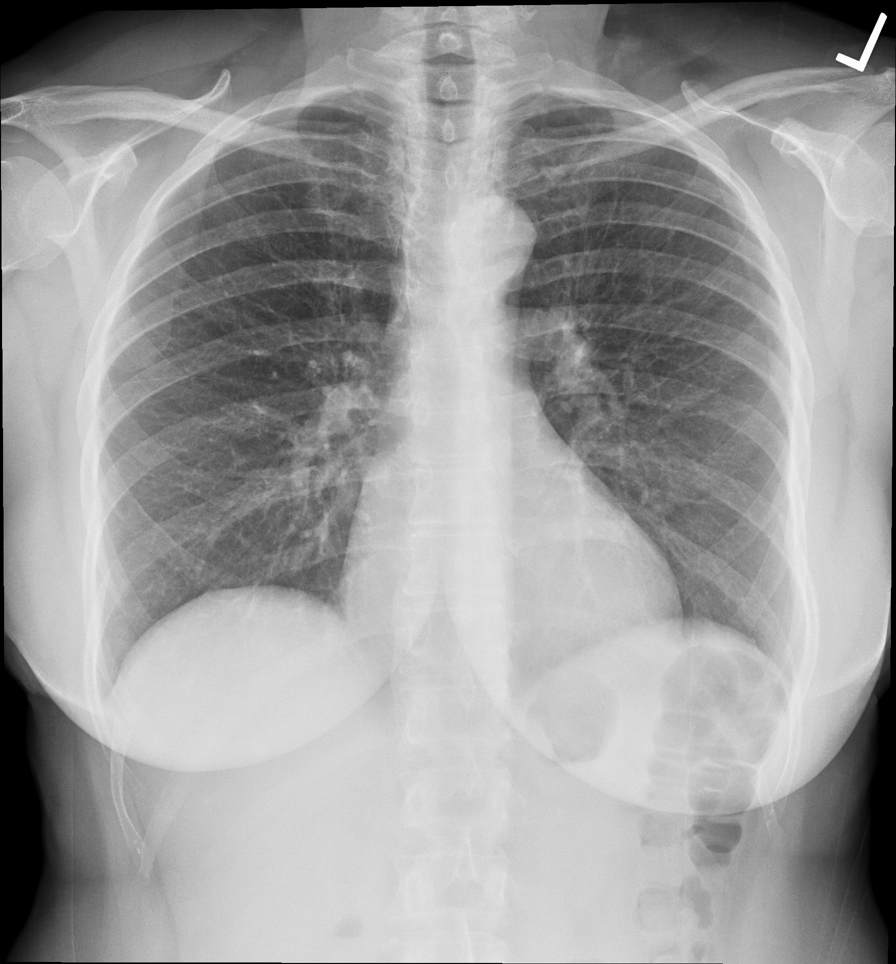
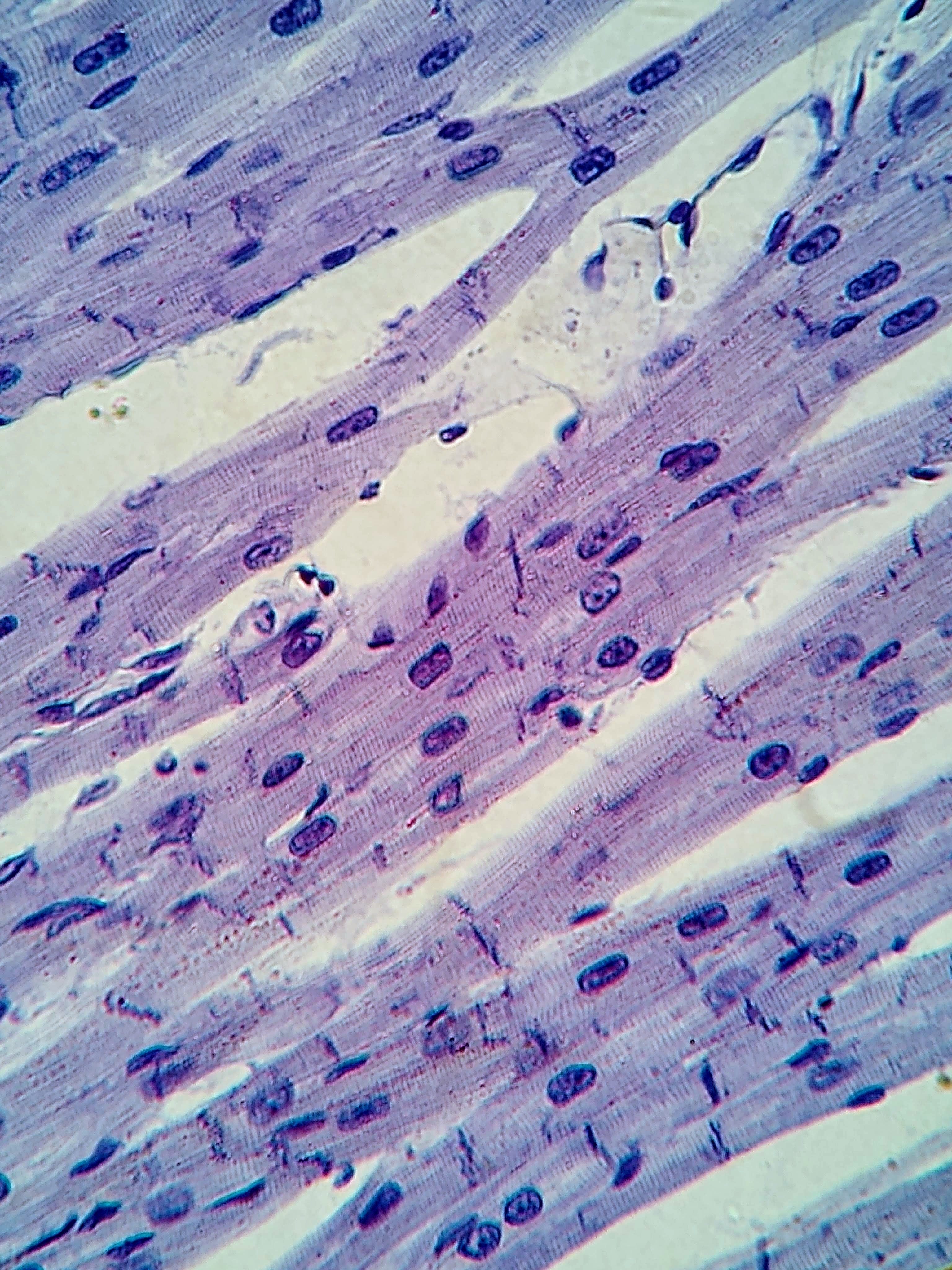
The chest radiograph below shows a projection of the thorax, including a cardiac silhouette among overlapping structures. It does not display individual heart chambers with the clarity of a cutaway illustration. The second image is a real light-microscope photograph of cardiac muscle. Its staining colors and magnification are properties of the preparation and image, not the natural appearance of the whole heart.
Compare the two images by stating their scale and purpose. One provides a broad thoracic relationship; the other provides a tissue pattern. The micrograph’s source does not specify a human specimen, so it is included as an example of cardiac tissue rather than identified as a sample from a human patient. The source describes the radiograph as normal; this article does not independently diagnose it.
10. Use circulation knowledge in team communication
For medical students, combine a vessel route with one mechanism: direction of flow, wall structure, or tissue exchange. For practice assistants and hospital teams, describe the measurement actually obtained and the circumstances around it. For physicians, a diagram can help explain the route being discussed without implying that a generic chart represents the individual’s anatomy in every detail.
In eldercare education, connect the overview with clear observations and the established care plan. Describe the location of an observation and how it differs from the person’s usual state, then communicate through the agreed local process. Do not convert a poster into an individual treatment instruction. The educational goal is a shared language for routes, measurements, and relationships that makes a clinical conversation easier to follow.
11. A short retrieval exercise
Close the labels and answer three questions in complete sentences: Where does blood go after leaving the right ventricle? Why is a pulmonary artery still an artery? What extra information is needed to calculate output from heart rate? Check the route, the definition, and the formula separately. Correct the type of error you made rather than simply repeating the whole diagram.
Finish by drawing the circulation as two linked loops and explaining it to a colleague who has not seen your drawing. If they can follow the route and identify the lung exchange step, your diagram is doing useful explanatory work. Return later with a regional chart and connect that local vessel pattern to the larger system.
Sources and image credits
Sources checked on 8 October 2026. Numerical examples, anatomical classification counts, and research estimates are identified separately in the text.
- OpenStax: vessel structure
- OpenStax: flow, pressure and resistance
- OpenStax: capillary exchange
- OpenStax: circulatory pathways
- OpenStax: cardiac physiology
- FIPAT: Terminologia Anatomica, second edition
Explanatory prose and simplified SVG graphics are original. Photographs retain their stated Creative Commons licenses; creator and source links appear beside each image. Catalog artwork is educational and decorative; no clinical authorship or peer-review credential is claimed.