A column that supports movement and protects neural tissue
The human spine is both an architectural support and a moving series of joints. Describing it as a stack of bones captures only one part of its organization. Vertebral bodies support axial loading, posterior elements surround a neural passage, and discs, ligaments, muscles, and small synovial joints influence how neighboring segments move. Medical students need to recognize these components together because a feature that looks simple in an isolated vertebra becomes more complex when the bone is placed within the complete column. Healthcare staff also benefit from distinguishing the vertebral column from the spinal cord, which occupies only part of the vertebral canal in adults.
A useful first approach is to follow the spine from the skull to the pelvis, identifying regional transitions rather than memorizing every projection at once. Next, examine a single typical vertebra and decide what faces anteriorly, posteriorly, superiorly, and inferiorly. Finally, add the soft tissues and the neural contents. This sequence helps explain why one diagram may emphasize bone while another emphasizes nerve roots or intervertebral discs. Both can be accurate, but they answer different anatomical questions. The objective is a connected three-dimensional model, not a collection of labels without spatial relationships.
Vertebral regions and counting conventions
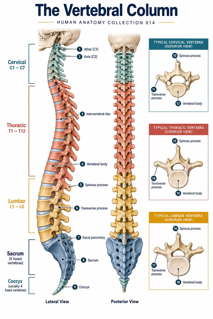
The standard regional pattern has seven cervical vertebrae, twelve thoracic vertebrae, and five lumbar vertebrae. The adult sacrum and coccyx are commonly counted as one bone each after fusion, giving twenty-six bones in the adult column under that convention. Before fusion, sacral and coccygeal components are counted separately; the familiar total of approximately thirty-three vertebrae therefore describes a different counting framework. Coccygeal number and transitional anatomy vary. These figures are teaching conventions, not a guarantee that every person's segmentation is identical. NCI SEER lists the adult regional bone counts.
In written communication, a regional prefix and a number identify a level: C refers to cervical, T to thoracic, and L to lumbar vertebrae. A phrase such as L4–L5 usually identifies the relationship or disc level between two adjacent vertebrae, rather than an additional bone. This distinction becomes especially important when learners compare an anatomical illustration with a report that describes several neighboring levels. Before discussing a numbered level, confirm what has been numbered: a vertebral body, an intervertebral space, a spinal nerve, or a spinal cord segment. These related structures do not share a universally identical position throughout the spine.
The typical vertebra in three dimensions
A typical vertebra includes a vertebral body anteriorly and a vertebral arch posteriorly. Pedicles link the body with the arch, while laminae complete its posterior portion. Together, body and arch enclose the vertebral foramen. When successive foramina align, they form the vertebral canal. Processes provide joint surfaces and attachment sites rather than simply decorating the bone. One spinous process projects posteriorly, two transverse processes project laterally, and four articular processes contribute to articulations with adjacent vertebrae. This familiar count of seven processes applies to the typical vertebral pattern; special vertebrae differ. NCBI describes typical vertebral components.
Imagine holding an isolated lumbar vertebra without an identifying label. Its large body should direct attention toward the anterior load-bearing portion. The opening behind that body locates the canal, and the spinous process establishes the posterior direction. Once those landmarks are fixed, the paired pedicles and articular processes become easier to interpret. Turning the specimen is more informative than repeatedly inspecting one view. A superior photograph can show the canal clearly while hiding the vertical relationship of the joint surfaces. Anatomical certainty improves when the same structure is identified from more than one perspective.
Cervical specialization: atlas, axis, and transverse foramina
The first cervical vertebra, the atlas or C1, supports the skull and has a ring-like arrangement rather than the body and spinous process of a typical vertebra. The axis, C2, bears the dens, a projection associated with rotational movement at the upper cervical region. These differences explain why a generic vertebral diagram is insufficient for studying the craniovertebral junction. Most cervical vertebrae also have transverse foramina, a distinctive regional feature. The vertebral artery generally enters the cervical transverse-foramen route at C6, although variation exists; the presence of a foramen does not mean that every cervical level carries the same contents. NCBI reviews cervical vertebral anatomy.
When comparing the atlas and axis, separate the supporting joint relationship from the motion it permits. The occipital condyles articulate with the atlas, whereas rotation involves the atlas–axis arrangement and associated ligaments. Nodding and turning the head are useful demonstrations of movement, but they distribute across multiple structures and should not be reduced to perfectly isolated hinges. A teaching model can exaggerate one motion to make a relationship visible; the accompanying explanation should identify that simplification. Students should also avoid interpreting the dens as a completely separate adult bone simply because it appears visually distinct in a diagram.
Thoracic vertebrae and the rib-bearing region
Thoracic vertebrae are identified partly by their relationships with ribs. Costal facets or demifacets on the vertebral bodies and, at many levels, facets on transverse processes contribute to rib articulations. The detailed pattern varies across the thoracic series, particularly near its ends. Read the anatomical reference on thoracic vertebral anatomy. A rib and a vertebra therefore should be studied as an interacting pair, not as unrelated specimens. The rib cage also influences regional movement and links vertebral anatomy with respiratory mechanics. A posterior skeletal photograph shows the arrangement of the ribs well, while a transverse diagram better explains the relationship between a rib head, a vertebral body, and the canal.
For practical learning, follow one rib from its vertebral attachment toward the anterior chest wall. Then reverse the route and identify which vertebral surfaces participate in its attachment. This exercise prevents an easy error: assuming that every rib is connected to a single vertebral body in exactly the same way. Regional descriptions summarize a common pattern, while individual levels refine it. In a clinical workplace, precise location language is more useful than calling the whole upper back the thoracic spine. Distinguish the vertebral level from the skin location where discomfort is reported or an external landmark is observed.
Lumbar support, the sacrum, and pelvic continuity
Lumbar vertebrae have comparatively substantial bodies consistent with their position in the load-bearing column. Their posterior elements and articular surfaces differ from those of the cervical and thoracic regions. The sacrum transfers forces between the vertebral column and the pelvic ring through the sacroiliac region. Its fused segments form a recognizable triangular structure with foramina and a sacral canal. The coccyx continues inferiorly and contributes attachment sites for pelvic soft tissues. These structures make the lower spine part of a larger mechanical system that includes the pelvis and lower limbs, rather than an unsupported endpoint. Read the anatomical reference on lumbar anatomy.
Consider standing up from a chair as an anatomical example. Movement involves the hips, knees, ankles, trunk muscles, and spinal joints together. A drawing that emphasizes the lumbar region should therefore not imply that all lifting or postural control belongs to lumbar bones alone. The pelvis provides a base whose orientation affects how the column is presented in a lateral image. This is why a discussion of lumbar curvature should include the position of the person or specimen. A fixed photograph captures one posture, whereas the living column responds continuously to movement, support, muscle activation, and changing external demands.
Intervertebral discs and paired facet joints
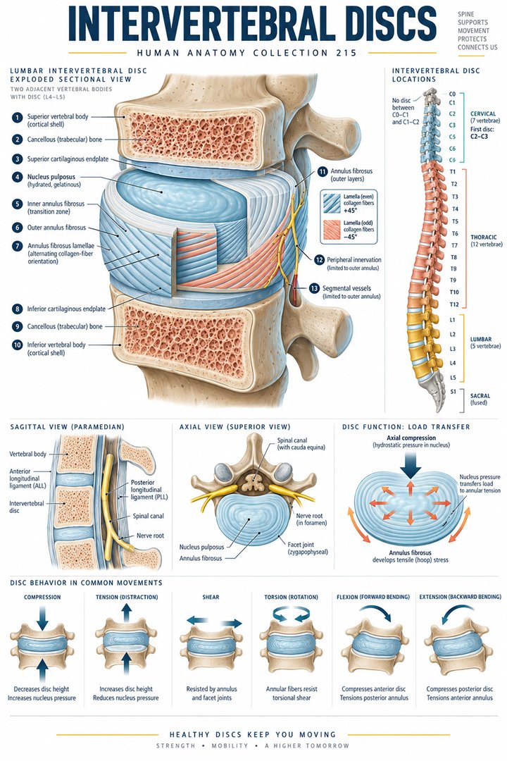
An intervertebral disc sits between neighboring vertebral bodies where discs are present. It comprises an outer annulus fibrosus and an inner nucleus pulposus, with cartilaginous endplates connecting the arrangement to the vertebral bodies. The annulus has a fibrous architecture suited to constraining the internal material and responding to multidirectional loading. Discs are not independent floating cushions. They operate with the vertebral endplates, surrounding ligaments, and posterior joints. Read the anatomical reference on disc anatomy. The joints between articular processes are zygapophysial, or facet, joints; these are synovial articulations and should be distinguished from the cartilaginous joint associated with the disc.
A spinal motion segment is useful to visualize as two neighboring vertebrae with their connecting disc, paired posterior joints, and supporting tissues. Viewing only the disc can conceal how posterior joint orientation influences movement. Viewing only the facet joints can conceal how forces pass through the bodies and disc. When a student compares two diagrams, the important question is whether each shows the same anatomical unit and orientation. Anatomical descriptions of loading are explanations of structure; they are not a prescription for an individual person's exercise, positioning, or treatment. Those decisions require information that an educational drawing cannot provide.
Curves, posture, and the difference between shape and diagnosis
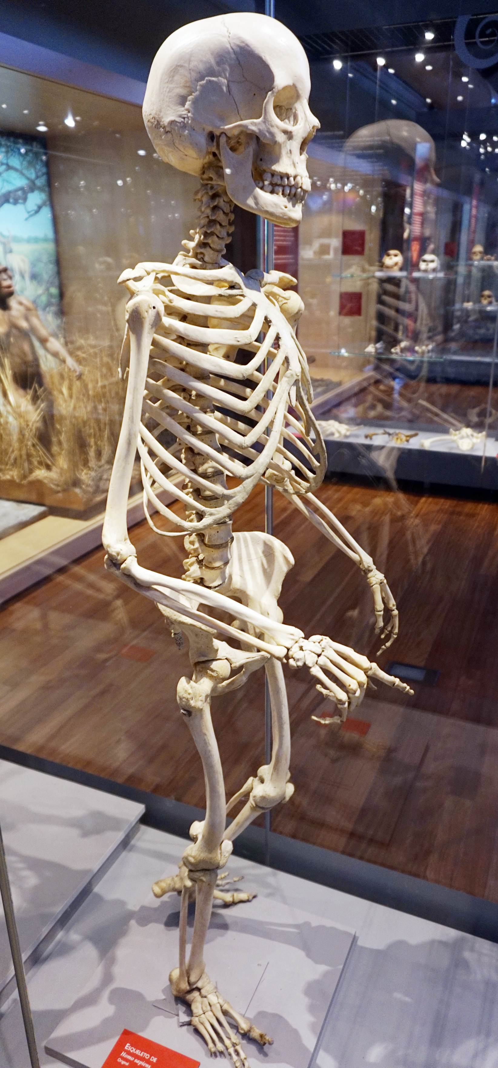
In the sagittal plane, the cervical and lumbar regions normally have lordotic curves, while thoracic and sacral regions have kyphotic curves. Lordosis and kyphosis describe directions of curvature and do not, by themselves, mean disease. Read the anatomical reference on spine structure. Normal spinal shape is not a perfectly straight line viewed from the side. Frontal alignment answers a different question from sagittal curvature. This distinction is important when illustrations place anterior, posterior, and lateral views next to each other. A learner may otherwise interpret any curve as abnormal because the reference image in memory was a frontal skeletal view.
To describe an image carefully, state the plane and region before judging the appearance. For example, a lateral illustration may emphasize the lumbar anterior convexity, whereas an anterior illustration emphasizes the continuity of the midline. Clothing, soft tissues, posture, and projection can alter the visible outline in a photograph. Surface appearance cannot define the precise state of every vertebral body or disc. In education, comparing several normal examples is often more helpful than treating one idealized outline as a universal template. Anatomical variation and age-related change should be discussed without assigning a diagnosis to an unlabeled photograph.
Ligaments, muscles, and distributed stability
Spinal stability involves passive tissues and active muscle control. Longitudinal ligaments run along the bodies, while additional ligaments connect posterior elements. Ligamenta flava join adjacent laminae; interspinous and supraspinous structures contribute to the posterior ligamentous arrangement. Muscles such as the erector spinae and multifidus have different positions and attachment patterns, supporting both movement and control. The exact contribution of one tissue depends on region and activity. Stability is therefore better understood as an integrated relationship than as the exclusive job of one named ligament or one muscle group.
A useful classroom comparison places a bare skeleton beside a muscle illustration. Ask what the skeleton alone cannot explain: control of movement, maintenance of posture, and the changing distribution of force. Then add the ligaments and identify which structures connect neighboring elements. The exercise shows why a visually small component can have an important role without encouraging exaggerated claims about a single structure. For assistants and nursing teams, this model supports clearer communication about body position and anatomical location. It does not replace locally approved moving-and-handling practice or assessment of a person's individual abilities.
Neural spaces are not interchangeable
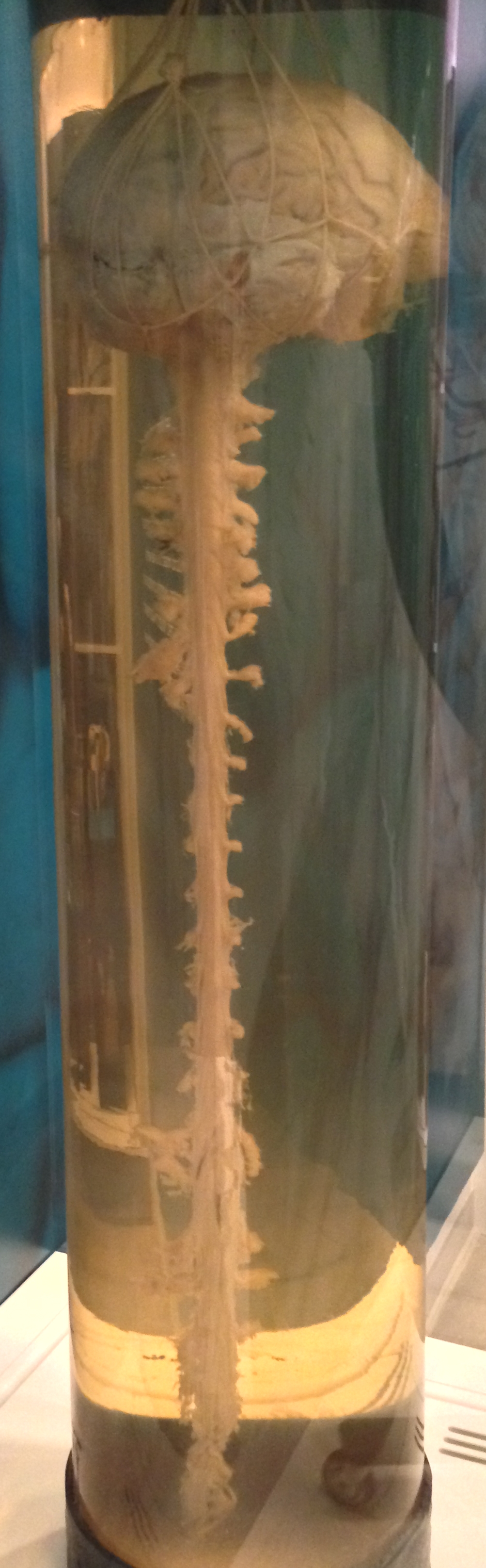
The vertebral canal is the longitudinal space within the column. Intervertebral foramina are lateral passages formed between adjacent vertebrae through which spinal nerves and related structures pass. The spinal cord, meninges, nerve roots, epidural tissues, and vessels occupy different relationships within these spaces. In adults, the cord usually ends near the upper lumbar vertebral region, while descending roots continue inferiorly as the cauda equina. Vertebral level and spinal cord segment are consequently different reference systems. Read the anatomical reference on cauda equina anatomy. The canal extending to the sacrum does not mean that the spinal cord itself extends continuously to the coccyx.
When teaching this distinction, draw the bones and canal first, then add the cord and descending roots in a second color. Label the endpoints explicitly rather than relying on the shape of a line. Next, follow a root toward a lateral exit and compare that route with the longitudinal canal. This makes the difference between central and foraminal spaces visible. For healthcare documentation, the practical lesson is to retain the terminology used in an examination or report and clarify its anatomical meaning. Replacing every neural-space term with “spinal cord” loses information and can create avoidable misunderstanding.
Latin vocabulary and an organized review method
Columna vertebralis names the vertebral column; vertebra cervicalis, vertebra thoracica, and vertebra lumbalis identify regional vertebrae. Corpus vertebrae refers to the body, arcus vertebrae to the arch, and foramen vertebrale to the opening within one vertebra. Processus spinosus and processus transversus identify spinous and transverse processes. Discus intervertebralis identifies a disc, while annulus fibrosus and nucleus pulposus distinguish its principal internal components. Os sacrum and os coccygis identify the fused inferior structures. Latin terms are most useful when each is attached to an actual landmark rather than learned as a disconnected translation list.
For a final review, take a blank regional diagram and explain five relationships aloud: how a body differs from an arch, how a disc differs from a facet joint, how a canal differs from a foramen, how a vertebral level differs from a cord segment, and how a normal sagittal curve differs from a perfectly straight side-view outline. These explanations test understanding more effectively than reciting regional numbers alone. Add counts only after the structures are understood, and state the counting convention. The resulting mental map supports further study of spinal nerves, regional movement, imaging, and the relationship between the axial skeleton and the pelvis.
Anatomical Latin: quick reference
| Latin term | English meaning |
|---|---|
| Columna vertebralis | Vertebral column |
| Corpus vertebrae | Vertebral body |
| Arcus vertebrae | Vertebral arch |
| Discus intervertebralis | Intervertebral disc |
Sources and image credits
Sources reviewed on 9 October 2026. Counts, estimates, study statistics and hypothetical calculations retain their context in the text.
- NCI SEER: axial skeleton
- NCBI: cervical vertebrae
- NCBI: head and neck cervical vertebrae
- NCBI: intervertebral discs
- NCBI: cauda equina
- NCBI: thoracic vertebrae
- NCBI: spinal organization and curves
- NCBI: lumbar spine
Explanatory prose and SVG graphics are original. Photograph authors, source records and licenses are credited beside each image. Catalog illustrations provide educational context; anatomical facts are supported by the linked sources.