1. The stomach is a muscular organ with a specialized lining
The human stomach connects the esophageal route with the duodenum while combining temporary storage, mixing, secretion, and regulated onward delivery. Its external shape is useful for orientation, but its wall and glandular organization explain much of its role. A complete introduction therefore moves from the organ outline to tissue layers and then to specialized cells.
For medical students and healthcare teams, distinguish the anatomical structure from the changing contents. A stomach picture does not show the same volume, shape, acidity, or activity under every condition. The organ’s relationships provide a framework, while a particular observation or measurement belongs to the circumstances in which it was obtained. This guide emphasizes those distinctions using a sectional poster and real endoscopic image.
2. Four commonly taught regions organize the outline
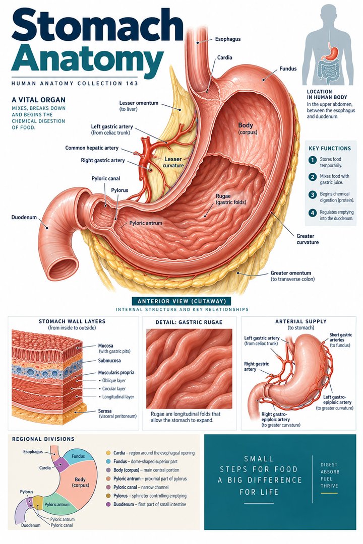
The cardia is associated with the esophageal entry, while fundus, body, and pyloric region provide other major subdivisions. The pyloric antrum leads toward the pyloric canal and outlet to the duodenum. These regional names identify relationships rather than four equally sized compartments. A diagram that changes the organ’s orientation should retain the entry and exit routes. [NCBI Bookshelf: stomach anatomy]
Trace the route from esophagus to duodenum before covering the regional labels. Then restart from the outlet and identify the structures leading back toward the body. This tests a connected map rather than a memorized list. It also helps learners understand why the pylorus is a specific region and junction rather than a name for every part of the lower stomach.
3. Curvatures and neighbors add spatial information
The greater and lesser curvatures describe different borders of the stomach. Their associated attachments and vascular relationships add another way to orient an isolated organ drawing. In the upper abdomen, the stomach’s position relates to neighboring structures, so an opened-organ poster that removes the surroundings should be interpreted as a selected anatomical view. [NCBI Bookshelf: stomach anatomy]
Compare a whole torso model with the stomach section. Name which neighboring structures establish orientation in the torso view and which internal details become visible in the section. This comparison prevents the isolated outline from being treated as a precise surface map for every person. For professional communication, retain the actual site or region named in an established report.
4. Wall coats and muscular orientations are different counts
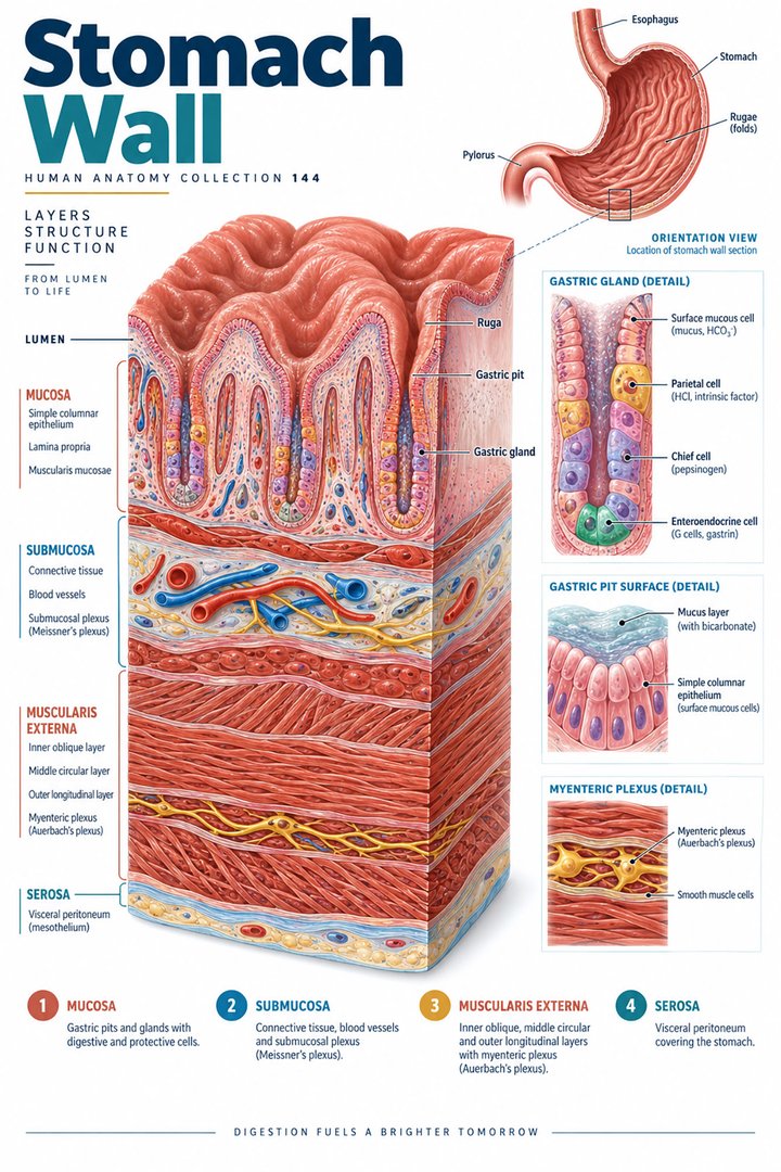
Four principal wall coats are commonly described: mucosa, submucosa, muscularis externa, and serosa. Within the muscularis externa, the stomach has oblique, circular, and longitudinal muscle orientations. These three orientations are subdivisions of a muscular coat, not replacements for the four whole-wall coats. Some oncological descriptions identify subserosal tissue separately, so the classification should be stated. [NCI SEER: gastrointestinal wall layers] [OpenStax: stomach structure and physiology]
A useful layered sketch begins at the lumen and moves outward. Label the whole coats first, then enlarge the muscular coat to show its orientations. The drawing becomes clearer when it distinguishes a hierarchical subdivision from another layer at the same level. This prevents a common counting error in which three muscle arrangements are presented as the stomach’s entire wall.
5. Rugae, pits, and glands describe different features
Gastric rugae involve mucosal and submucosal folds and flatten with distension. Gastric pits lead into glands within the mucosal organization. These features belong to different scales, so a large fold in an endoscopic image should not be labeled as one microscopic gland. The stomach does not have the absorptive villi characteristic of the small intestine. [OpenStax: stomach structure and physiology]
Place an internal surface view beside a microscopic diagram. Ask which feature can be seen at each scale and which requires an explanatory enlargement. This makes a poster’s inset more useful and prevents similar-looking protrusions from being treated as interchangeable structures. A descriptive word such as fold does not by itself identify the tissue composition or microscopic function.
6. Specialized cells contribute different secretions
Surface mucous cells contribute protective mucus, parietal cells produce acid and intrinsic factor, chief cells produce pepsinogen, and G cells release gastrin. These are distinct roles within the glandular and regulatory arrangement. A single arrow labeled gastric secretion conceals the difference between protective material, digestive precursors, and endocrine signaling. [OpenStax: stomach structure and physiology]
For revision, connect each named cell with its product before adding the product’s purpose. Then distinguish release into the luminal environment from a hormonal relationship. This prevents every secreted substance from being assigned the same route. It also makes the organ’s lining easier to understand as specialized tissue rather than a passive surface that simply contains acid.
7. Intrinsic factor links the stomach with ileal absorption
Intrinsic factor is produced by gastric parietal cells, but vitamin B12 absorption associated with it occurs in the distal ileum. Food-bound B12 is released during digestion, initially associates with haptocorrin, and later binds intrinsic factor after pancreatic enzymes act in the duodenum. The stomach’s contribution and the final absorption site are therefore different parts of a connected route. [NIH Office of Dietary Supplements: vitamin B12 absorption]
This is an excellent cross-system teaching example. Draw the source organ and the later absorption region separately, then name the connecting digestive steps. Avoid using “absorbed in the stomach” as a shortcut because intrinsic factor originates there. A source of a required substance need not be the destination at which its eventual physiological effect is completed.
8. Vessels and neural pathways support the organ
Gastric arterial networks derive predominantly from the celiac-trunk arrangement. Vagal and enteric relationships contribute to regulation of secretion and movement. These routes are not directly visible in an ordinary endoscopic view of the mucosal surface. A detailed poster can add them, but should preserve the distinction between luminal observation and deeper supporting organization. [NCBI Bookshelf: stomach anatomy]
Use a wall diagram to place neural and vascular relationships at the appropriate scale. Name what the illustration omits rather than trying to place every pathway in one crowded image. For healthcare discussion, an anatomical account of regulation does not determine the cause of a person’s symptoms or the meaning of a single test. Those interpretations require the established clinical context.
9. Physiological numbers are not fixed properties of every image
A textbook gastric-content pH range of approximately 1.5 to 3.5 can provide an introductory description of an acidic environment. Meals and other conditions affect the contents, so this should not be treated as a fixed measurement that applies whenever a stomach is observed. The pH number describes hydrogen-ion activity on a logarithmic scale, not a percentage of acid. [OpenStax: stomach structure and physiology]
When discussing numbers, name the compartment, method, and condition. A wall image does not measure luminal pH, and a pH value does not reveal all structural layers. Similarly, avoid setting one universal maximum stomach capacity or meal-emptying time from a general anatomy drawing. Quantitative teaching is most useful when the assumptions remain attached to the number.
10. Latin terms keep regions and layers precise
Gaster and ventriculus are selected names for the stomach. Fundus gastricus, corpus gastricum, and pylorus retain regional distinctions. Curvatura major and curvatura minor identify the curvatures. Tunica mucosa and tela submucosa help name wall relationships. Learn these expressions with the view and scale in which they are best demonstrated. [FIPAT: Terminologia Anatomica, second edition]
Create a vocabulary table with separate categories for regions, borders, and wall layers. Add a short location sentence rather than a translation alone. This makes terminology easier to use when a clinical report describes the gastric body, antral region, or mucosal surface. For patient-facing discussion, ordinary English can explain the term while retaining the specific part being discussed.
| English | Selected Latin |
|---|---|
| Stomach | Gaster / ventriculus |
| Fundus / body | Fundus gastricus / corpus gastricum |
| Greater / lesser curvature | Curvatura major / curvatura minor |
| Mucosa / submucosa | Tunica mucosa / tela submucosa |
11. A real endoscopic photograph has a defined viewpoint
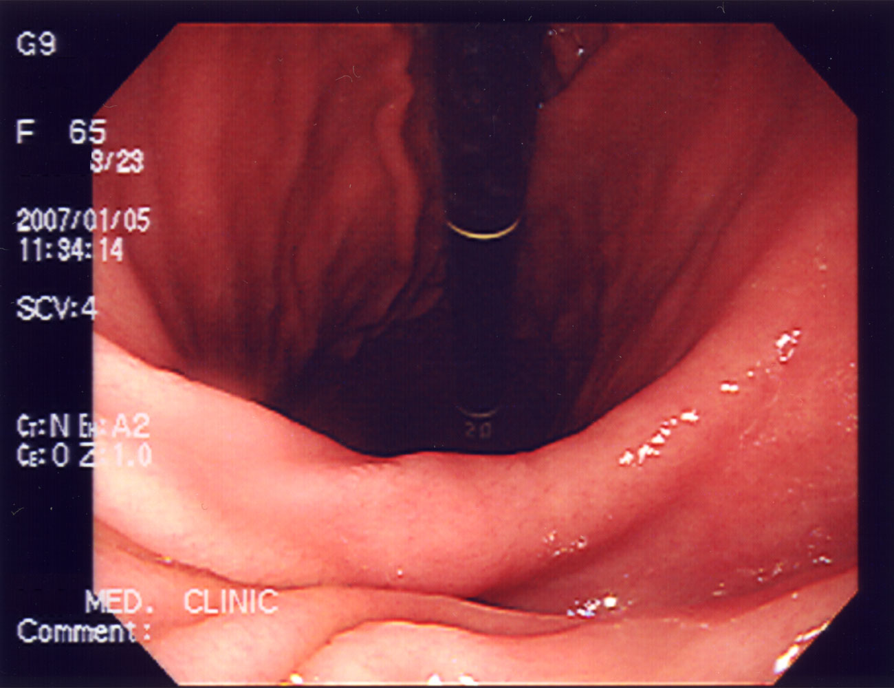
The endoscopic image below shows the stomach body from within its lumen. The source describes the view as normal, but no independent clinical interpretation is made here. Illumination, viewing direction, and the instrument’s perspective influence the appearance. The image does not expose the entire muscular wall, external curvatures, or all neighboring organs visible in a sectional teaching illustration.
Medical students can compare visible folds with the diagram while retaining the difference in scale and method. Practice assistants, hospital staff, physicians, and eldercare teams can use the regional vocabulary to clarify established findings. Finish revision by naming a region, distinguishing a whole-wall coat from a muscular orientation, and explaining the intrinsic-factor connection to the ileum. Those relationships provide a strong structural foundation.
Sources and image credits
Sources checked on 8 October 2026. Numerical examples, anatomical classification counts, and research estimates are identified separately in the text.
- FIPAT: Terminologia Anatomica, second edition
- NCBI Bookshelf: stomach anatomy
- OpenStax: stomach structure and physiology
- NCI SEER: gastrointestinal wall layers
- NIH Office of Dietary Supplements: vitamin B12 absorption
Explanatory prose and simplified SVG graphics are original. Photographs retain their stated Creative Commons licenses; creator and source links appear beside each image. Catalog artwork is educational and decorative; no clinical authorship or peer-review credential is claimed.