An anatomical map of chemical communication
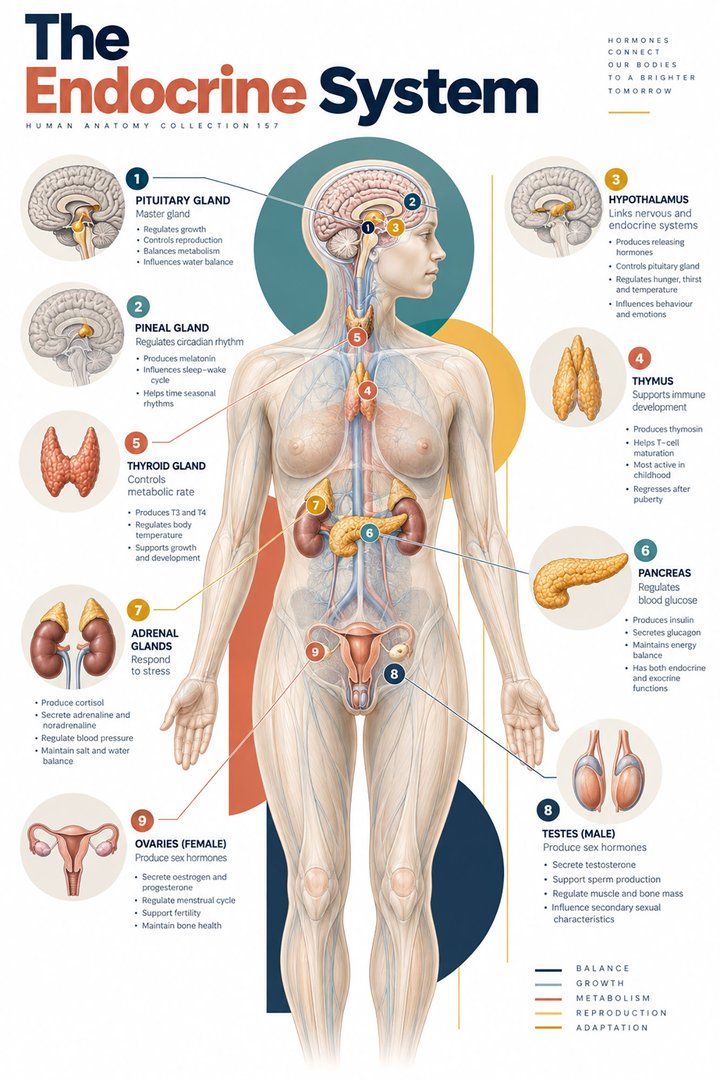
Endocrine glands release hormones into the internal environment, allowing chemical information to reach responsive tissues through the circulation. A map of these glands spans the head, neck, abdomen, and pelvis. This distribution is useful to learn, but a gland's position is only the first part of the story. Its cells, blood supply, hormone products, and relationship to other structures determine how it functions. A useful endocrine atlas therefore connects a location to a microscopic organization and then to a physiological pathway. It should make clear which tissue produces the signal, how that signal leaves the tissue, and which cells can respond. NIDDK: Endocrine diseases introduces the system's broad roles.
The endocrine system is not a set of isolated control boxes. A hormone-producing organ can also have digestive, reproductive, or other functions, and endocrine cells can be scattered within organs that are not usually called glands. The word system groups related communication processes; it does not imply that all the tissues look alike. For medical students, this is an opportunity to combine gross anatomy with histology. For healthcare staff, the same framework helps explain why a small anatomical structure can influence several observable functions. Throughout this guide, gland names refer to typical anatomy, while individual position, shape, size, and developmental variation remain important.
Endocrine versus exocrine: follow the exit route
The fundamental distinction between endocrine and exocrine tissue concerns the route by which a secretion reaches its destination. Exocrine secretions pass through ducts to an epithelial surface or a lumen, while endocrine hormones enter the interstitial fluid and circulation. A pancreatic digestive enzyme delivered to the duodenum and pancreatic insulin released into blood therefore leave the same organ by different pathways. This distinction is clearer when shown as two separate arrows rather than a single label saying pancreatic secretion. It also prevents the misconception that every gland must have a visible duct. The general organization is outlined in OpenStax: Endocrine overview.
A duct is an anatomical conduit, not merely a line joining two names in a chart. Its epithelial lining, connections, and final opening matter. A capillary network is a different kind of route with different barriers and pressures. When reviewing a specimen, look for the organization of secretory cells around ducts, follicles, or blood vessels, while recognizing that a routine stain does not identify every hormone. The same cell shape can support different functions in different organs. Begin with the organ and its arrangement, then use the appropriate specialized evidence to identify a particular cell population. Structure guides interpretation without replacing the methods needed to establish function.
The pituitary has two different anatomical relationships
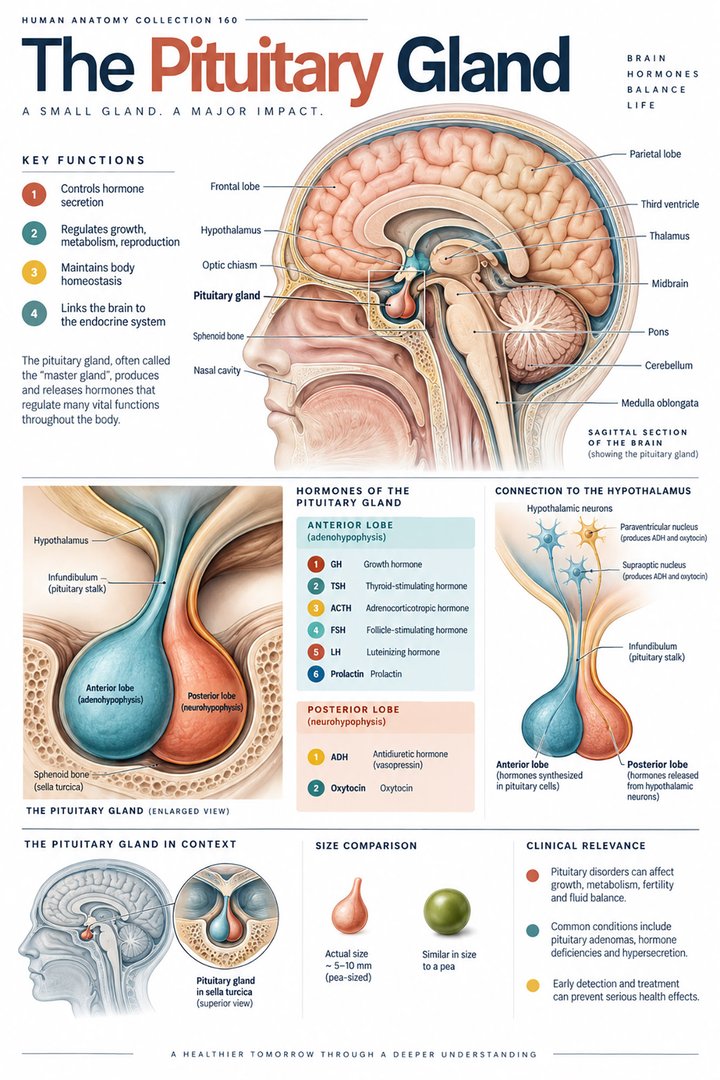
The pituitary gland, hypophysis, lies in the sella turcica of the sphenoid bone and connects with the hypothalamus through the pituitary stalk. Its anterior component, the adenohypophysis, contains glandular cells that synthesize several hormones. Its posterior component, the neurohypophysis, is closely connected to hypothalamic neurons and stores and releases oxytocin and vasopressin made in the hypothalamus. Saying that the posterior pituitary manufactures these two hormones is therefore inaccurate. A two-color diagram should show the difference between hormone synthesis and the site of release. The relevant anatomical relationship is described in OpenStax: Pituitary and hypothalamus.
The anterior pituitary receives hypothalamic regulatory signals through a portal blood vessel arrangement. This allows a signal to travel between nearby tissues through a specialized vascular route before joining the wider circulation. The posterior route instead involves neuronal axons reaching the release region. These two connections explain why the pituitary should not be taught as a single bag of identical cells. The familiar expression master gland is a convenient introduction, but it can hide regulation by the hypothalamus, feedback from target tissues, and signals independent of the pituitary. A better learning exercise asks students to draw the source, connecting route, release site, and target separately for one anterior and one posterior pituitary hormone.
Thyroid follicles organize synthesis and storage
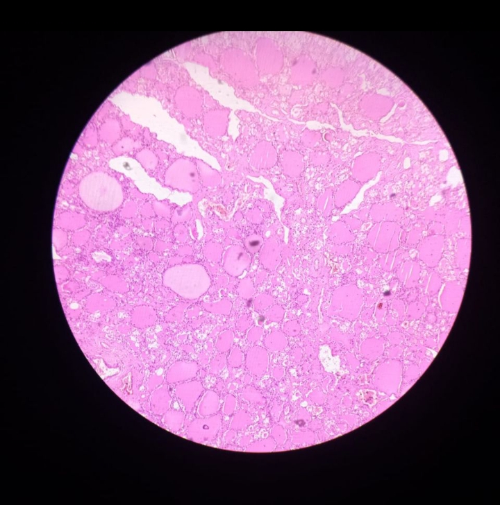
The thyroid gland, glandula thyroidea, usually has right and left lobes connected by an isthmus in the anterior neck. Its microscopic hallmark is the follicle: a ring of epithelial cells surrounding a lumen containing colloid. Follicular organization is important because thyroid hormone synthesis and storage are closely associated with this extracellular compartment. The two familiar hormone abbreviations, T3 and T4, refer to molecules with three and four iodine atoms respectively. This is a molecular count, not a count of gland lobes or a measure of a patient's thyroid activity. The follicular arrangement is explained in OpenStax: Thyroid gland.
A real thyroid micrograph gives students a chance to connect a whole organ with many small functional units. First identify the follicular outline, then distinguish the epithelial border from the central material and the spaces between follicles. Sectioning can make one three-dimensional follicle appear as circles or irregular profiles of different sizes. The appearance alone does not provide a hormone concentration or establish a diagnosis. A useful caption identifies the tissue and stain and avoids implying that every visible cell has been individually verified. This disciplined approach also helps staff explain why ultrasound anatomy and blood tests answer related but distinct questions about the gland.
Parathyroid glands are separate organs with a separate role
The parathyroid glands, glandulae parathyroideae, are usually small structures associated with the posterior thyroid region. Four is the typical teaching count, but their number and position can vary. They are not simply posterior parts of the thyroid. Their parathyroid hormone participates in calcium regulation, linking the gland with bone, kidney, and intestinal physiology. The proximity of two endocrine tissues in the neck does not imply that they produce the same hormones or share the same regulatory pathway. A regional illustration should give the thyroid and parathyroids separate labels and should note anatomical variation rather than promising four identical dots in every patient. See OpenStax: Parathyroid glands.
This anatomical relationship is a good example of why names can mislead when learned without context. Para describes proximity, not equivalence. A student who understands the separate tissues can follow a discussion of calcium regulation without confusing it with the regulation of energy metabolism by thyroid hormones. In a learning session, compare what remains the same and what changes when switching from an anterior to a posterior view of the neck. The airway and gland relationship stays three-dimensional even when a page presents only one surface. For professional communication, keep the named gland and the physiological issue aligned, and avoid using a thyroid label for an unexplained calcium-related finding.
Adrenal cortex and medulla share an organ but differ in organization
The paired adrenal glands, glandulae suprarenales, sit in relation to the upper kidneys. Their outer cortex and inner medulla have different cellular organizations and hormone products. The cortex is conventionally divided into three zones: zona glomerulosa, zona fasciculata, and zona reticularis. These regions are associated with mineralocorticoids, glucocorticoids, and adrenal androgens respectively. The medulla contains chromaffin cells associated with catecholamine secretion and sympathetic regulation. The count of three cortical zones describes histological regions, not three independent glands. The broad arrangement is detailed in OpenStax: Adrenal glands.
On a histological section, first establish which side is the capsule and which is the medullary region, then compare cell arrangements between them. A teaching chart often uses sharply colored bands, whereas real boundaries can be less crisp. An anatomical photograph may reveal external shape and position without showing the internal layers at all. Pairing these views helps learners avoid expecting the microscopic organization to be visible on the surface of a preserved organ. The adrenal example also shows that being next to a kidney does not make a gland part of the urinary collecting system. Anatomical proximity can matter clinically while physiological function remains distinct.
Pancreatic islets are endocrine tissue within a mixed organ
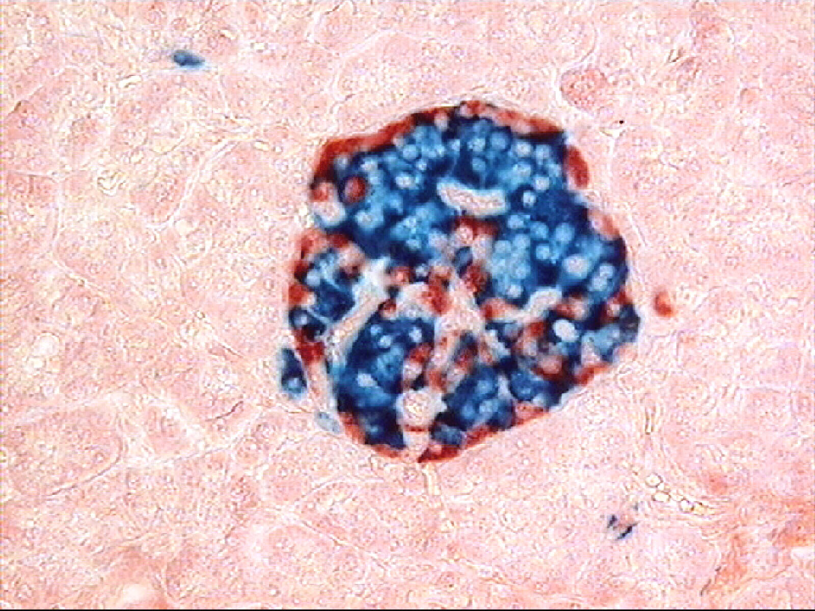
The pancreas contains exocrine tissue and endocrine cell groups called pancreatic islets, insulae pancreaticae. Beta cells produce insulin and alpha cells produce glucagon; other islet populations contribute additional regulatory signals. These cell groups are associated with a blood supply that supports endocrine communication. A pancreatic illustration should therefore show islets beside a vascular route and exocrine tissue connected with ducts. Depicting all pancreatic products entering the intestine would erase the endocrine function, while showing all products entering blood would erase the digestive function. OpenStax: Endocrine pancreas outlines these different populations and their roles.
The islet is a useful bridge between microscopic anatomy and coordinated whole-body metabolism. A hormone can be released from a small tissue region yet influence liver, muscle, and adipose processes elsewhere. That does not mean every tissue responds in the same way, or that one hormone alone explains all glucose regulation. In a real immunostained image, colors may identify selected proteins or cell groups according to the method used; they are not the natural colors of the living organ. A caption should preserve this distinction. Students can ask which structures are demonstrated directly and which physiological arrows have been added to explain the image.
Pineal and gonadal endocrine tissues add time and reproductive context
The pineal gland, glandula pinealis, is an intracranial endocrine structure associated with melatonin secretion and circadian signaling. Its inclusion on an endocrine map demonstrates that glands can be closely integrated with neural information. A simple light-dark illustration can show timing relationships without claiming that one hormone is a complete explanation of sleep. Sleep and wakefulness depend on many connected processes, and a hormone concentration is only one part of that system. The gland's location and its association with melatonin are introduced in OpenStax: Pineal gland. This section should be read as anatomy and physiology rather than advice about using melatonin products.
The testes and ovaries also contain endocrine tissue, alongside their roles in gamete production. Gonadal endocrine activity interacts with pituitary and hypothalamic signals, but the organ's structure is not reduced to that single axis. A testis contains seminiferous tubules and interstitial tissue with different functions; an ovary contains follicles, supporting tissue, and structures that change during reproductive cycles. A useful map names the gonads while recognizing variation associated with age, developmental history, pregnancy, and other physiological states. Hormone names should not be used as rigid labels for identity: several so-called sex hormones are present in people across sexes and serve functions beyond reproduction. The anatomical question concerns tissue and signaling, not assumptions about a person.
Endocrine cells also belong to familiar non-gland organs
The heart, kidneys, gastrointestinal tract, and other tissues have endocrine functions even though their primary names often suggest something else. Examples include renal erythropoietin, cardiac natriuretic peptides, and gastrointestinal hormones involved in digestive coordination. The placenta is another important temporary endocrine organ during pregnancy. These examples make a closed list of a fixed number of endocrine glands less useful than a map that distinguishes major dedicated glands from endocrine cells within other organs. A claim such as there are exactly eight endocrine organs can depend on how the list is grouped. The wider distribution is described in OpenStax: Secondary endocrine functions.
This distinction matters in multidisciplinary care. A discussion of kidney function may involve fluid handling, hormone activation, and blood cell production, not just urine formation. A discussion of pregnancy may involve an endocrine organ that is absent outside that physiological context. For learning, add an endocrine arrow to a familiar organ diagram and label the specific signal rather than recoloring the whole organ as an endocrine gland. This preserves its other roles and encourages an integrated view. Staff can then explain why blood tests and clinical observations may be relevant to several systems at once without suggesting that every result arises from one isolated gland.
Feedback, receptors, and the limits of a gland diagram
Hormones act through receptors and intracellular mechanisms in responsive cells. A circulating signal can pass many tissues, yet the presence and regulation of receptors help determine where and how it has an effect. Negative feedback means that a downstream change reduces an upstream drive; it is not equivalent to a gland switching off permanently. The thyroid axis provides a familiar example linking hypothalamic TRH, pituitary TSH, and thyroid hormone. Other systems use different combinations of feedback, neural influence, nutrient signals, and local regulation. OpenStax: Hormones explains the general principles. A diagram should distinguish stimulatory and inhibitory arrows rather than using one arrow style for every relationship.
A gland map cannot by itself explain a person's laboratory result. Time of sampling, physiological state, medications, binding proteins, receptor response, and the analytical method can influence interpretation. Nor does a normal external shape prove normal hormone activity. For students, the strongest learning sequence is location, cell architecture, secretion route, target, and feedback. For practice and hospital staff, clear terminology improves preparation, documentation, and communication with the clinician responsible for interpretation. For teams supporting older adults, recording new functional changes without attributing them prematurely to age or one gland remains essential. Endocrine anatomy becomes most useful when it helps people ask precise questions about a connected physiological system.
A practical way to read an endocrine atlas
When opening an endocrine atlas, identify the viewing plane before interpreting gland position. A midsagittal head image can show a pituitary relationship that an anterior body outline cannot, while a posterior neck image reveals relationships hidden in a familiar thyroid silhouette. Next ask whether the picture represents external anatomy, a cut surface, or a microscopic field. These are different scales of evidence. The final step is to trace one hormone route in words, naming the producing tissue and the responsive tissue separately. If a map includes a connection without explaining whether it represents a blood vessel, a nerve, or feedback, add that distinction in a learning note. This small exercise prevents a common problem: knowing the colored gland names but being unable to explain how the diagram relates to a physiological process. For a teaching team, comparing two views of the same structure can be more informative than adding more labels to an already crowded page.
Anatomical Latin: quick reference
| Latin term | English meaning |
|---|---|
| Hypophysis | Pituitary gland |
| Glandula thyroidea | Thyroid gland |
| Glandula suprarenalis | Adrenal gland |
| Insulae pancreaticae | Pancreatic islets |
Sources and image credits
Sources reviewed on 9 October 2026. Counts, estimates, study statistics and hypothetical calculations retain their context in the text.
- Endocrine diseases
- Hormones
- Pituitary and hypothalamus
- Thyroid
- Parathyroid glands
- Adrenal glands
- Pineal gland
- Endocrine pancreas
- Secondary endocrine organs
- OpenStax: Endocrine overview
Explanatory prose and SVG graphics are original. Photograph authors, source records and licenses are credited beside each image. Catalog illustrations provide educational context; anatomical facts are supported by the linked sources.