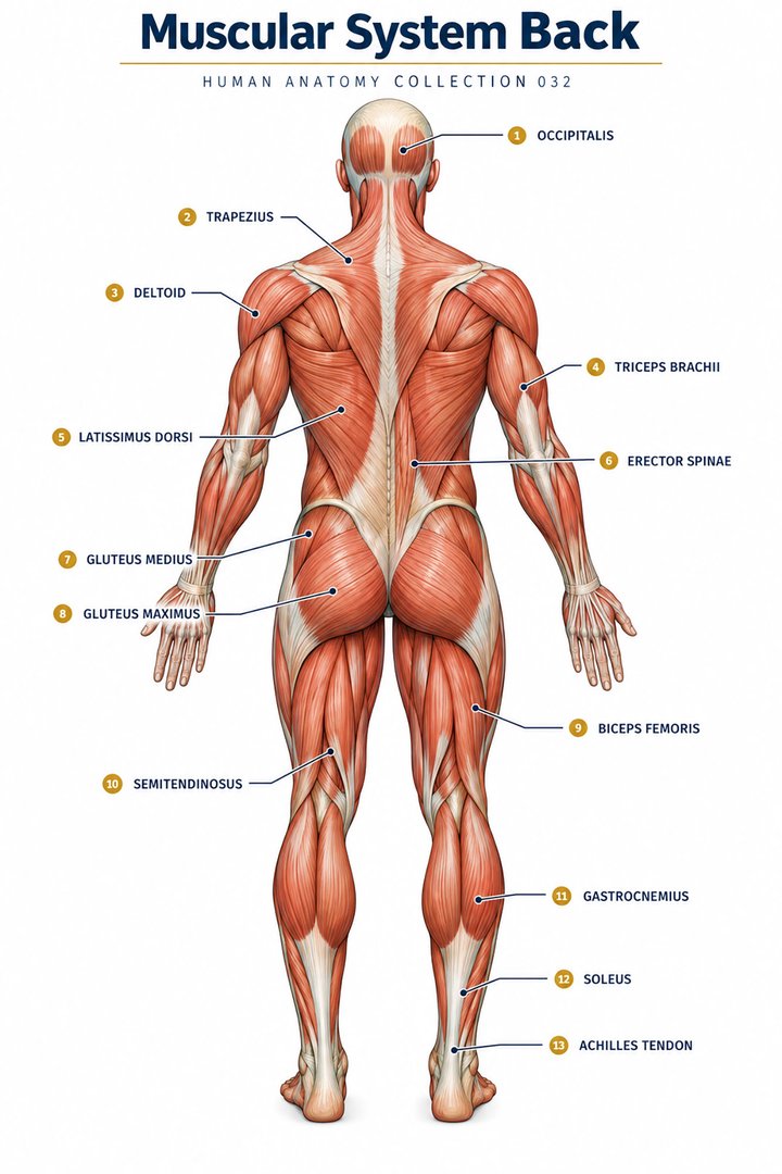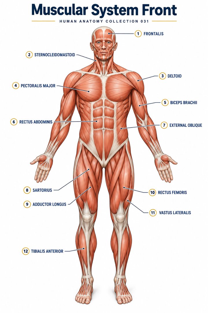
1. Muscles connect anatomy with action
A muscular system overview should explain more than the visible outlines of a fit body. Muscles generate movement, support posture, contribute to joint stability, and produce heat. Their work ranges from deliberate limb motion to the movement of blood and contents within hollow organs. For medical students and healthcare professionals, the most useful framework connects tissue type, anatomical arrangement, and observed action. [muscle intro]
A superficial muscle poster is a starting map. It cannot show every deep muscle, attachment, nerve, or variation. When learning a region, establish the viewpoint and the layer shown before memorizing names. An anterior view and a posterior view may show different structures rather than two sides of an identical set of labels.
Ask three questions about each muscle: Where is it attached? Which joint or structure does it act on? How does its action contribute to a task? This makes later learning about detailed origins, insertions, and innervation easier to organize.
2. Three muscle tissue types
Skeletal muscle is striated and usually associated with voluntary control, although many of its activities are automatic or reflexive. Cardiac muscle is striated tissue within the heart, with distinctive cellular connections and involuntary activity. Smooth muscle lacks the obvious striations of the other two types and participates in functions such as movement within hollow organs and changes in vessel caliber. “Striated” describes appearance; “voluntary” describes control. They are different classifications. [muscle types]
| Tissue type | Typical location | Learning distinction |
|---|---|---|
| Skeletal muscle | Limb and trunk muscles | Striated; movement and support |
| Cardiac muscle | Myocardium | Striated; specialized heart tissue |
| Smooth muscle | Walls of many hollow organs and vessels | No obvious striations; regulated involuntary activity |
The three categories are a useful structural count, not three equal shares of the body. A whole muscular system chart often concentrates on skeletal muscles, whereas physiology teaching includes all three tissue types. Read the title and caption to discover which meaning of “muscle” the resource is using. [muscle types]
3. From whole muscle to sarcomere
A skeletal muscle contains fascicles, and each fascicle contains muscle fibers. A muscle fiber is a cell containing myofibrils. Repeating sarcomeres organize the contractile machinery within myofibrils. Connective-tissue layers associated with this hierarchy include epimysium around the whole muscle, perimysium around fascicles, and endomysium around individual fibers. These tissues help connect microscopic force production with the organ’s mechanical output. [muscle structure]
During study, move down this hierarchy slowly. A fiber and a fascicle are different levels; a myofibril is not another word for a whole muscle. Connecting each name to its level prevents a common kind of confusion when moving between a body poster and a micrograph.
4. Contraction depends on signaling, calcium, and energy
Activation at the neuromuscular junction initiates a sequence that leads to calcium release within the skeletal muscle fiber. Calcium permits interactions between the contractile proteins, while ATP supports cross-bridge cycling and other processes needed for contraction and relaxation. In the sliding-filament model, thin and thick filaments change their overlap; the filaments themselves do not simply shrink. [contraction]
The shortening of a sarcomere is therefore different from saying that a protein filament becomes shorter. Also distinguish activation from visible limb movement: force can be generated without an obvious change in joint position. A diagram of molecular events explains one level of muscle function, while a regional illustration explains another. [contraction]
For patient education, a simple explanation can focus on coordinated nerve signals, muscle force, and the movement they produce. For examination revision, return to the detailed sequence and explain where calcium and ATP participate. Matching the explanation to the audience preserves accuracy without overwhelming the discussion.
5. Muscles cooperate through roles and leverage
An agonist contributes to a particular action, an antagonist opposes it, and synergists assist or reduce unwanted motion. Fixators help stabilize a structure during an action. These are functional roles in a task, not permanent personality labels attached to a muscle. A muscle’s role can differ with the movement and circumstances being described. [muscle actions]
Fascicle arrangement influences how muscle fibers are organized, while attachments and joint geometry affect leverage. A visible line on a poster cannot by itself explain the full force or movement of a muscle. Follow the attachment across the joint and consider which movement the arrangement can support. [muscle actions]
6. Important regions and Latin muscle names
The trunk includes muscles that move and stabilize the vertebral region, abdominal wall, and thorax. The rectus abdominis, external oblique, internal oblique, and transversus abdominis provide a useful framework for the anterolateral abdominal wall. The diaphragm and intercostal muscles participate in breathing. Their functions remind learners that skeletal muscle activity is not limited to deliberate exercise. [trunk]
In the upper limb, the deltoid and muscles acting around the shoulder participate in positioning the arm. Biceps brachii and brachialis contribute to elbow flexion, while triceps brachii contributes to extension. The rotator cuff includes supraspinatus, infraspinatus, teres minor, and subscapularis. Learning the region alongside its actions is more useful than treating each name as an isolated label. [upper limb]
In the lower limb, muscles around the hip, knee, and ankle coordinate support and locomotion. Gluteal muscles, the quadriceps femoris group, hamstrings, and the calf musculature provide a broad introductory map. The gastrocnemius and soleus contribute to plantar flexion; their arrangement differs, so a single “calf muscle” label can hide important detail. [lower limb]
| Name | Meaning or cue | Region |
|---|---|---|
| Musculus deltoideus | Deltoid muscle | Shoulder |
| Musculus biceps brachii | Two-headed muscle of the arm | Anterior arm |
| Musculus triceps brachii | Three-headed muscle of the arm | Posterior arm |
| Musculus rectus abdominis | Straight abdominal muscle | Anterior trunk |
| Musculus gluteus maximus | Gluteus maximus muscle | Posterior hip |
| Musculus gastrocnemius | Gastrocnemius muscle | Posterior lower leg |
Anatomical Latin names encode information about location, form, or arrangement. The singular musculus means muscle. Use the complete name when needed to avoid confusing muscles that share a descriptive word. [terminology]
7. Numerical anchors without misleading comparisons
Useful counts include three muscle tissue types, four muscles in the rotator cuff, and four principal components in the quadriceps femoris group. These are different categories of organization, so the counts should be used as learning anchors rather than compared as measures of size or importance. [muscle types] [upper limb] [lower limb]
The National Cancer Institute’s SEER training module attributes nearly 85% of body heat production to muscle contraction. This is an introductory teaching estimate, not a fixed measurement for every person or circumstance. It highlights the thermal role of muscle, but conditions such as activity and the metabolic state influence heat production. [muscle intro]
Be cautious with rankings such as “the strongest muscle.” Force, endurance, leverage, and force relative to size are different measurements. Before accepting a striking claim, ask which outcome was measured and under what conditions. A memorable statistic should improve understanding, not replace a clear definition.
8. Real images reveal structure at different scales
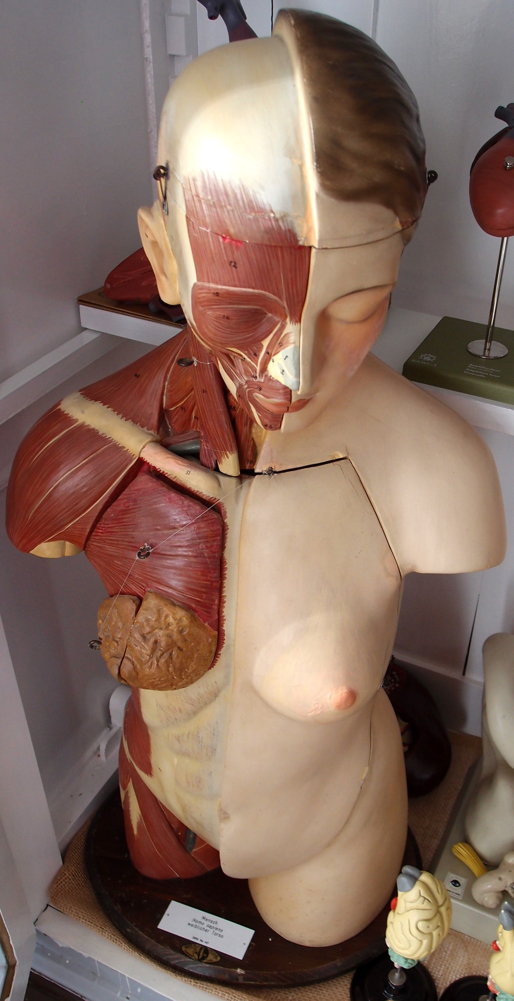
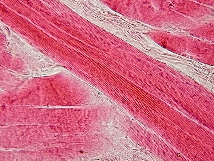
The torso photograph records a real teaching model with exposed superficial layers. Its colors and removable pieces were chosen for learning. The micrograph shows actual skeletal muscle in longitudinal section; repeated transverse striations can be seen within elongated tissue profiles. Staining and section orientation influence appearance, so the pink tones should not be interpreted as natural muscle colors.
Use these images as a scale exercise. Describe the visible regional arrangement on the model, then the tissue pattern in the micrograph. Neither gives the full set of attachments and nerve relationships. A complete understanding requires the whole-muscle map, a regional atlas, and the microscopic organization to be connected.
9. Clinical relevance and a practical study method
For practice assistants and hospital teams, describe an observed task before assigning it to one muscle: reaching, rising, walking, or maintaining a position. A task recruits several structures. Difficulty with that task cannot be explained solely by pointing to the largest muscle on a poster.
For eldercare teaching, connect the anatomical overview with the person’s actual abilities and established care plan. Use clear language about position, movement, and assistance. A diagram can support a conversation, but a generalized overview does not determine an individual exercise prescription or transfer method.
For students and physicians, choose one action and trace the participating muscles, joint, and nerve relationships. Then explain the same action in patient-friendly language. This tests whether the knowledge forms a connected model or remains a collection of names.
A repeatable sequence is to establish the viewpoint, identify the muscle, locate its attachments, describe an action, and compare a deeper or alternate view. End by naming one partner muscle and one stabilizing relationship. This keeps the muscular system grounded in coordinated function, from sarcomere activity to movement in everyday life.
Sources and image credits
Sources checked on 8 October 2026. Links next to claims open the relevant publication. Text is independently written; numerical estimates and teaching classifications are identified separately.
- Terminology
- Muscle Intro
- Muscle Types
- Muscle Structure
- Contraction
- Muscle Actions
- Trunk
- Upper Limb
- Lower Limb
Original schematics are conceptual teaching graphics, not patient images or diagnostic maps. External photographs and clinical images retain the licenses linked in their captions. No clinical author or peer-review credential is claimed.