1. Organs are living structures, not isolated labels
The human body is often introduced as a collection of familiar organs: the heart, brain, lungs, liver, and kidneys. In clinical work, that list becomes useful only when it is connected to location, tissue structure, blood supply, and function. Medical students need a framework for learning details; practice assistants and hospital teams need precise language; physicians and eldercare professionals need explanations that connect observations with the wider person.
An organ is an anatomically distinct structure built from at least two tissue types. Organs cooperate within organ systems, but a single organ may contribute to several systems. This is why asking for one definitive organ count is less useful than stating which structures are included and how they are classified. Introductory anatomy commonly describes eleven organ systems. [organization]
A practical starting question is: “What reaches this organ, what happens inside it, and what leaves?” For the kidney, think about blood and urine. For the lung, think about air and blood. For an endocrine gland, think about the hormone entering the circulation. These questions turn a flat poster into a map of relationships.
2. Locate the organ before describing its job
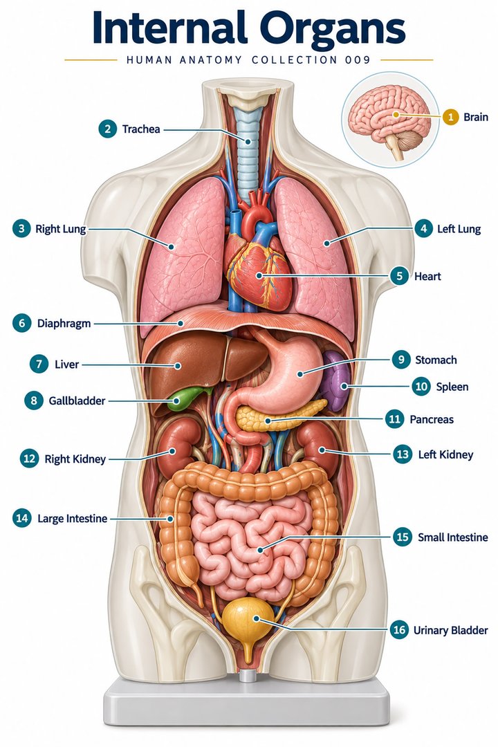
Right and left refer to the patient, not the viewer. An anterior illustration therefore places the patient’s right on the viewer’s left. A diagram also needs depth: a structure may be behind another organ even when both are displayed side by side in a teaching image. Use the diaphragm as a major boundary between the thorax and abdomen, then distinguish the pelvis and posterior abdominal region. [organization]
When teaching placement, describe one neighbor as well as one region. “Heart in the thorax, between the lungs” communicates more than “heart in the chest.” If a patient’s description differs from an atlas position, record their account accurately rather than forcing it to fit the illustration. Anatomy helps organize an assessment; it does not replace it.
3. The heart and lungs: a coordinated exchange network
The heart, or cor, is a muscular pump with four chambers: two atria and two ventricles. Its right side sends blood toward the lungs; its left side supplies the systemic circulation. Valves support one-way flow. Coronary vessels serve the heart muscle itself. When reading a diagram, follow the direction of blood instead of assuming that every red vessel is an artery or every blue vessel a vein. [heart]
The lungs, pulmones, bring air close to blood across thin exchange surfaces. Air travels through branching conducting passages before reaching the respiratory region. Gas exchange transfers oxygen into the blood and carbon dioxide toward exhaled air. The diaphragm helps change thoracic volume during breathing. A useful explanation therefore connects airway, lung, blood, and respiratory muscles rather than treating “breathing” as the work of one organ. [lungs]
At handover, location and function can be kept separate: a diagram shows where a structure lies, while observations describe what the person is experiencing. This distinction is especially useful when discussing breathlessness, fatigue, or activity tolerance without implying a diagnosis from the illustration alone.
4. Liver, pancreas, and kidneys: processing and regulation
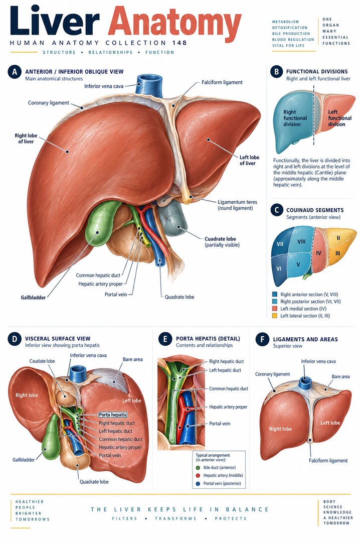
The liver, hepar, produces bile and processes substances arriving in the blood. Bile reaches the intestine through ducts, with the gallbladder providing storage and concentration. The pancreas has an exocrine component that delivers digestive secretions through ducts. Food does not normally pass through either the liver or pancreas; these organs support the alimentary tract from outside its lumen. [accessory]
The endocrine pancreas releases hormones into the bloodstream. Insulin and glucagon contribute to blood glucose regulation through different actions. This offers a memorable example of one organ participating in two systems: digestive secretions use ducts, whereas endocrine signals use blood. The route of a secretion is as important as the name of the organ producing it. [pancreas]
The kidneys, renes, remove wastes and excess fluid while regulating the internal environment. Each contains about one million nephrons, which combine filtration with tubular processing. NIDDK describes adult urine output as roughly one to two US quarts daily, approximately 0.95–1.89 litres by unit conversion. This is an educational approximation, not a patient-specific target; output depends on circumstances. Most filtered water is returned to the circulation rather than excreted. [kidneys]
5. Brain and skin: integration and protection
The entire brain is the encephalon. The cerebrum is only one major component, alongside structures including the cerebellum and brainstem. Surface lobes, internal nuclei, white-matter pathways, and ventricular spaces represent different ways of organizing the same complex organ. The spinal cord, medulla spinalis, connects central processing with peripheral pathways. A colored brain map can introduce organization, but its borders do not imply that complex activities occur within completely independent compartments. [brain]
Skin is also an organ. Its epidermis provides an outer epithelial layer, while the dermis contains connective tissue and structures such as blood vessels and sensory components. The underlying hypodermis is associated with the skin but is not usually counted as one of its two principal layers. This distinction matters when a diagram combines skin and subcutaneous tissue into a single layered view. [skin]
For eldercare teaching, connecting the external surface with underlying tissues helps colleagues explain why the visible appearance is only part of the story. Describe what is seen, where it is seen, and which additional assessment is needed within the person’s care plan.
6. Latin vocabulary and memorable numerical facts
| English | Latin designation | Useful distinction |
|---|---|---|
| Heart | Cor | Four chambers, two linked circulations |
| Brain | Encephalon | The cerebrum is a component, not a synonym for the whole |
| Lung | Pulmo | Plural: pulmones |
| Liver | Hepar | Produces bile |
| Kidney | Ren | Plural: renes |
| Stomach | Gaster | A hollow gastrointestinal organ |
| Urinary bladder | Vesica urinaria | Distinguish from vesica biliaris, the gallbladder |
These selected Latin designations support comparison between English-language resources and international anatomical nomenclature. Some Latin anatomical words have Greek origins. Learn the preferred term alongside common synonyms rather than assuming that every clinical word comes from the same language. [terminology]
Numbers become useful when their meaning is stated. Four heart chambers is a structural count. Eleven systems is a teaching classification. Approximately one million nephrons per kidney is a biological estimate. None should be used as a universal measure of health or as proof that every individual matches a textbook pattern.
7. Real visual material: models and clinical sections
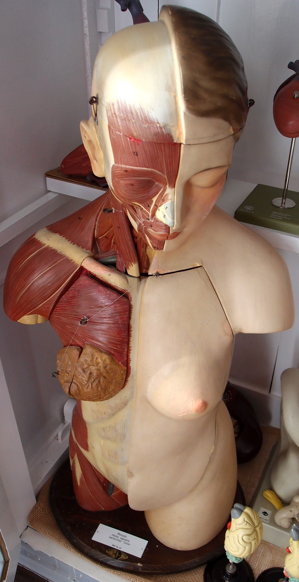
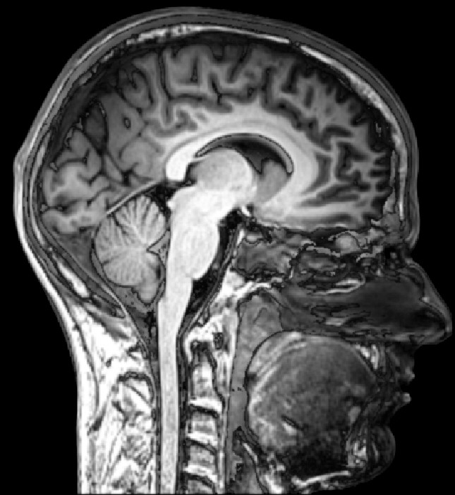
The torso image is a real photograph of a teaching model. Its exposed layers and colors were chosen for instruction. The MRI is a real sectional image of a head; the face points toward the image’s right. Neither is a complete map of every organ. Compare what the representation reveals and what it omits before drawing conclusions.
A useful exercise is to describe the same structure at three levels: its region, its immediate neighbors, and its internal organization. For the brain, begin with the cranial region, then identify broad relationships on the sagittal MRI, then return to a labeled atlas. Treat the image as a bridge between vocabulary and three-dimensional understanding.
8. Apply organ knowledge to communication and learning
For practice assistants, a body chart can help clarify the side and site of a symptom. For hospital teams, a shared diagram can improve the clarity of an explanation. For physicians, it can help distinguish a structural problem from a functional discussion. For students, it can connect a lecture on tissues with the organ they later study in a laboratory.
Try a brief learning sequence: select an organ, write its English and Latin names, locate it, name two neighbors, and trace one input and one output. Then describe its main function in a sentence that a patient could understand. This exposes gaps that memorizing a list may hide.
Finally, revisit the organ within a system. The lung exchanges gases, but useful gas exchange depends on airflow, circulation, and respiratory muscle activity. The kidney produces urine, but its function also connects to fluid balance and endocrine regulation. A strong organ overview remains a guide to relationships: the detail becomes easier to remember when it belongs to a coherent map.
Sources and image credits
Sources checked on 8 October 2026. Links next to claims open the relevant publication. Text is independently written; numerical estimates and teaching classifications are identified separately.
Original schematics are conceptual teaching graphics, not patient images or diagnostic maps. External photographs and clinical images retain the licenses linked in their captions. No clinical author or peer-review credential is claimed.