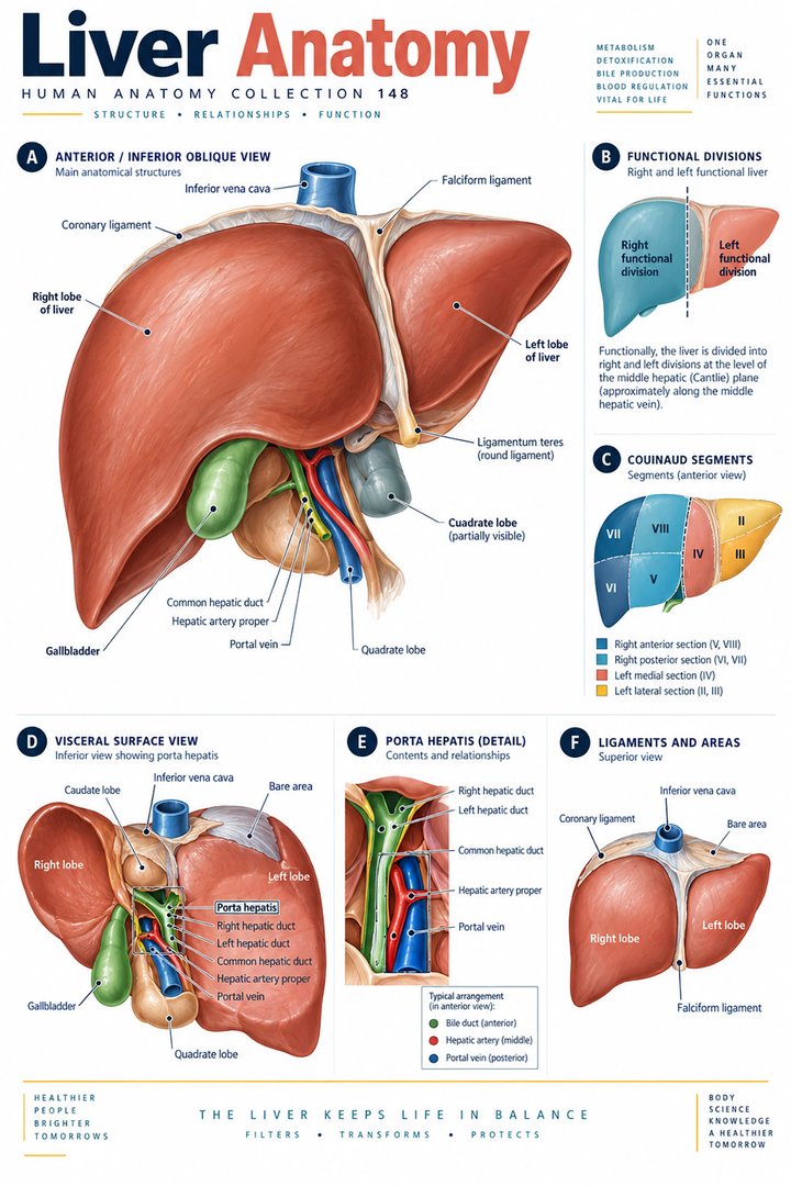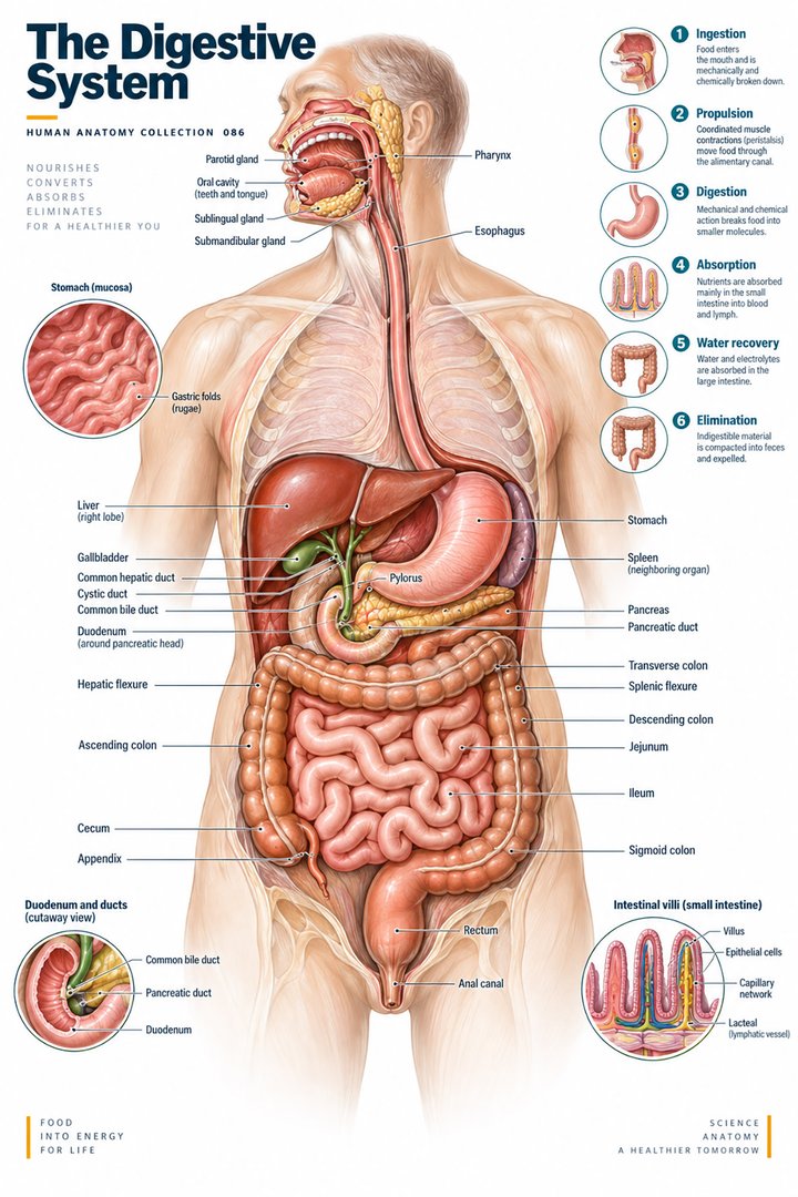
1. Digestion is a coordinated journey
The human digestive system connects the outside environment with the body’s internal nutrition. It takes in food, moves it, breaks it into usable components, absorbs nutrients, and removes remaining waste. For medical students and healthcare teams, the value of learning this system lies in linking the route through the organs with the processes occurring along that route. A diagram can clarify the journey, but it should also show the supporting organs and control networks.
The gastrointestinal tract is a continuous series of hollow organs extending from the mouth toward the anus. The liver, pancreas, and gallbladder support digestion through secretions; they are not chambers in the normal path of swallowed food. The small intestine comprises the duodenum, jejunum, and ileum, while the large intestine includes regions such as the cecum, colon, and rectum. [digestive]
A practical learning question is: “What changes between the entrance and exit of this region?” In the mouth, a meal becomes a prepared bolus. In the stomach, contents are mixed with secretions. In the intestine, digestion and absorption interact. This approach organizes details around a process rather than a disconnected list.
2. Trace the alimentary pathway
Before reading a full-body poster, establish the viewpoint and the patient’s right and left. The large intestine may be drawn as a frame around small-intestinal loops; the simplified layout does not mean that every loop sits in an identical position in every individual. Where the diagram uses arrows, check whether they represent food, secretions, blood, or neural information.
| Structure | Latin designation | Learning cue |
|---|---|---|
| Oral cavity | Cavitas oris | Entry and preparation |
| Esophagus | Oesophagus | Transport |
| Stomach | Gaster | Mixing chamber |
| Small intestine | Intestinum tenue | Includes duodenum, jejunum, ileum |
| Large intestine | Intestinum crassum | Includes colon and rectum |
| Liver | Hepar | Bile production |
| Gallbladder | Vesica biliaris | Bile storage and concentration |
These selected designations connect the English labels with standardized anatomical Latin. Some resources also use accepted synonyms, so compare terms rather than assuming that a different label necessarily identifies a different organ. [terminology]
3. Mouth, pharynx, and esophagus
Chewing provides mechanical processing, while saliva lubricates the contents and begins aspects of chemical digestion. The tongue helps gather food into a bolus and position it for swallowing. Swallowing coordinates several structures so that material enters the esophagus while the airway is protected. The esophagus then propels the bolus toward the stomach through muscular activity. [mouth]
An adult complete permanent dentition is conventionally described as 32 teeth, including third molars. The three pairs of major salivary glands are the parotid, submandibular, and sublingual glands. These are structural counts, not guarantees about an individual’s dentition or oral health. [mouth]
For patient explanations, distinguish preparation from transport. A problem described as “food sticking” needs careful clarification of the experience and location rather than an immediate label from a poster. For learning, compare the muscular route of swallowing with a separate airway diagram so that the shared region of the pharynx becomes understandable.
4. The stomach is a mixing organ with a protective lining
The stomach has regions including the cardia, fundus, body, and pyloric part. Gastric secretions and muscular mixing help process the meal, producing chyme that is delivered into the duodenum. Gastric glands contain specialized cells with different secretory roles. Mucus and other protective mechanisms help defend the lining against the conditions needed for digestion. [stomach]
A stomach illustration often shows a simplified outline, but its appearance changes with filling and contraction. A labeled cavity is therefore a learning convention rather than a fixed size. Also distinguish the lumen—the internal space containing the meal—from the tissues making up the stomach wall.
This distinction helps when discussing endoscopy. An endoscope presents the mucosal surface from inside the lumen. A sectional drawing can show wall layers, while a broad external diagram can show regional relationships. These are complementary views, not competing versions of the anatomy.
5. The intestinal wall: structure for movement and absorption
Much of the gastrointestinal wall follows a layered organization: mucosa, submucosa, muscularis externa, and an outer serosa or adventitia according to location. The mucosa faces the lumen; deeper layers provide support and contractile activity. Neural plexuses are associated with the wall and contribute to local regulation. A layered diagram should be read from the lumen outward before the labels are memorized. [digestive overview]
Small-intestinal folds, villi, and microvilli provide different scales of surface organization. The small intestine is a major site of digestion and nutrient absorption. The large intestine processes the remaining contents, including recovery of water and electrolytes. “Small” and “large” refer primarily to relative diameter, rather than telling you which structure is longer. [intestines]
When comparing a full-body diagram with a microscopic view, follow the scale deliberately: whole tract, organ wall, mucosal projection, and cell surface. The route remains connected even though the appearance changes dramatically.
6. Accessory organs and the control of digestion
The liver produces bile, and the gallbladder stores and concentrates it. Pancreatic exocrine secretions provide enzymes and bicarbonate to the small intestine. Bile supports fat digestion by helping emulsify lipid droplets; it is not itself a digestive enzyme. These secretions meet the intestinal contents through ducts, which should be shown as a different route from the alimentary pathway. [accessory]
Digestive activities include ingestion, propulsion, mechanical digestion, chemical digestion, absorption, and defecation. Neural and hormonal regulation coordinates these activities across regions. Peristalsis helps propel contents, while mixing movements serve a different mechanical purpose. A contraction should therefore not automatically be described as transport in one direction. [digestive processes]
An instructive diagram uses separate arrow styles for different kinds of flow. In a teaching session, ask colleagues to trace the food route in one color and the secretion routes in another. This reveals why a liver label alone does not explain how the organ supports the intestine.
7. Useful figures and facts, with their limitations
OpenStax gives a teaching estimate of approximately one to one-and-a-half litres of saliva produced daily. The actual amount varies with physiological circumstances and stimulation. The estimate is valuable because it highlights how much secretion contributes to routine oral function, but it should not be interpreted as an individual daily target. [mouth]
The small intestine has three named regions, whereas the stomach is commonly described through four main regions. Counts like these provide a revision framework. They are not measures of how much digestion each part performs, and they do not imply that all regions have the same length or function. [intestines] [stomach]
Avoid repeating a single total gut length as though it were an exact universal measurement. Definitions, measurement technique, muscle tone, and individual variation influence reported lengths. For an introductory clinical guide, understanding the route and the level being described is usually more useful than memorizing an unqualified number.
8. Real images: the view inside the stomach
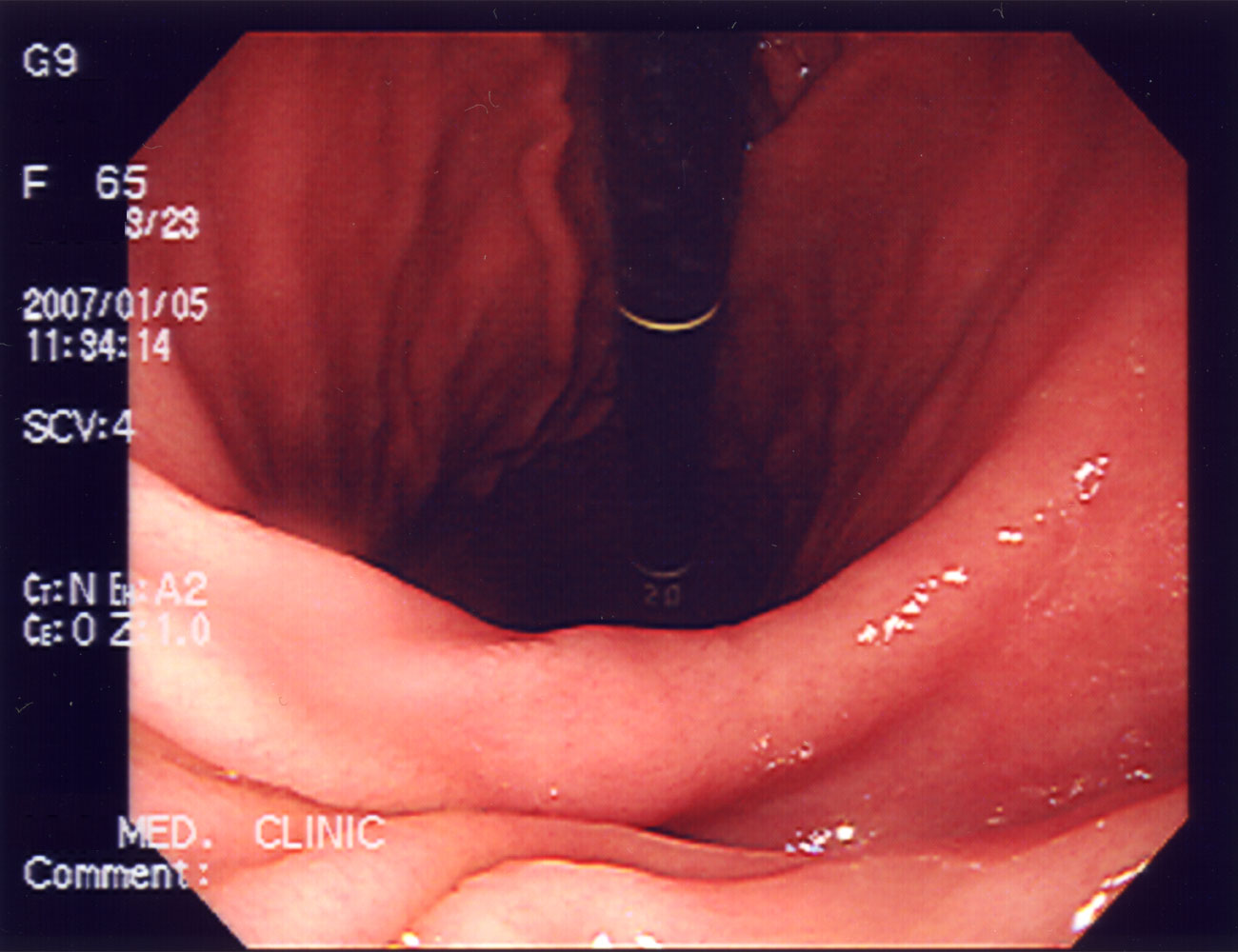
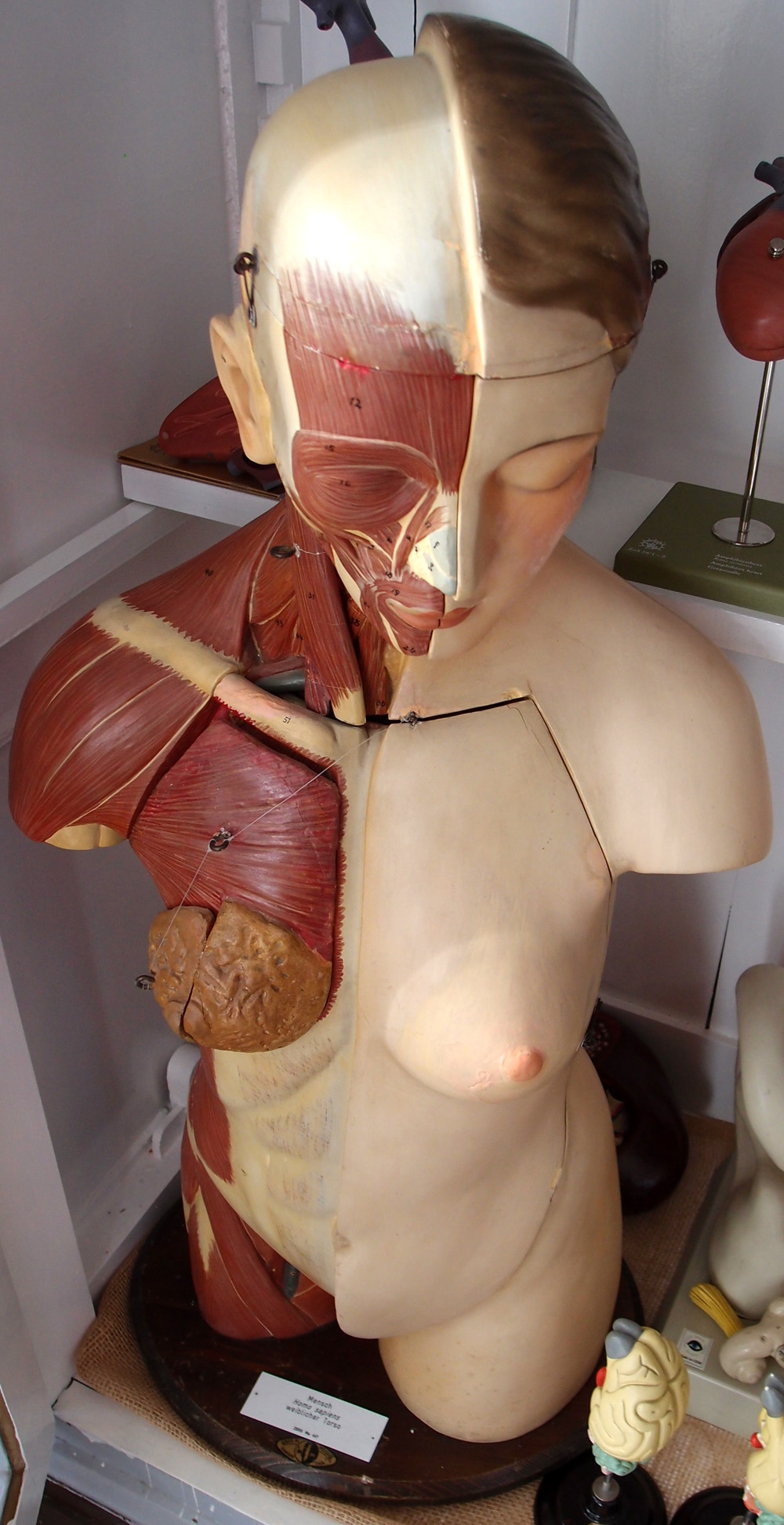
The endoscopic image shows an actual mucosal surface inside the stomach. The source describes it as a normal examination. This article uses it to illustrate viewpoint, without independently diagnosing the subject. The dark instrument and perspective differ from the clean outlines of a textbook diagram. The second image is a real photograph of a physical teaching model, whose exposed layers are artificial.
Ask what is directly visible in each representation. Endoscopy can show the luminal surface, but it does not provide the full external arrangement of organs. A torso model can explain regional layers, but its colors are instructional. Use a caption to identify the representation before interpreting a feature.
9. Connect anatomy with careful clinical communication
Practice assistants can use the anatomical route to clarify a patient’s description. Hospital teams can use a body chart to make the side and region explicit. Physicians can explain how an investigation samples a particular view. Eldercare teams can connect a discussion about eating, swallowing, or elimination with the person’s existing assessment and care plan.
Document the observation separately from its interpretation. “Patient reports discomfort after eating” records an experience; a digestive diagram cannot establish its cause. Similarly, an educational map of the bowel does not determine the origin of a person’s abdominal pain. Use the map to improve the clarity of the conversation.
For revision, trace a meal through the tract, then revisit each stage with three questions: What moves? What is secreted? What is absorbed? Add the Latin term only after the relationship is clear. This creates a durable understanding of the digestive system as a coordinated process that supports nutrition and internal balance.
Sources and image credits
Sources checked on 8 October 2026. Links next to claims open the relevant publication. Text is independently written; numerical estimates and teaching classifications are identified separately.
Original schematics are conceptual teaching graphics, not patient images or diagnostic maps. External photographs and clinical images retain the licenses linked in their captions. No clinical author or peer-review credential is claimed.