1. The skeleton is a living support system
Skeletal system anatomy is about more than naming bones. The skeleton supports the body, protects vulnerable structures, provides attachments for muscles, and participates in mineral storage and blood-cell formation. Understanding these roles makes a bone diagram relevant to movement, clinical communication, imaging, and everyday care. Medical students learn its details; practice assistants and hospital teams use its landmarks; physicians and eldercare professionals connect those landmarks with function. [bone functions]
Begin with relationships. A bone has neighboring bones, articulating surfaces, soft-tissue attachments, and internal architecture. A skeleton displayed without muscles offers a clear framework, but it cannot explain movement alone. At each region, ask which joints permit motion and which tissues help control or limit that motion.
A useful study rule is to pair every name with a practical reference: femur with the thigh, tibia with the medial lower leg, and scapula with the posterior shoulder region. These connections help prevent confusion between a bone, an entire limb segment, and a joint.
2. Axial and appendicular divisions
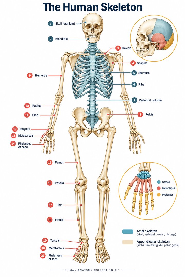
The conventional adult skeleton contains 206 bones. The axial division contains 80, including the skull, vertebral column, and thoracic cage. The appendicular division contains 126, including the limbs and their girdles. Calculated from these teaching counts, approximately 38.8% are axial and 61.2% appendicular. The proportions describe a conventional classification; they are not population prevalence figures. [skeleton]
This division offers a route through a crowded poster. First identify the central framework, then trace the attachment of each limb. Next move from the girdle to the proximal segment, the distal segment, and the hand or foot. A consistent sequence is easier to retrieve than jumping randomly between labels.
3. Bone shapes and anatomical vocabulary
Long bones are longer than they are wide; the term describes shape rather than absolute size. Short bones have more compact proportions. Flat bones provide broad surfaces, while irregular bones have complex forms that do not fit the other groups. Sesamoid bones develop within tendons; the patella is a familiar example. These categories help organize anatomy without implying that every bone is identical within a group. [bone classes]
| Bone or structure | Latin designation | Regional cue |
|---|---|---|
| Skull | Cranium | Head |
| Vertebral column | Columna vertebralis | Central trunk framework |
| Breastbone | Sternum | Anterior thorax |
| Rib | Costa | Thoracic cage |
| Shoulder blade | Scapula | Posterior shoulder |
| Upper arm bone | Humerus | Arm |
| Thigh bone | Femur | Thigh |
| Kneecap | Patella | Anterior knee |
| Shin bone | Tibia | Medial lower leg |
| Heel bone | Calcaneus | Posterior foot |
International anatomical nomenclature uses standardized Latin designations. Learn a regional cue with the name, then add a recognizable feature or relationship. “Scapula: posterior shoulder blade” provides an anchor for later learning about its surfaces, processes, and muscle attachments. [terminology]
4. Inside a bone: compact and trabecular architecture
Compact bone forms a dense structural component. Trabecular, or spongy, bone contains a lattice of supporting trabeculae and intervening spaces. In a typical long bone, the diaphysis is the shaft and the epiphyses are the ends. The medullary cavity lies within the shaft, and the periosteum covers external bone surfaces except where articular cartilage is present. Internal surfaces are associated with the endosteum. [bone structure]
Osteoblasts form bone matrix, osteocytes occupy the mature tissue, and osteoclasts resorb bone. This cellular activity makes the skeleton a changing biological system. A cutaway illustration should therefore be read as a combination of architecture and living tissue rather than a picture of inert material. [bone structure]
When comparing a dry specimen with a cutaway, identify what has been removed or simplified. A specimen may show external landmarks clearly while concealing marrow spaces and cellular detail. The image and specimen answer different questions.
5. The vertebral column: regions, counts, and curves
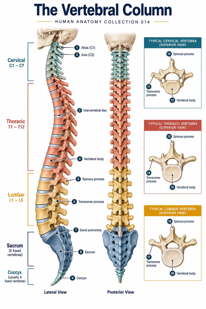
The mobile vertebral sequence comprises seven cervical, twelve thoracic, and five lumbar vertebrae. In conventional adult counting, the sacrum and coccyx bring the vertebral column to 26 bones. Developmental vertebral segments are a different count because fusion changes how they are represented. The spinal cord is neural tissue; the vertebral column is its surrounding bony framework. They are closely related but not interchangeable terms. [spine]
The column normally has regional curves rather than a straight profile. Intervertebral discs and supporting ligaments contribute to its mechanical organization. A lateral diagram is useful for curves; an anterior view is useful for regional alignment. Learning both viewpoints prevents a single flat image from becoming the whole mental model. [spine]
6. Joints connect structure with movement
Joints can be classified structurally as fibrous, cartilaginous, or synovial. Functional classifications describe how much movement they permit. These are different questions, so a learner should not substitute one vocabulary for the other. A skull suture and a freely moving limb joint demonstrate why the skeleton needs more than one type of connection. [joints]
A synovial joint has a cavity, an articular capsule, synovial components, and articular cartilage covering opposing surfaces. Ligaments and surrounding tissues contribute to stability. Synovial shapes include plane, hinge, pivot, condyloid, saddle, and ball-and-socket forms. Their geometry helps explain potential movement, but actual movement also depends on surrounding restraints and muscle control. [synovial]
During revision, trace the surfaces that meet, name the joint, and describe a permitted movement. Avoid treating every articulation in a region as one joint. The shoulder complex, for example, needs a broader regional explanation than a single label on the humeral head.
7. Growth, adaptation, and clinical context
Bone develops through intramembranous or endochondral ossification. Longitudinal growth involves the epiphyseal growth plate before closure. Bone also changes in response to remodeling throughout life. A child’s skeleton should therefore not be described simply as a smaller adult skeleton: growth regions and fusion stages affect its organization. [bone growth]
For clinical communication, describe the location without assuming a cause. “Pain over the lateral right ankle” gives a region; it does not establish that a particular bone is fractured. An atlas helps locate structures, while the person’s history, examination, and appropriate investigations establish the finding.
For eldercare teams, use anatomical knowledge to make location descriptions clear during mobility and skin discussions. Connect a surface landmark with the underlying structure and the person’s existing care plan. Practical knowledge includes recognizing when a diagram’s idealized alignment cannot represent the individual’s posture, surgical history, or mobility.
8. Real skeletal images: specimen and radiograph
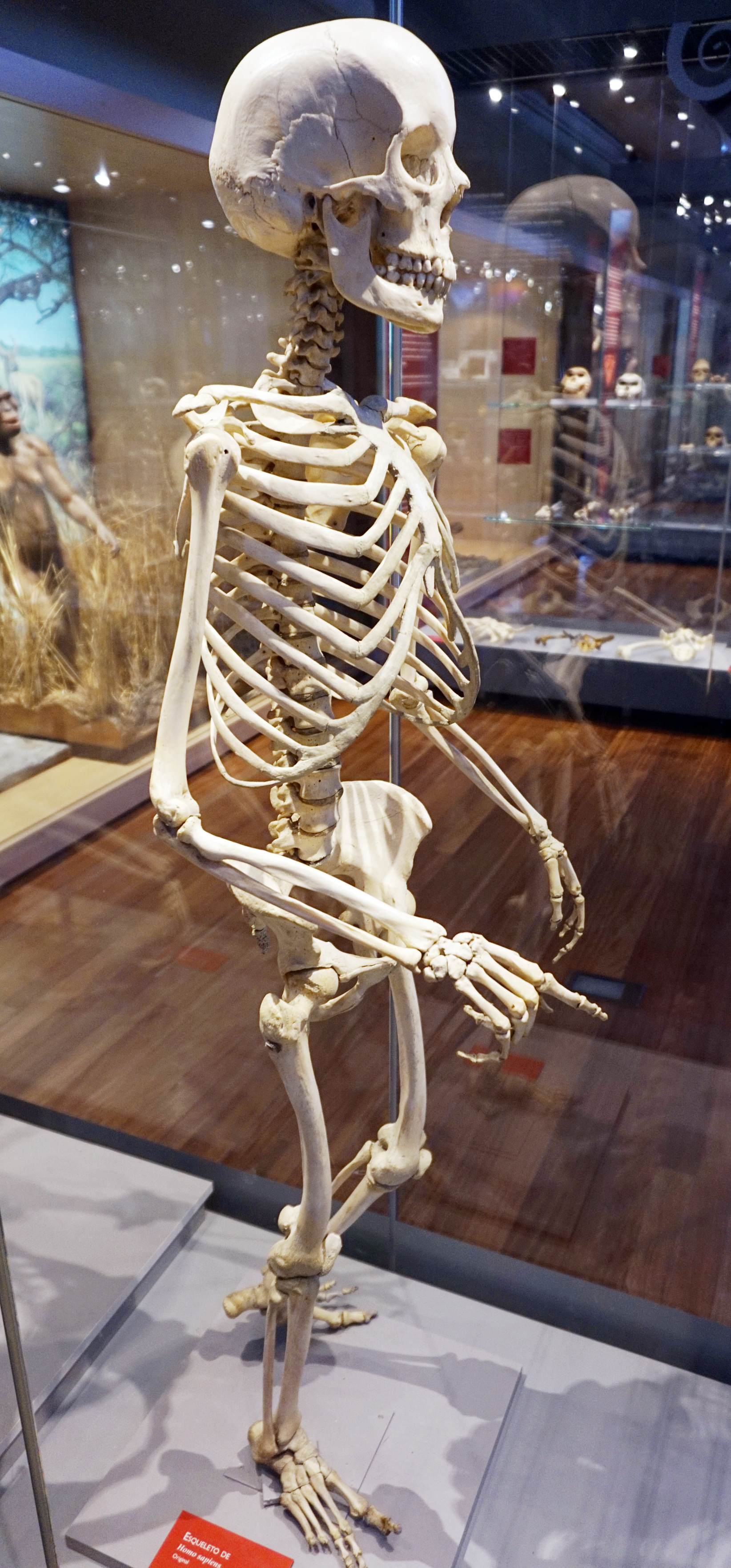
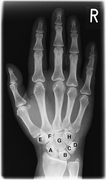
The museum photograph shows a displayed human skeleton, whereas the hand image is a projection radiograph. The R marker identifies the right hand. On the radiograph, several structures overlap because the image projects depth onto one plane. The visible letters label the carpal region in the source image; they should not be interpreted without its legend.
Use these images to compare representation rather than to make a diagnosis. Find a broad landmark on the specimen, then describe what a radiograph reveals or hides. A sharp edge in a photograph and a bright outline in an X-ray do not encode the same information.
9. Build a reliable skeletal study routine
Choose one region at a time. Start with the English and Latin names, establish right and left, locate the neighboring joint, and identify one landmark. Then cover the labels and explain the route through the region aloud. A second viewpoint tests whether you understand the arrangement or have memorized only the first picture.
For team teaching, ask one colleague to describe a landmark while another points to it on a chart. Discuss disagreements about depth or laterality. This exercise can reveal ambiguous words before they are used in a record or handover.
The aim is a coherent framework: bones support and protect, joints connect, and muscles act through these relationships. Counts and Latin names are useful anchors, but the strongest understanding is the ability to describe where a structure is and how it participates in the living body.
Sources and image credits
Sources checked on 8 October 2026. Links next to claims open the relevant publication. Text is independently written; numerical estimates and teaching classifications are identified separately.
Original schematics are conceptual teaching graphics, not patient images or diagnostic maps. External photographs and clinical images retain the licenses linked in their captions. No clinical author or peer-review credential is claimed.