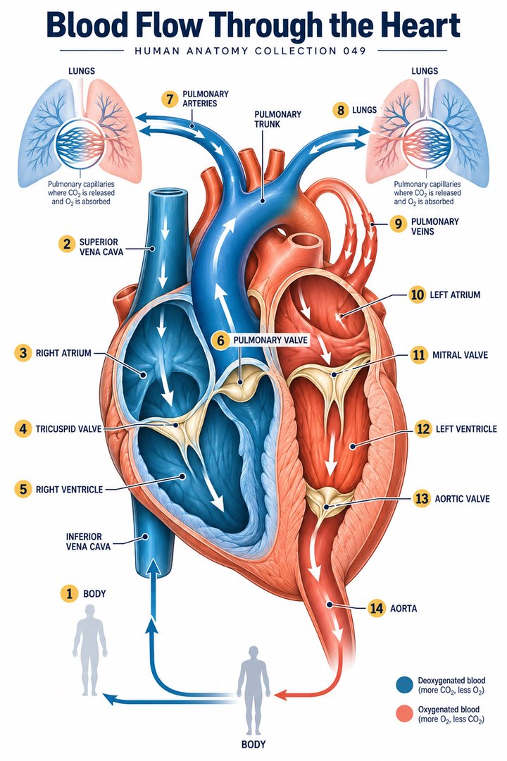
Blood Flow Through the Heart
Individual poster · 300 DPI print files
Regular $9.90
Explore poster ↗$4.90USD
Offer price · Save $5.00Individual poster · 300 DPI print files
Reduced web preview. Separate high-resolution print files are supplied with the digital download.
ANATOMY 049
A modern anatomy illustration exploring blood flow through the heart, designed for educational and decorative use.
Purchases are not available yet. Checkout and download delivery are being prepared.
Sold by Kunstolymp.de · Germany
Compare prices & the complete collection ↗This is a DIGITAL DOWNLOAD. No physical product will be shipped. After purchase, download your files and print them at home, at a local print shop, or through an online professional printing service.
The separate 300 DPI print files are ideal for 24×36-inch posters (approximately 60×90 cm). ISO and 5×7 formats are included. The white border lets you adjust the layout or trim to fit other frame sizes; check the preview before printing to keep the illustration and labels visible.
The planned bundle delivery includes a PDF information sheet with a link to the relevant Google Drive folder and download instructions. The store is currently in preview and purchases are not yet available.
A science-inspired choice for a student’s study corner, an educator’s classroom, or an anatomy enthusiast’s wall. Final licensing terms will apply.
CLINICAL CONTEXT
Blood flow through the heart is a practical framework for understanding symptoms, examination, investigations, and treatment. Deoxygenated blood travels from the venae cavae to the right atrium, through the tricuspid valve to the right ventricle, and via the pulmonary valve and pulmonary arteries to the lungs. Oxygenated blood returns through pulmonary veins to the left atrium, crosses the mitral valve into the left ventricle, and leaves through the aortic valve to the aorta. Pressure gradients, valve competence, ventricular relaxation and contraction, pulmonary vascular resistance, and circulating volume all influence this route. A disruption at one point may create congestion upstream and reduced flow downstream.
Patients do not describe pressure-volume loops; they describe shortness of breath, ankle swelling, exercise intolerance, chest pain, palpitations, dizziness, fatigue, cough, or reduced ability to work and care for others. Left-sided pump dysfunction may lead to pulmonary congestion and breathlessness; right-sided dysfunction may contribute to peripheral oedema, abdominal fullness, or raised jugular venous pressure. Valve stenosis or regurgitation can alter flow and impose chronic chamber load. Pulmonary embolism, severe anaemia, sepsis, arrhythmia, pericardial disease, and acute coronary syndromes can also compromise effective circulation. A focused history should establish timing, triggers, positional symptoms, fluid change, bleeding, infection, thromboembolic risk, medicines, and baseline functional capacity.
Assessment begins with airway, breathing, circulation, observations, appearance, pulse, blood pressure, oxygenation, perfusion, lung examination, oedema, and focused cardiac findings. ECG can identify rhythm and ischaemic clues; chest radiography, echocardiography, biomarkers, natriuretic peptides, blood tests, and imaging of coronary or pulmonary vessels may answer specific questions. Treatment targets the cause and physiology: oxygen or ventilatory support when clinically indicated, fluid management, diuretics, anticoagulation, antimicrobials, rate or rhythm treatment, disease-modifying heart-failure medicines, valve intervention, coronary revascularisation, or management of anaemia and endocrine disease. Students should distinguish the flow diagram from a complete diagnostic pathway: the same symptom can arise from different lesions and several problems may coexist.
Cardiac rehabilitation, appropriately prescribed activity, tobacco cessation, nutrition support, medication adherence, and shared symptom monitoring can improve recovery and long-term risk. Breathing exercises, relaxation, and psychological support may help distress but must not be used to explain away possible cardiorespiratory disease. Supplements and herbs require interaction checks. New severe breathlessness, chest pressure, cyanosis, collapse, fainting, confusion, coughing blood, or rapidly worsening oedema requires urgent local assessment. This guide is educational; follow current local emergency and specialty protocols.
Further reading: MedlinePlus: understanding cardiovascular disease · NHS: coronary heart disease
Explain the working diagnosis, important alternatives, planned tests, expected course, and specific safety-netting in language the patient can use. Invite questions about treatment goals, capacity, cost, culture, and complementary therapies. Use a professional interpreter when language access could affect consent or understanding, and confirm clear arrangements for timely clinical review in writing. Document the reasoning, reassess response to treatment, and coordinate care across professions when illness affects more than one system. Teaching anatomy is most useful when it supports this partnership: accurate localisation, respectful listening, timely escalation, and a plan that remains responsive to the individual patient.
HUMAN ANATOMY COLLECTION · 001—300
A closer look at what makes us human. Discover anatomy posters for your study space, classroom, and everyday curiosity.
DIGITAL DOWNLOADS · 300 DPI PRINT FILES
EXPLORE BY SYSTEM
LEARN & EXPLORE
Detailed English guides with Latin terminology, diagrams, real images, and clinical learning connections.
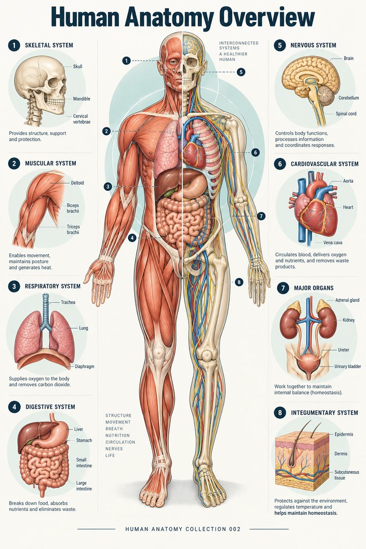 Human Anatomy Diagrams ↗
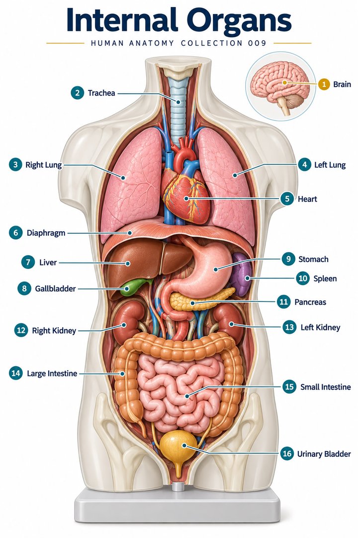 Human Body Organs ↗
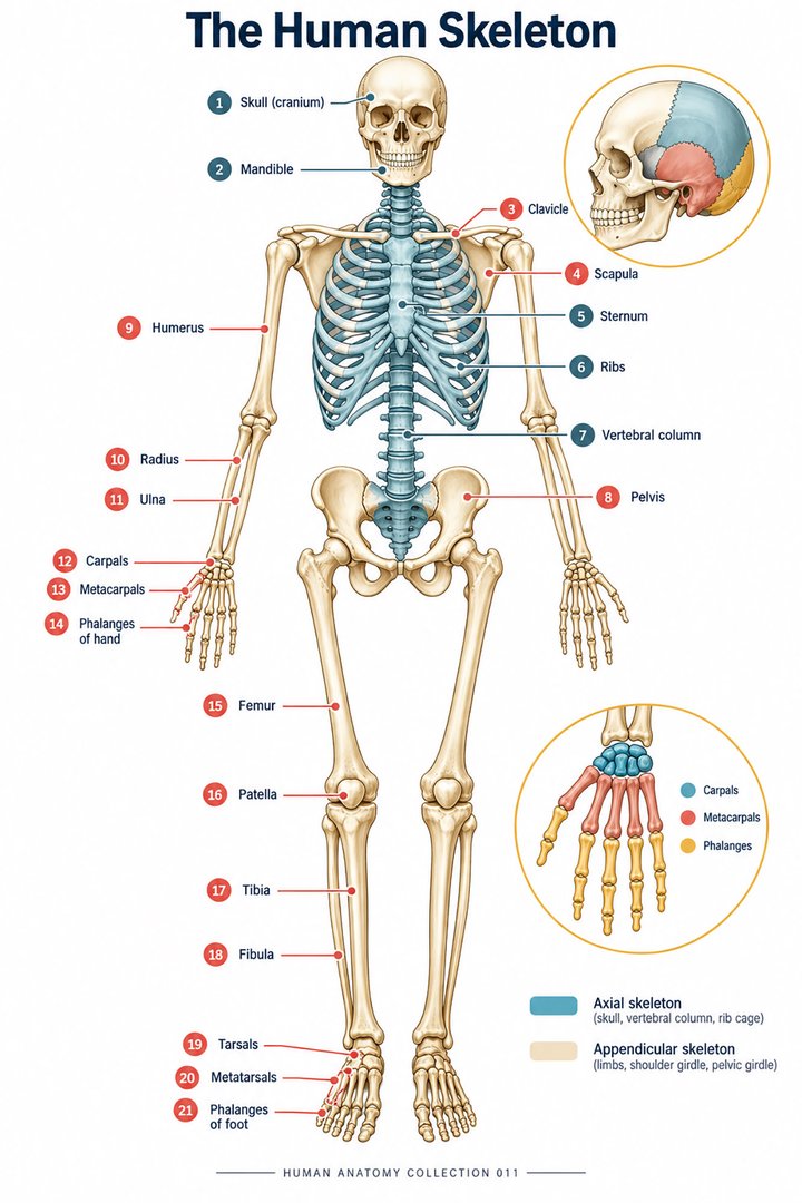 Skeletal System Anatomy ↗
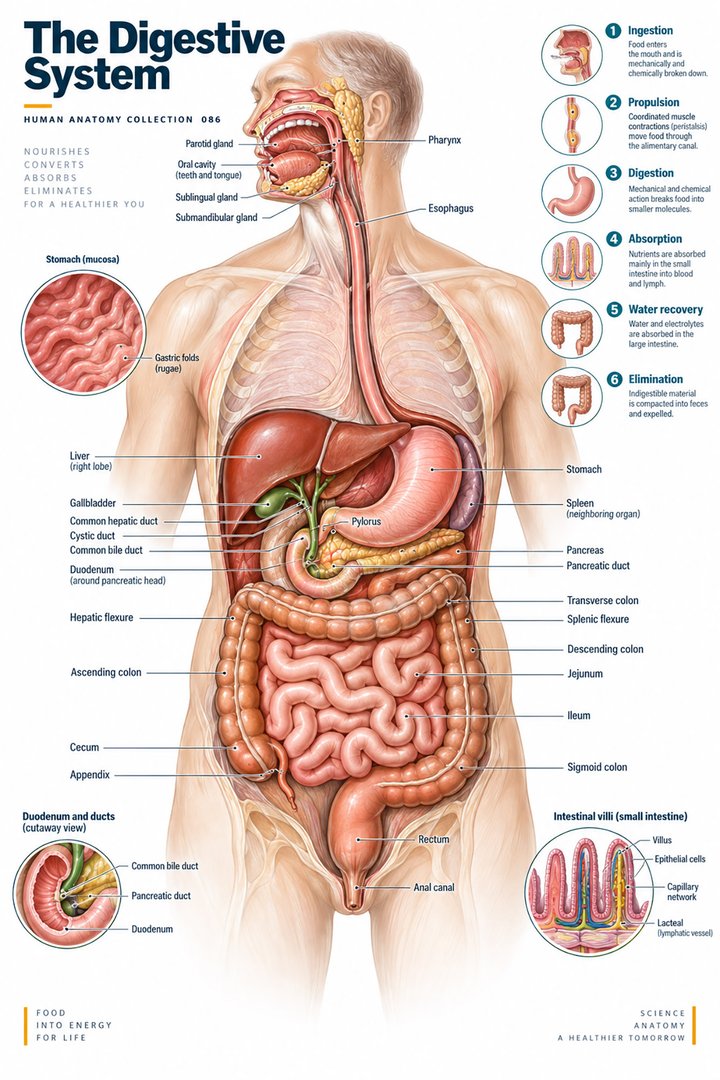 Human Digestive System ↗
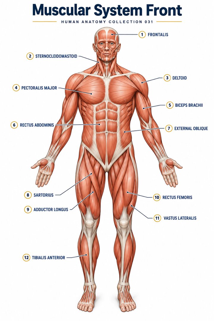 Muscular System Overview ↗
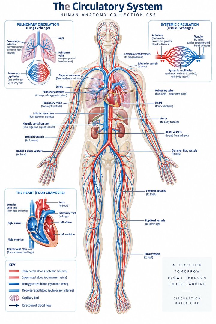 Human Circulatory System ↗
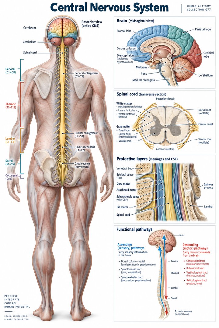 Human Nervous System ↗
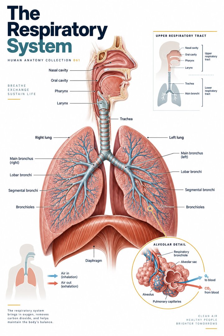 Respiratory System Functions ↗
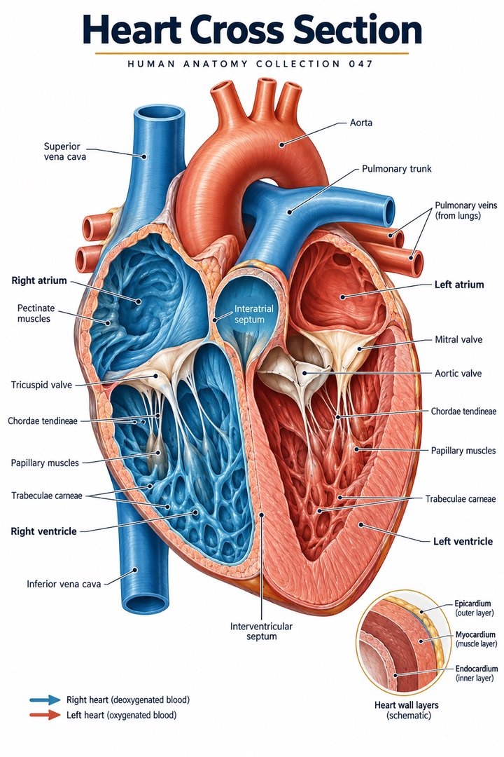 Human Heart Structure ↗
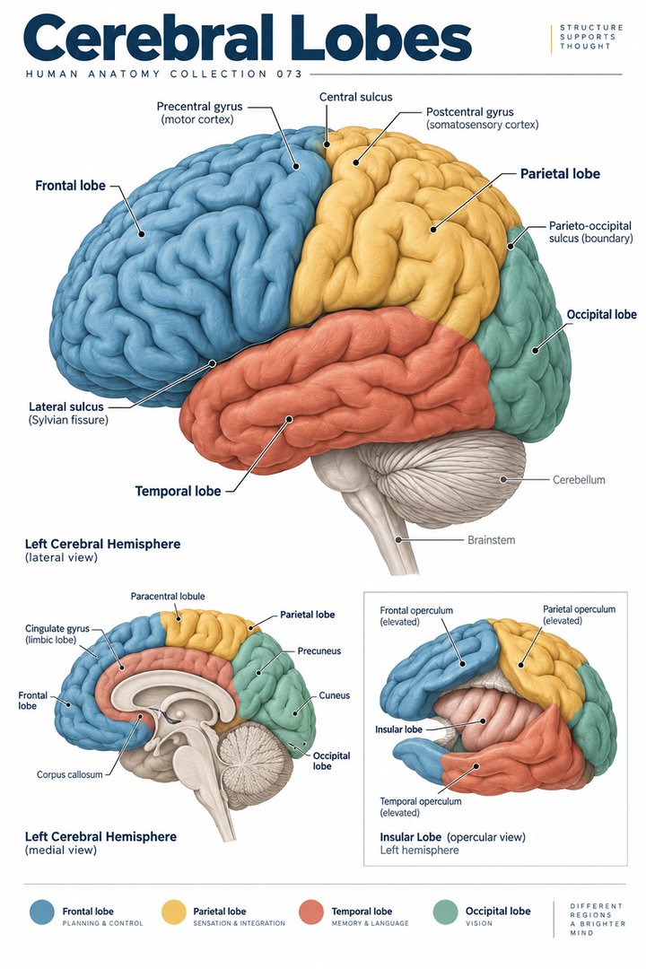 Human Brain Lobes ↗
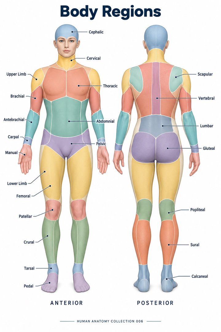 Human Body Parts ↗
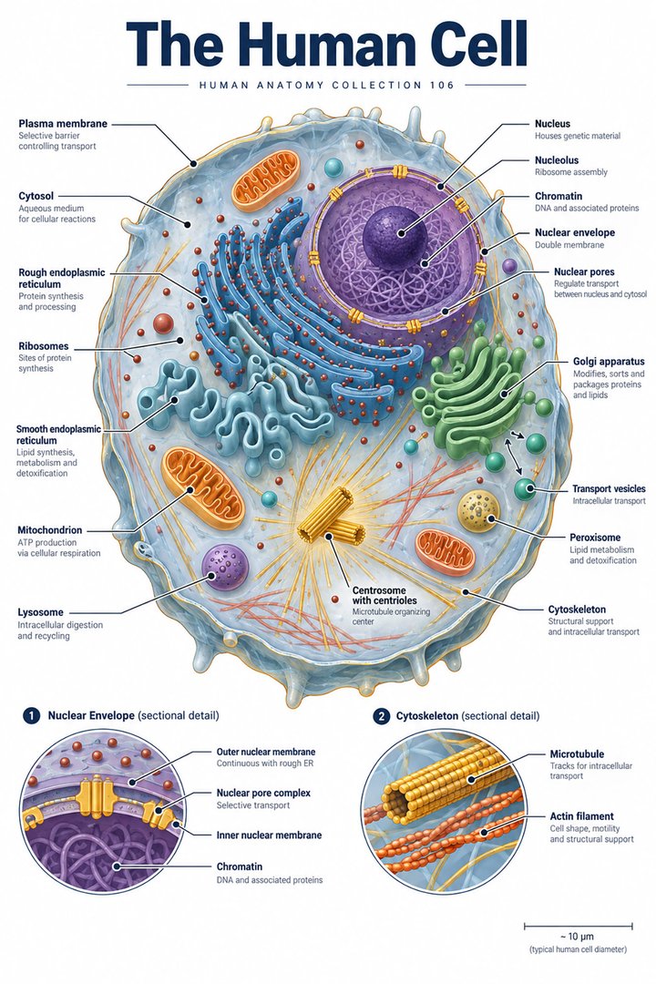 Human Cell Structure ↗
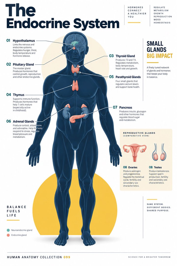 Human Endocrine System ↗
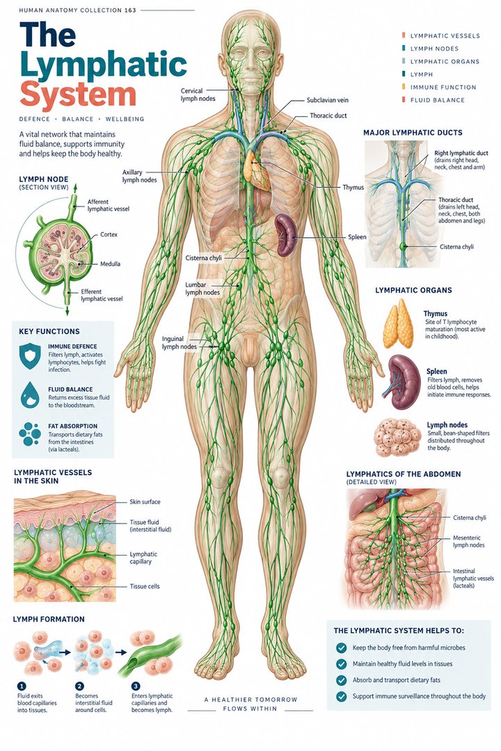 Human Lymphatic System ↗
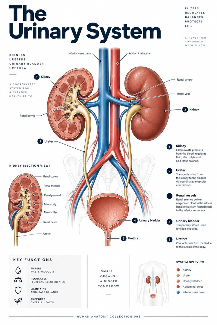 Human Urinary System ↗
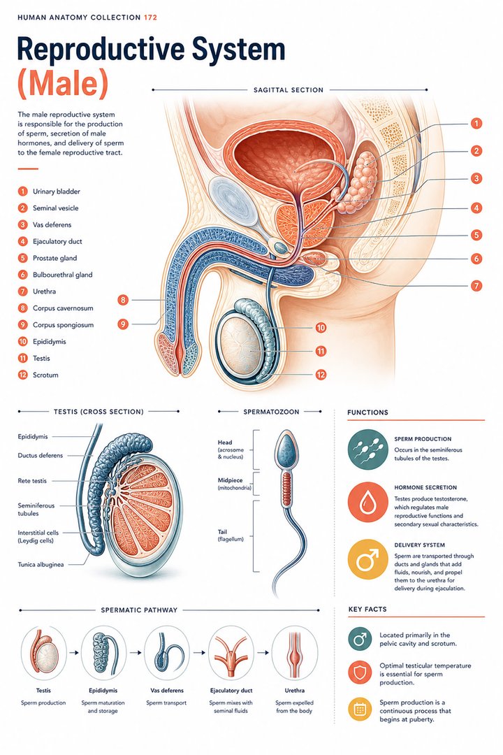 Male Reproductive System ↗
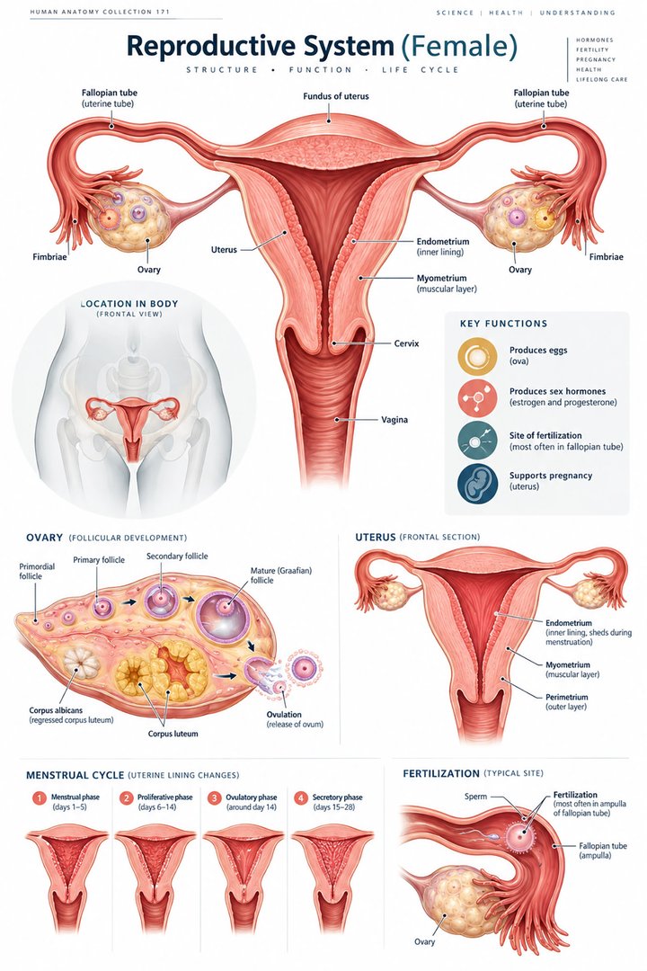 Female Reproductive System ↗
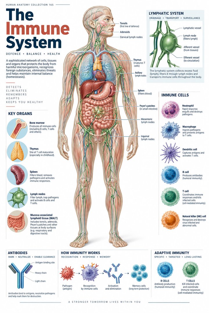 Human Immune System ↗Central Nervous System ↗
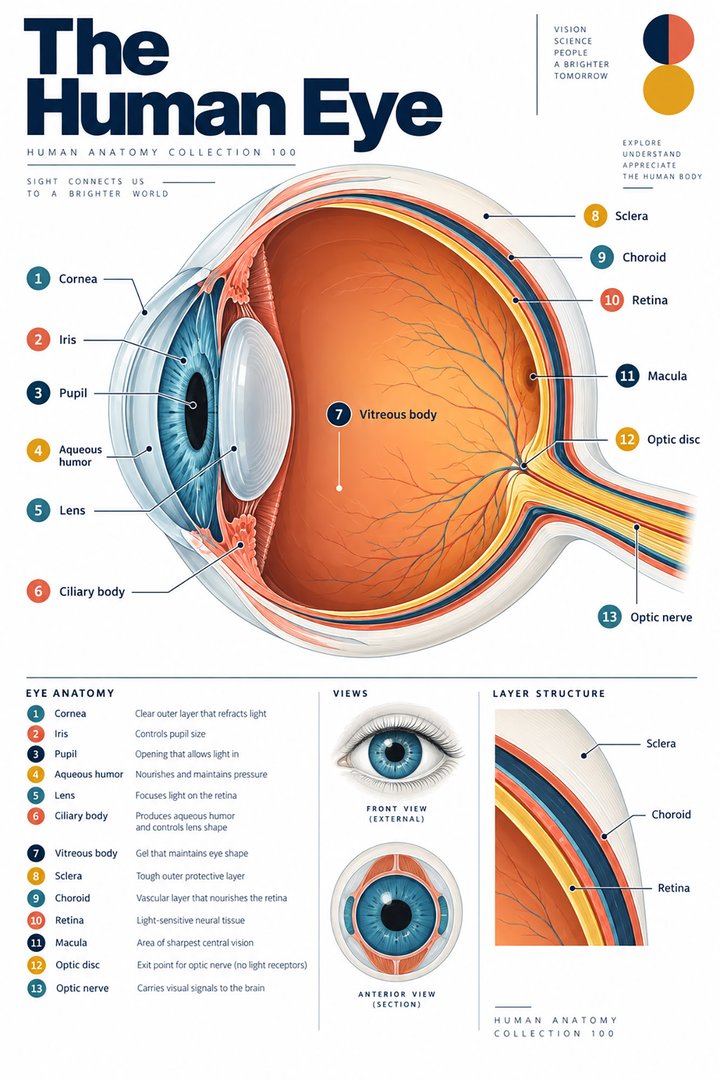 Human Eye Structure ↗
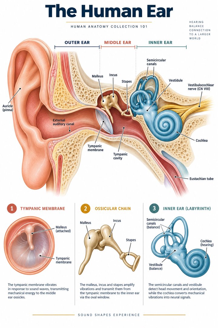 Human Ear Structure ↗
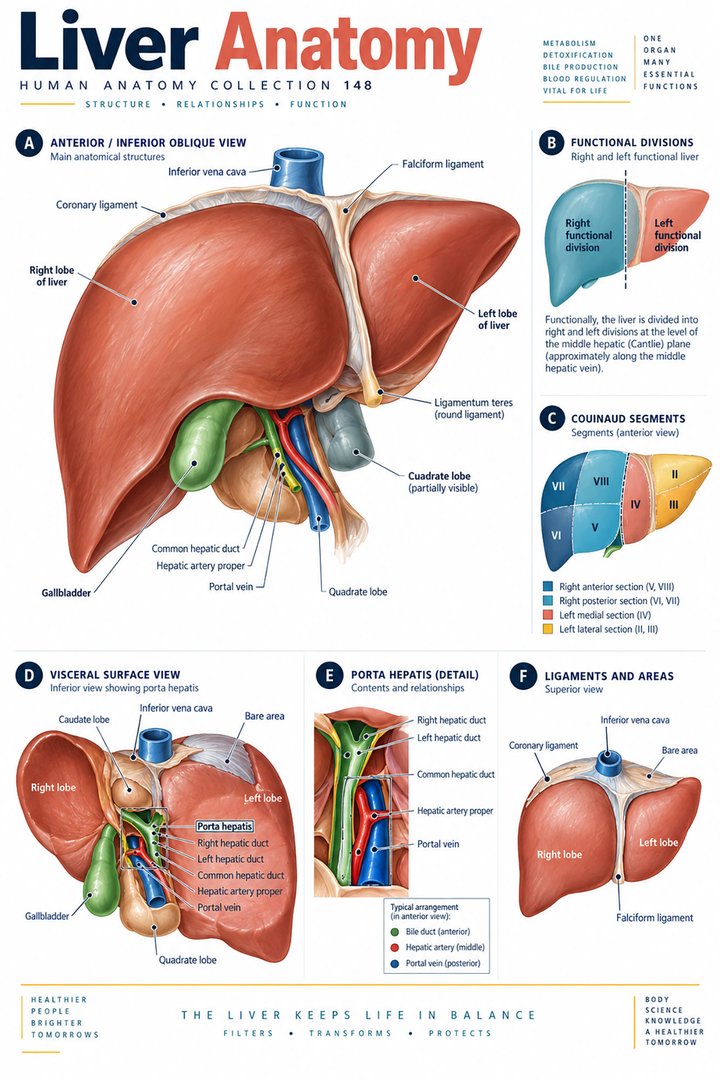 Human Liver Function ↗
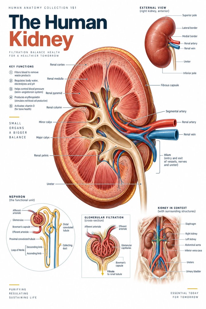 Human Kidney Anatomy ↗
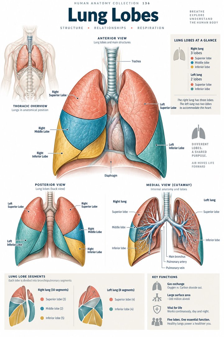 Human Lung Anatomy ↗
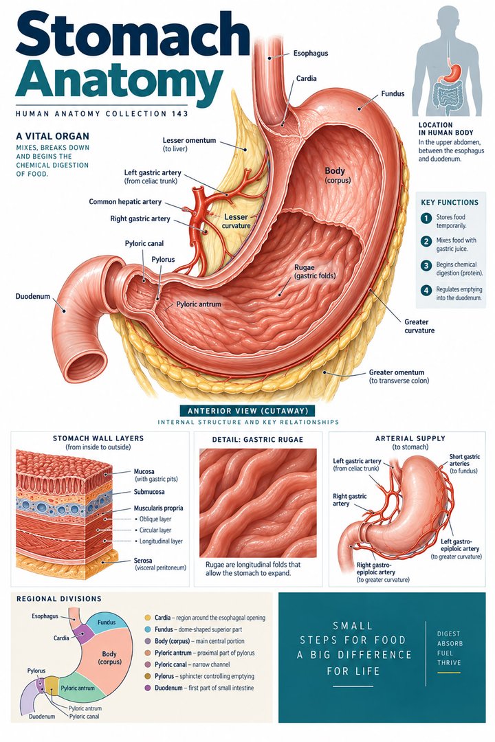 Human Stomach Structure ↗
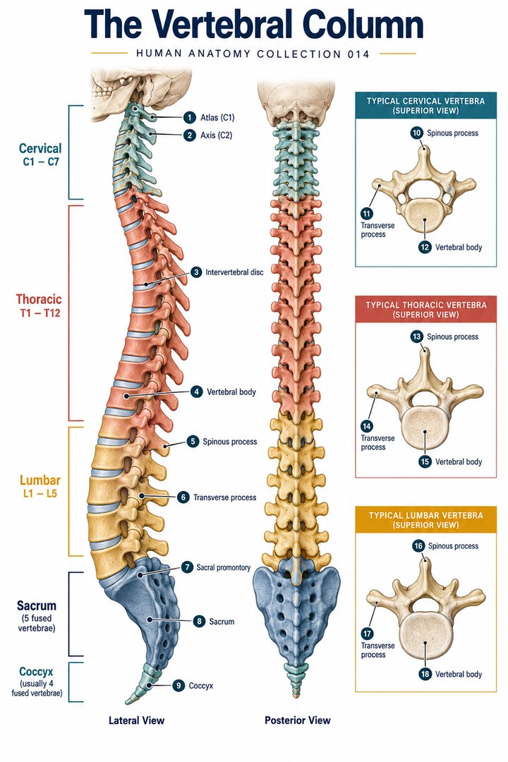 Human Spine Anatomy ↗
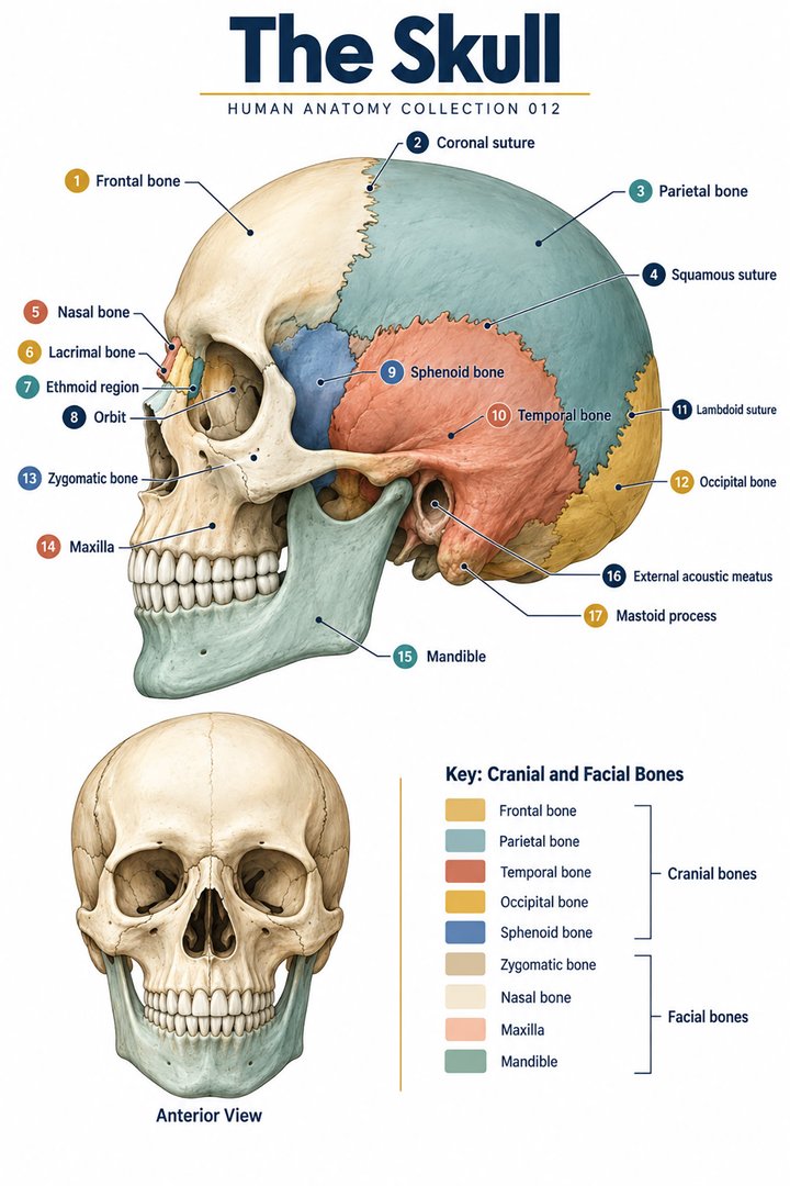 Human Skull Anatomy ↗
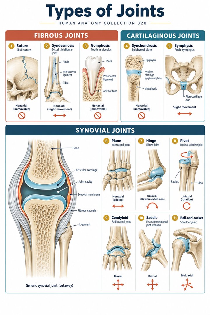 Human Joint Types ↗
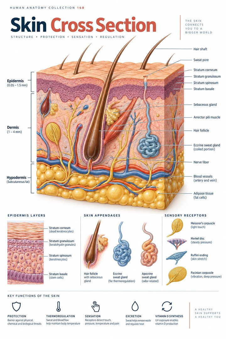 Human Skin Layers ↗
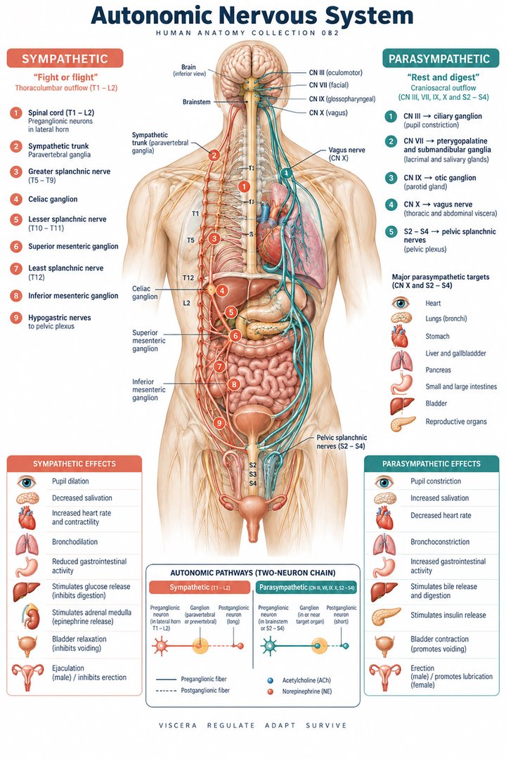 Autonomic Nervous System ↗
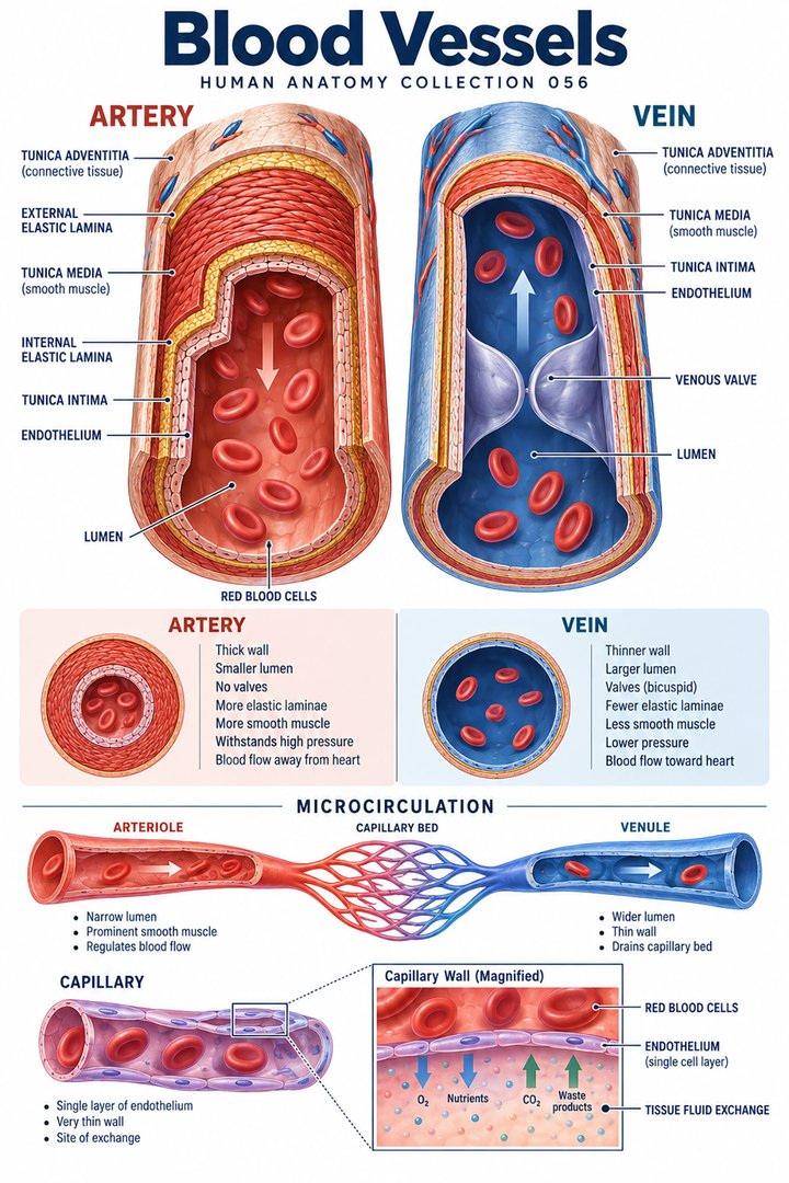 Human Blood Vessels ↗
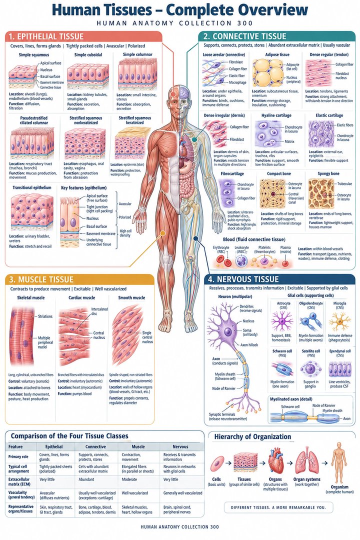 Human Body Tissues ↗
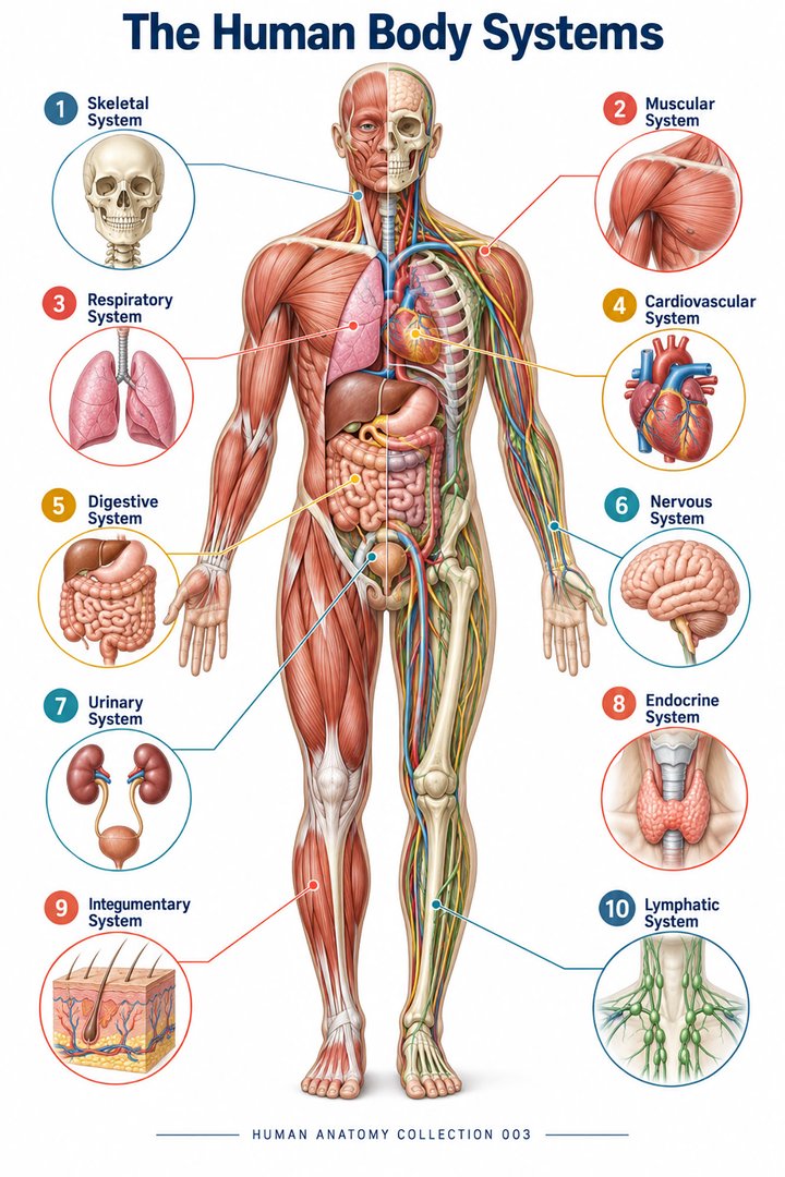 Human Body Systems ↗
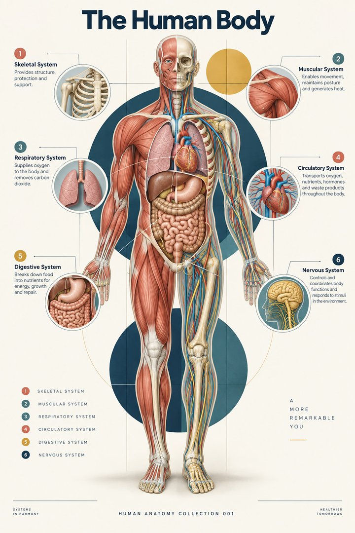 Human Body Facts ↗Human Anatomy and Physiology ↗
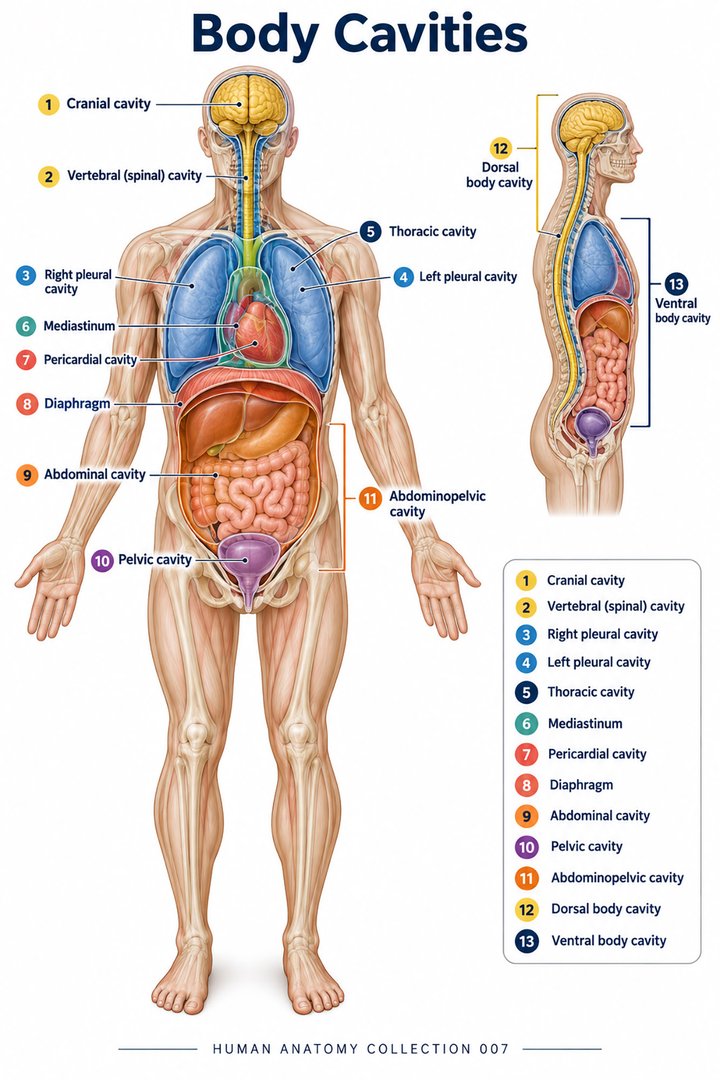 Human Body Cavities ↗Human Torso Anatomy ↗Human Digestive Tract ↗
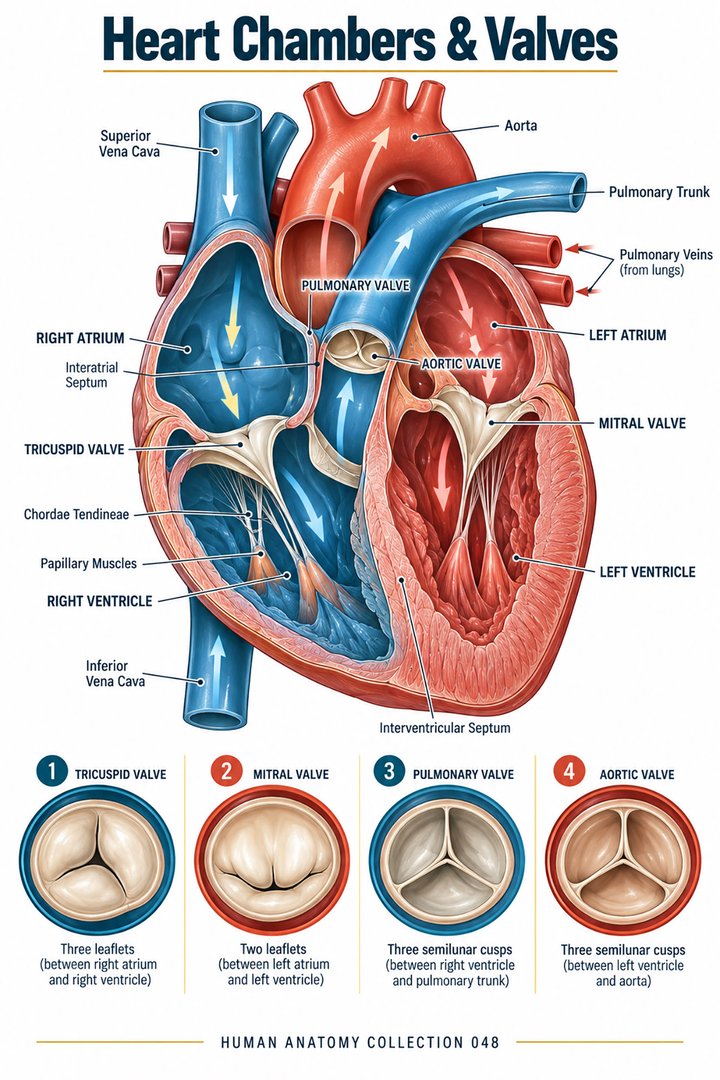 Human Heart Chambers ↗Human Organ Placement ↗
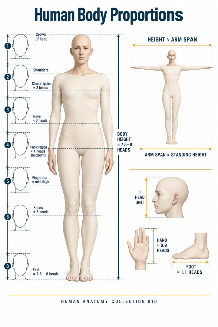 Human Body Proportions ↗
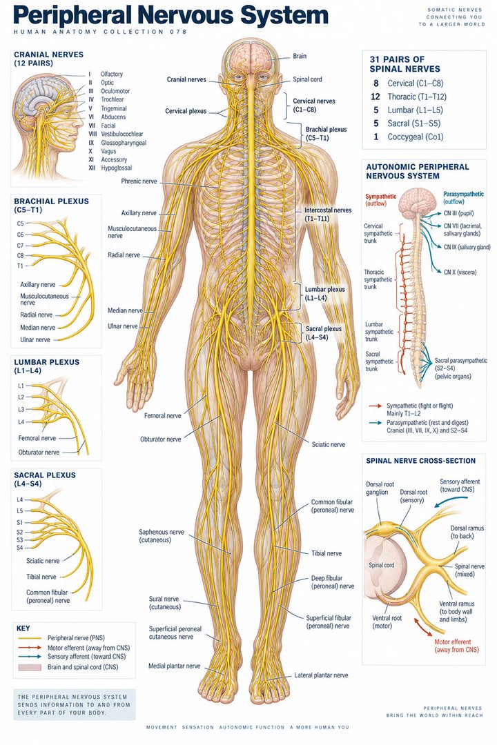 Peripheral Nervous System ↗
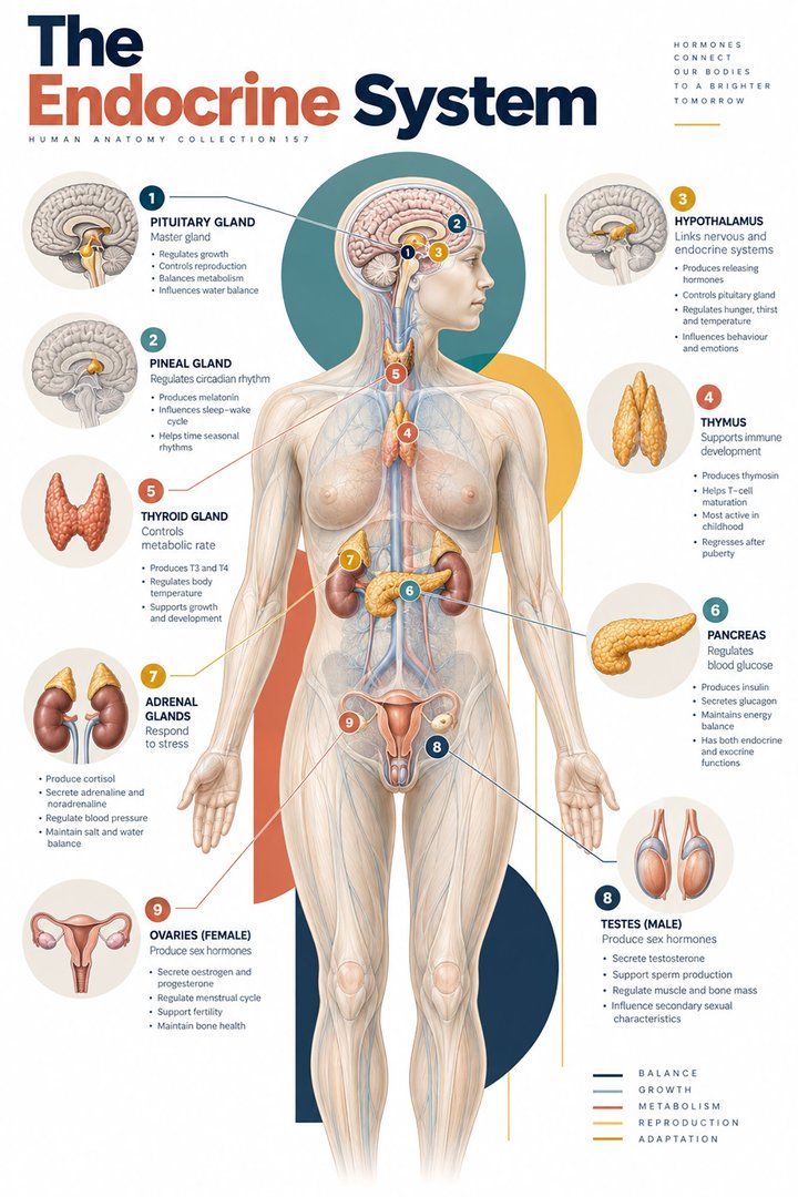 Human Endocrine Glands ↗
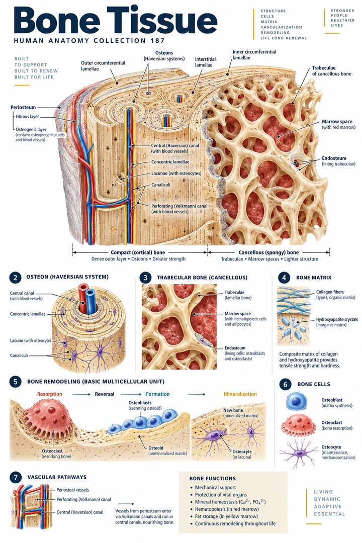 Human Bone Density ↗Human Blood Circulation ↗
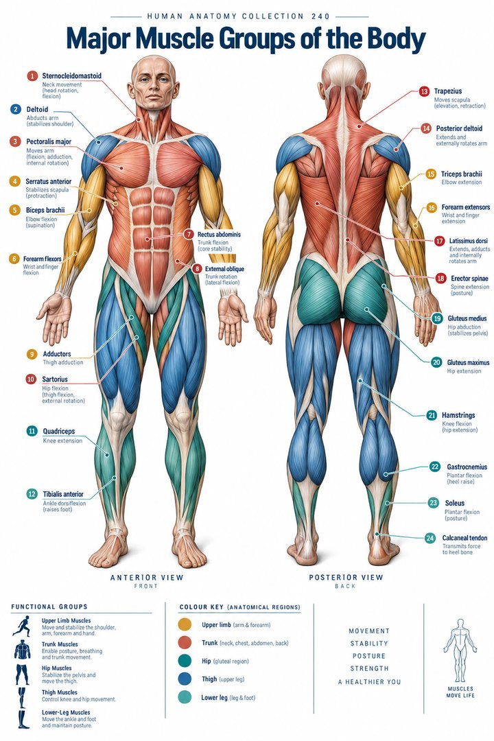 Human Muscle Groups ↗
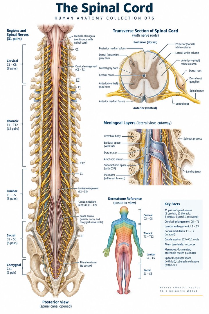 Human Spinal Cord ↗
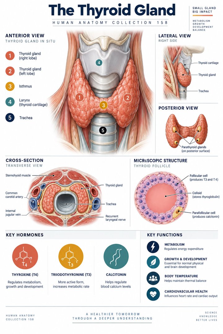 Human Thyroid Gland ↗
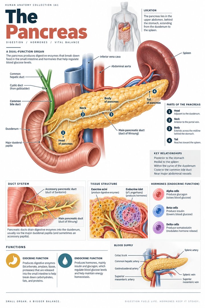 Human Pancreas Function ↗
 Human Gallbladder Anatomy ↗