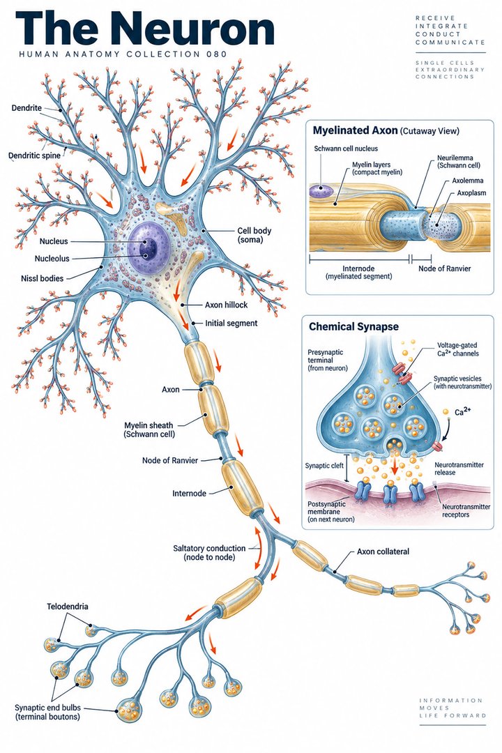
The Neuron
Individual poster · 300 DPI print files
Regular $9.90
Explore poster ↗$4.90USD
Offer price · Save $5.00Individual poster · 300 DPI print files
Reduced web preview. Separate high-resolution print files are supplied with the digital download.
ANATOMY 080
A modern anatomy illustration exploring the neuron, designed for educational and decorative use.
Purchases are not available yet. Checkout and download delivery are being prepared.
Sold by Kunstolymp.de · Germany
Compare prices & the complete collection ↗This is a DIGITAL DOWNLOAD. No physical product will be shipped. After purchase, download your files and print them at home, at a local print shop, or through an online professional printing service.
The separate 300 DPI print files are ideal for 24×36-inch posters (approximately 60×90 cm). ISO and 5×7 formats are included. The white border lets you adjust the layout or trim to fit other frame sizes; check the preview before printing to keep the illustration and labels visible.
The planned bundle delivery includes a PDF information sheet with a link to the relevant Google Drive folder and download instructions. The store is currently in preview and purchases are not yet available.
A science-inspired choice for a student’s study corner, an educator’s classroom, or an anatomy enthusiast’s wall. Final licensing terms will apply.
CLINICAL CONTEXT
A neuron is an excitable cell specialised to receive, integrate, and transmit information. Dendrites and the cell body receive synaptic input; the axon initial segment helps initiate action potentials; the axon conducts signals, often faster when insulated by myelin; and presynaptic terminals release neurotransmitters across synapses. Ion gradients, membrane channels, transport proteins, glia, and vascular support all contribute to normal function. This cellular map connects symptoms to mechanism: altered conduction can produce weakness or numbness, abnormal firing can contribute to seizures or neuropathic pain, and selective neuronal injury may impair movement, memory, language, sensation, or autonomic control.
Peripheral neuropathy can cause burning pain, pins and needles, reduced sensation, weakness, or balance problems. Diabetes, alcohol exposure, nutritional deficiency, medicines, autoimmune disease, infection, compression, and inherited conditions are among possible causes. Multiple sclerosis involves immune-mediated central nervous system injury and can produce episodes of visual disturbance, sensory change, weakness, or impaired coordination. Epilepsy reflects a tendency to recurrent unprovoked seizures, while stroke, trauma, infection, tumour, and neurodegenerative disease can injure neurons through different mechanisms. Patients may describe fatigue, falls, altered dexterity, pain, cognitive change, or loss of independence rather than a clear anatomical deficit. History should establish onset, progression, distribution, associated symptoms, exposures, medicines, family history, and effect on daily life.
Neurological examination tests mental status, cranial nerves, motor power, tone, reflexes, sensation, coordination, gait, and relevant autonomic function. Localisation guides differential diagnosis and focused testing. Depending on the presentation, clinicians may use blood tests, nerve-conduction studies and electromyography, MRI or CT, electroencephalography, cerebrospinal fluid analysis, or genetic testing. Treatable causes such as vitamin deficiency, endocrine disease, medication toxicity, infection, or nerve compression should be sought. Management is cause-specific and may include disease-modifying therapy, antiseizure medicines, immunotherapy, surgery, pain management, assistive devices, and multidisciplinary rehabilitation. Physiotherapy, occupational therapy, speech and language therapy, psychological support, and adaptations at home or work help preserve function and participation.
Sleep routines, pacing, graded exercise prescribed to ability, mindfulness, and peer support may help symptom coping and quality of life, but they do not regenerate injured neurons or replace disease-directed treatment. Evidence for acupuncture, supplements, and herbal products varies; some products affect bleeding, sedation, liver enzymes, or antiseizure drug levels. Review all therapies and supplements with the clinical team, particularly during pregnancy or when taking multiple medicines. Sudden facial droop, one-sided weakness, new speech or vision loss, a first seizure, rapidly progressive weakness, severe new headache, fever with neck stiffness, or altered consciousness requires urgent emergency assessment. This educational article does not diagnose an individual.
Further reading: MedlinePlus: neurological diseases · WHO: brain health
Explain the working diagnosis, important alternatives, planned tests, expected course, and specific safety-netting in language the patient can use. Invite questions about treatment goals, capacity, cost, culture, and complementary therapies. Use a professional interpreter when language access could affect consent or understanding, and confirm clear arrangements for timely clinical review in writing. Document the reasoning, reassess response to treatment, and coordinate care across professions when illness affects more than one system. Teaching anatomy is most useful when it supports this partnership: accurate localisation, respectful listening, timely escalation, and a plan that remains responsive to the individual patient.
HUMAN ANATOMY COLLECTION · 001—300
A closer look at what makes us human. Discover anatomy posters for your study space, classroom, and everyday curiosity.
DIGITAL DOWNLOADS · 300 DPI PRINT FILES
EXPLORE BY SYSTEM
LEARN & EXPLORE
Detailed English guides with Latin terminology, diagrams, real images, and clinical learning connections.
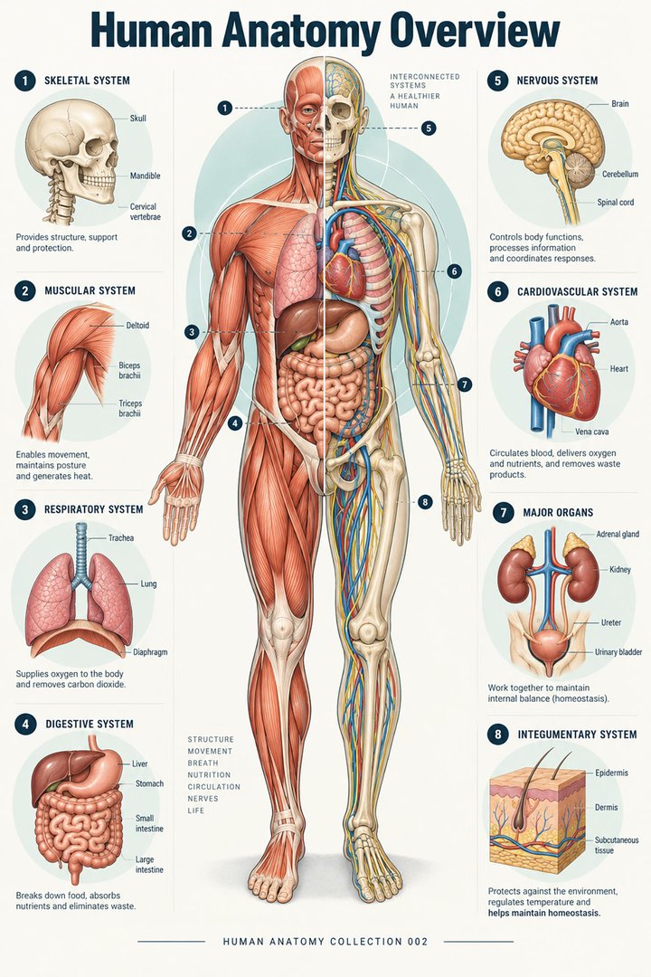 Human Anatomy Diagrams ↗
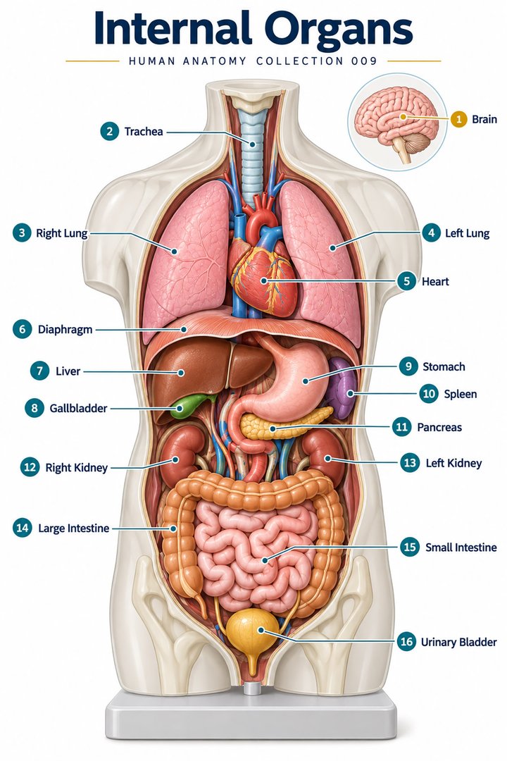 Human Body Organs ↗
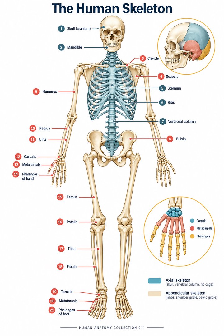 Skeletal System Anatomy ↗
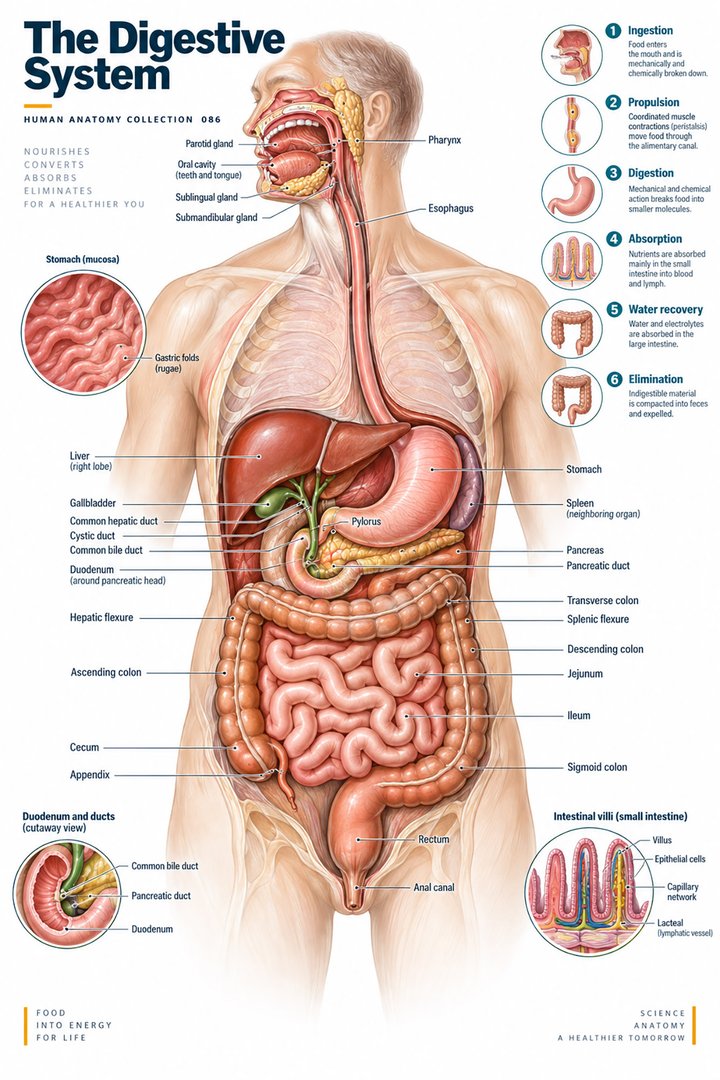 Human Digestive System ↗
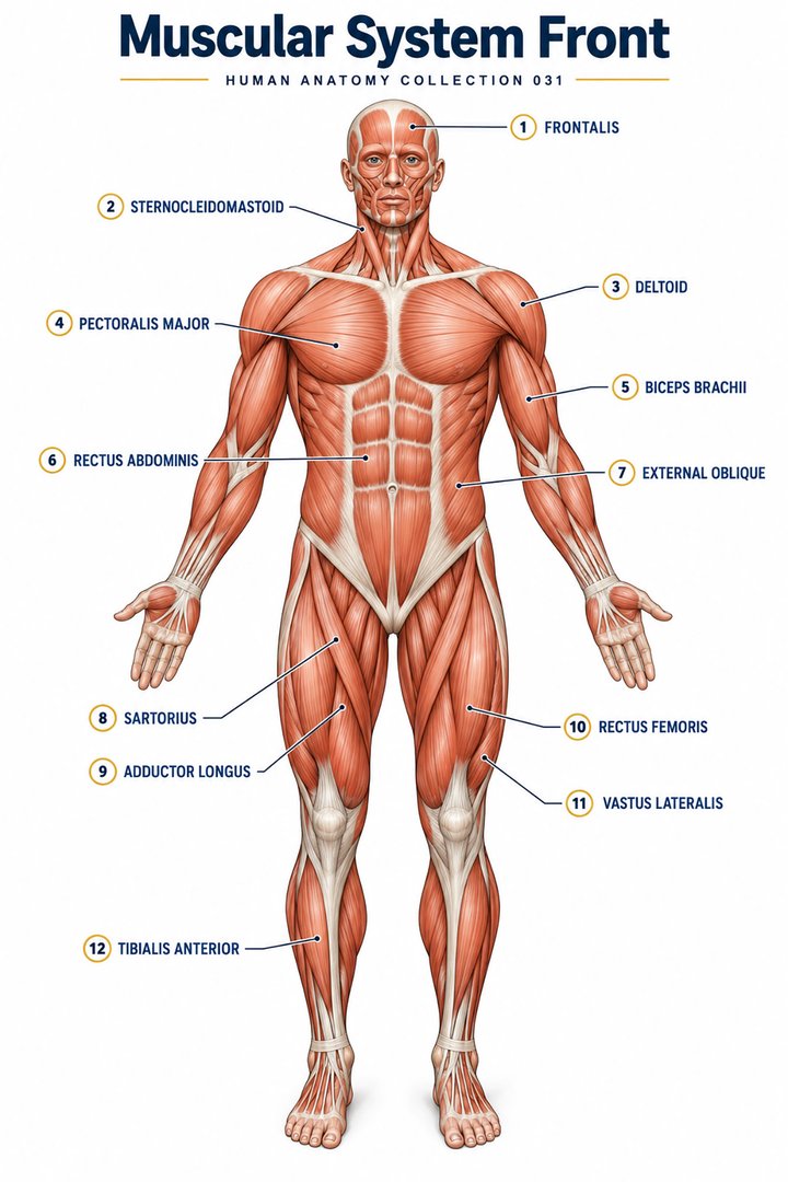 Muscular System Overview ↗
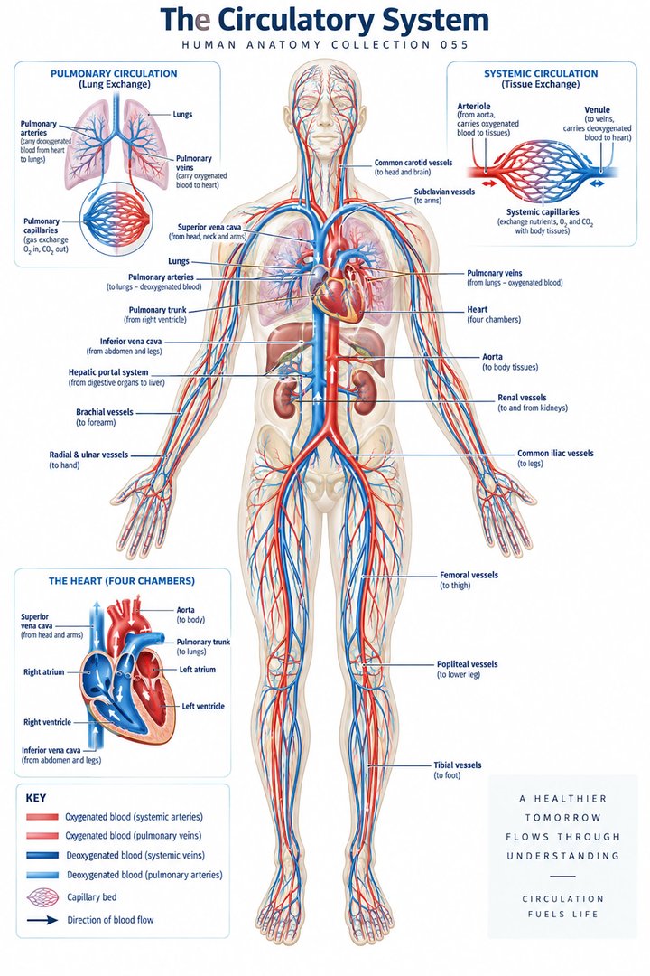 Human Circulatory System ↗
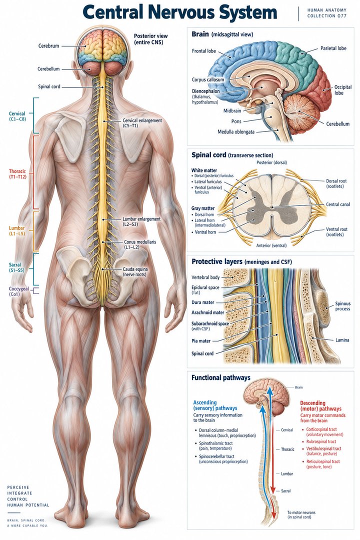 Human Nervous System ↗
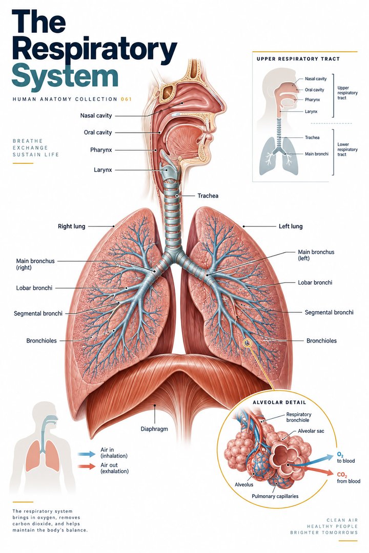 Respiratory System Functions ↗
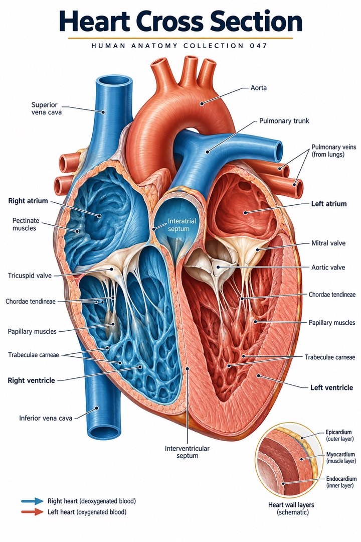 Human Heart Structure ↗
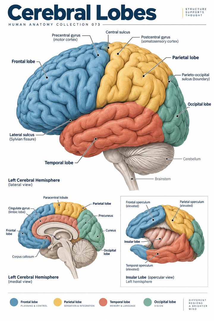 Human Brain Lobes ↗
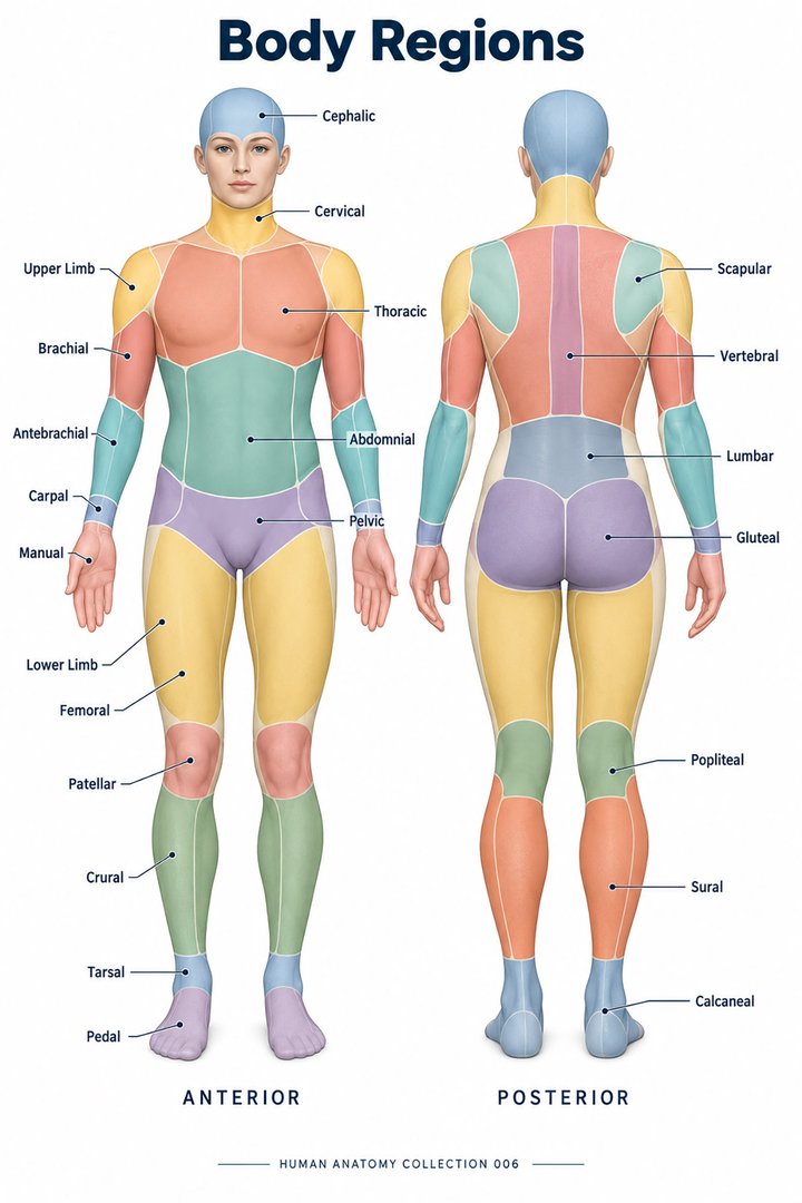 Human Body Parts ↗
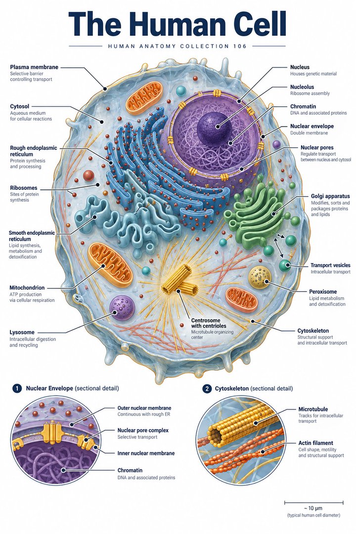 Human Cell Structure ↗
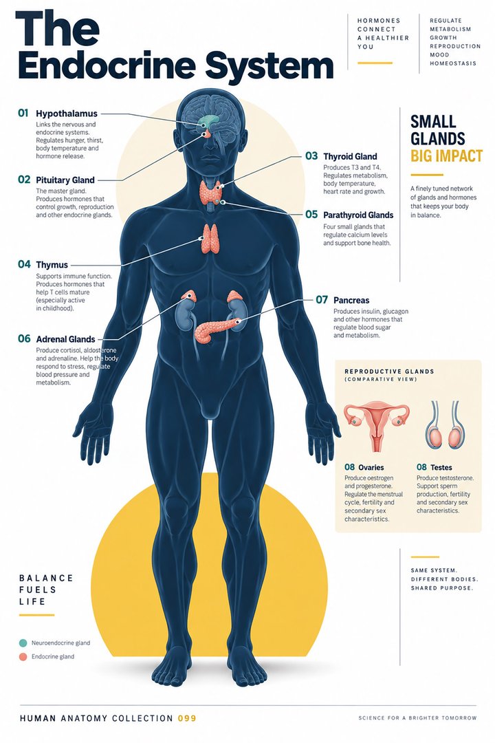 Human Endocrine System ↗
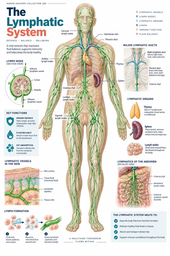 Human Lymphatic System ↗
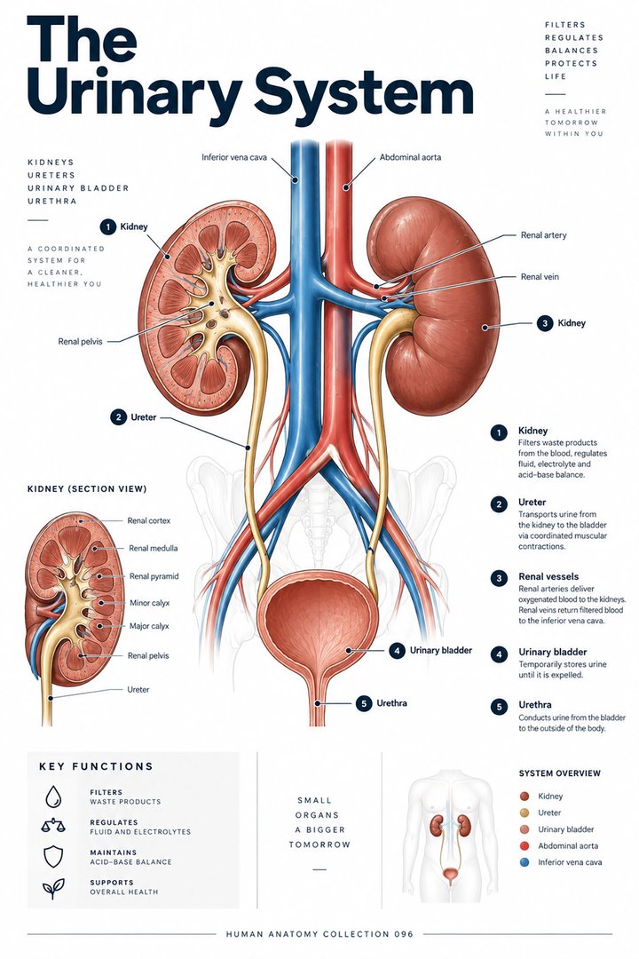 Human Urinary System ↗
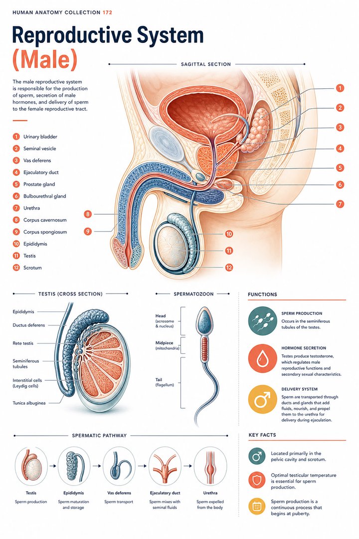 Male Reproductive System ↗
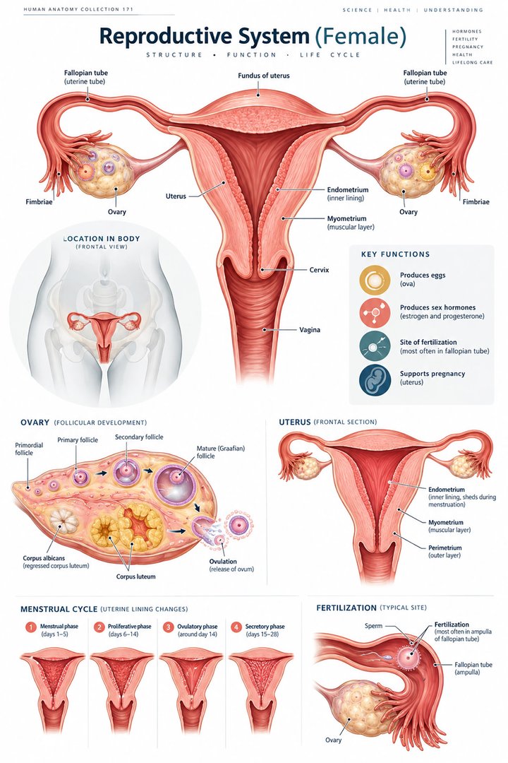 Female Reproductive System ↗
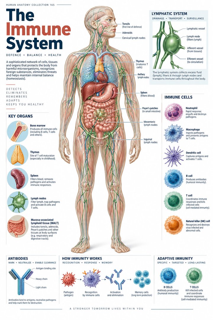 Human Immune System ↗Central Nervous System ↗
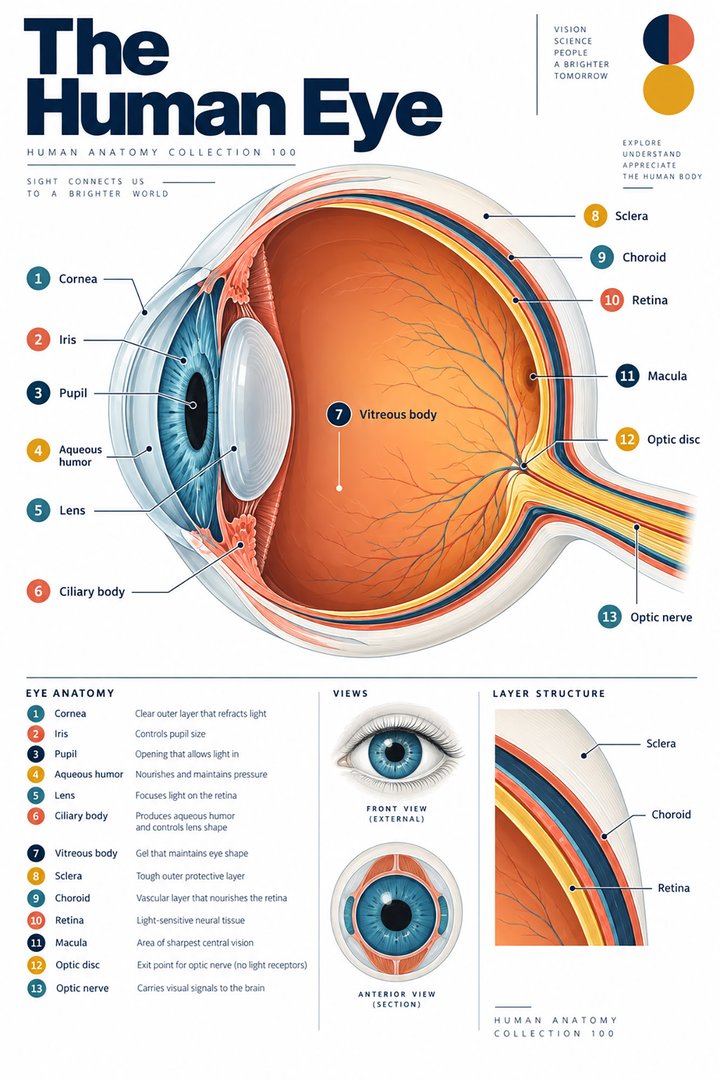 Human Eye Structure ↗
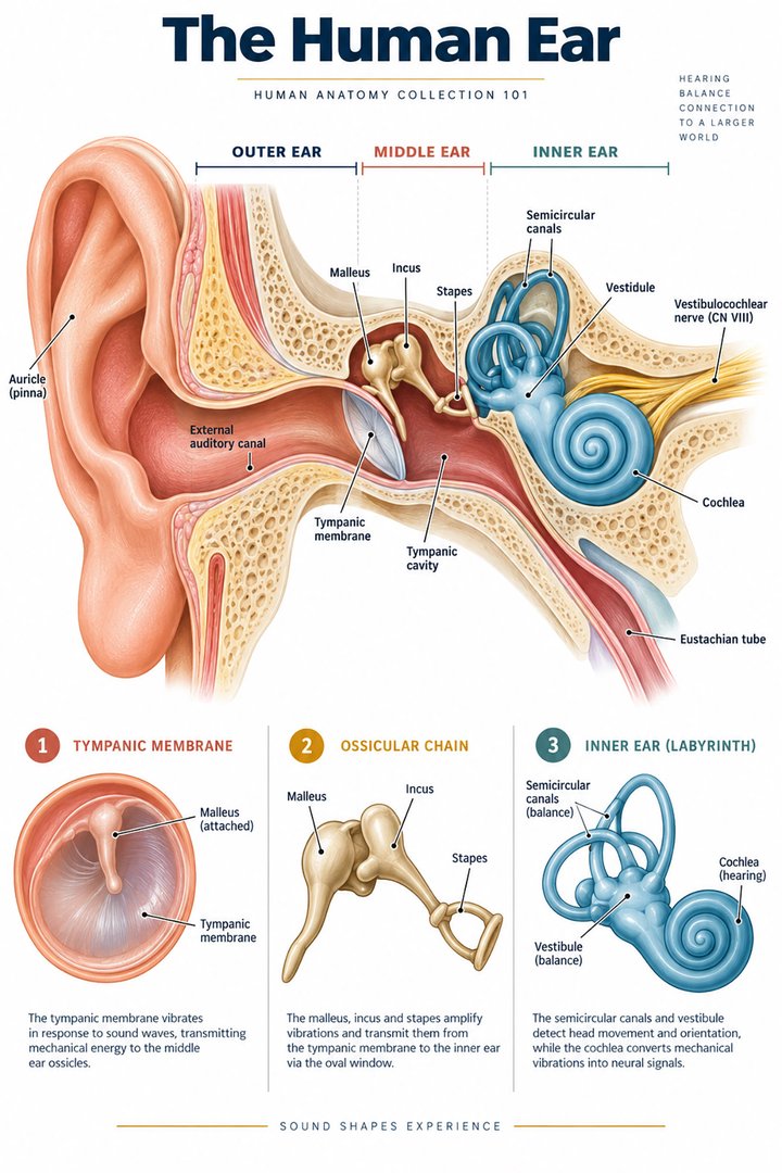 Human Ear Structure ↗
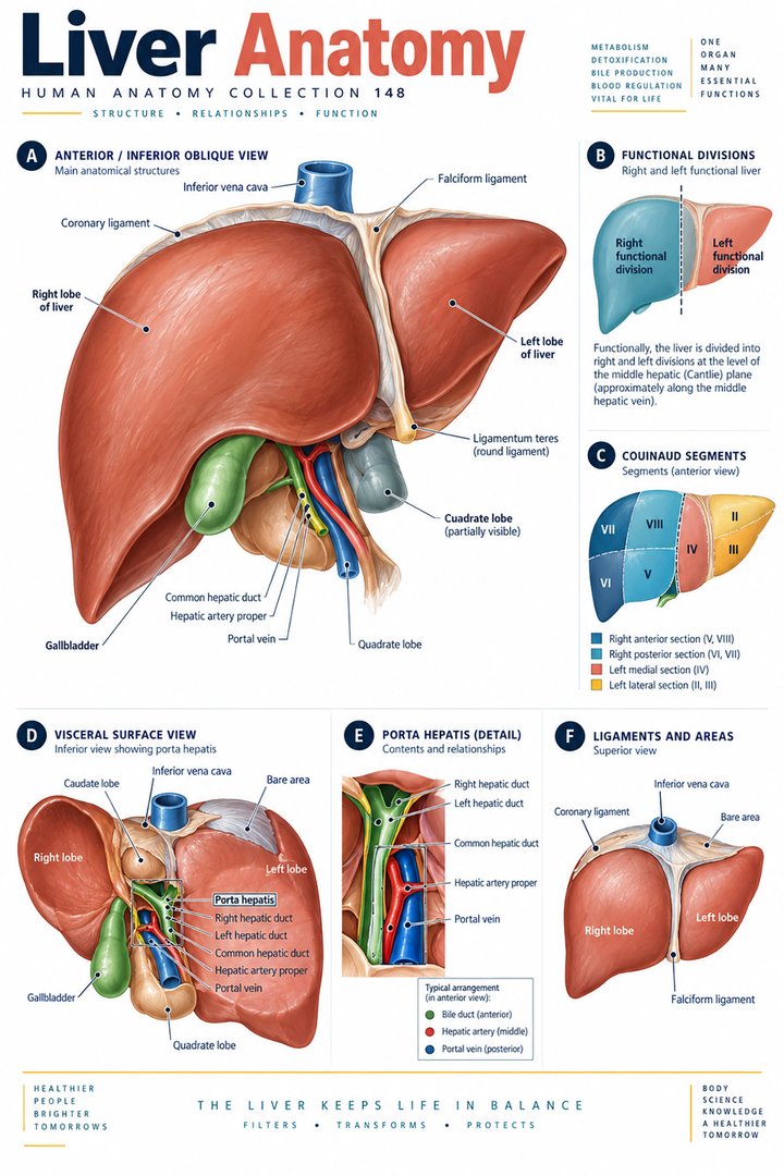 Human Liver Function ↗
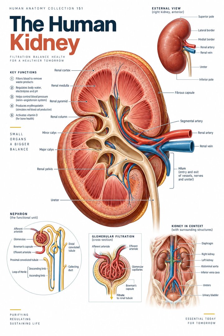 Human Kidney Anatomy ↗
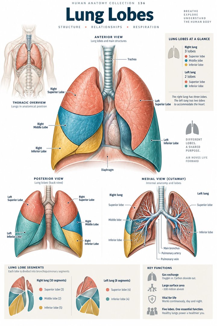 Human Lung Anatomy ↗
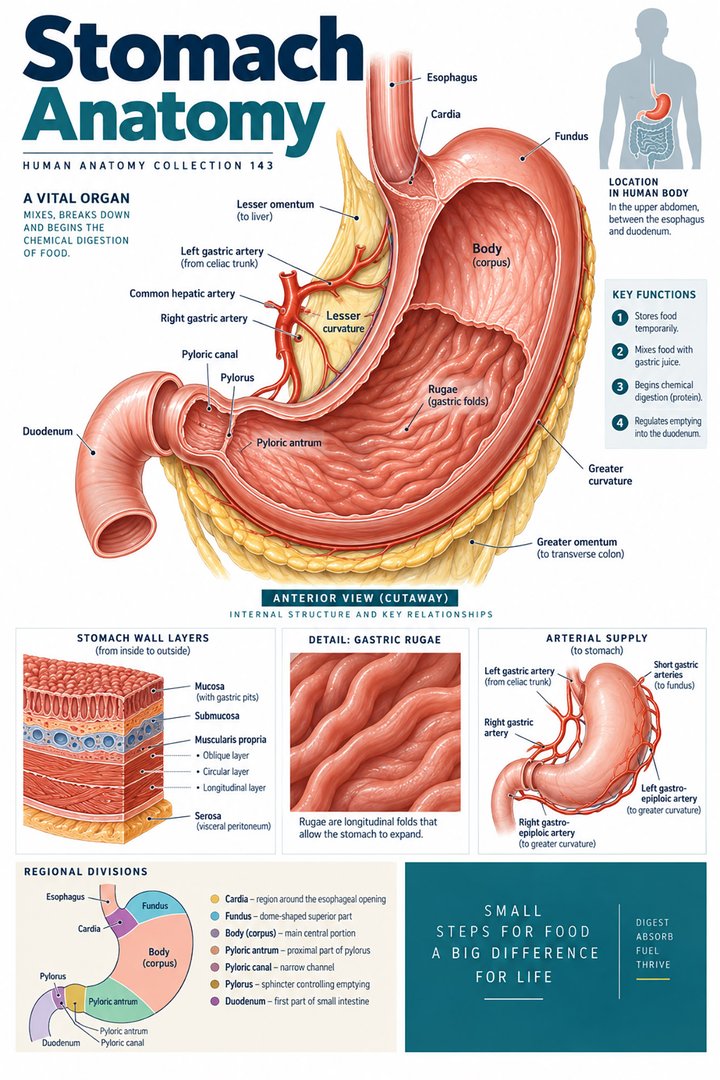 Human Stomach Structure ↗
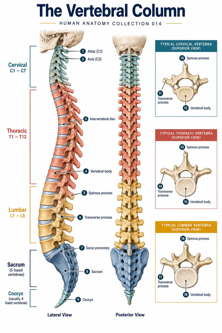 Human Spine Anatomy ↗
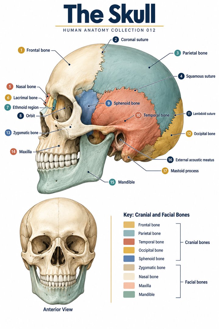 Human Skull Anatomy ↗
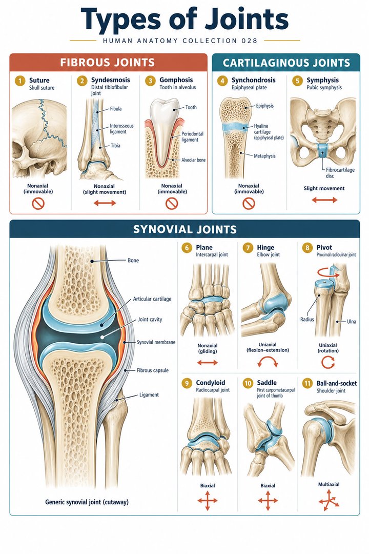 Human Joint Types ↗
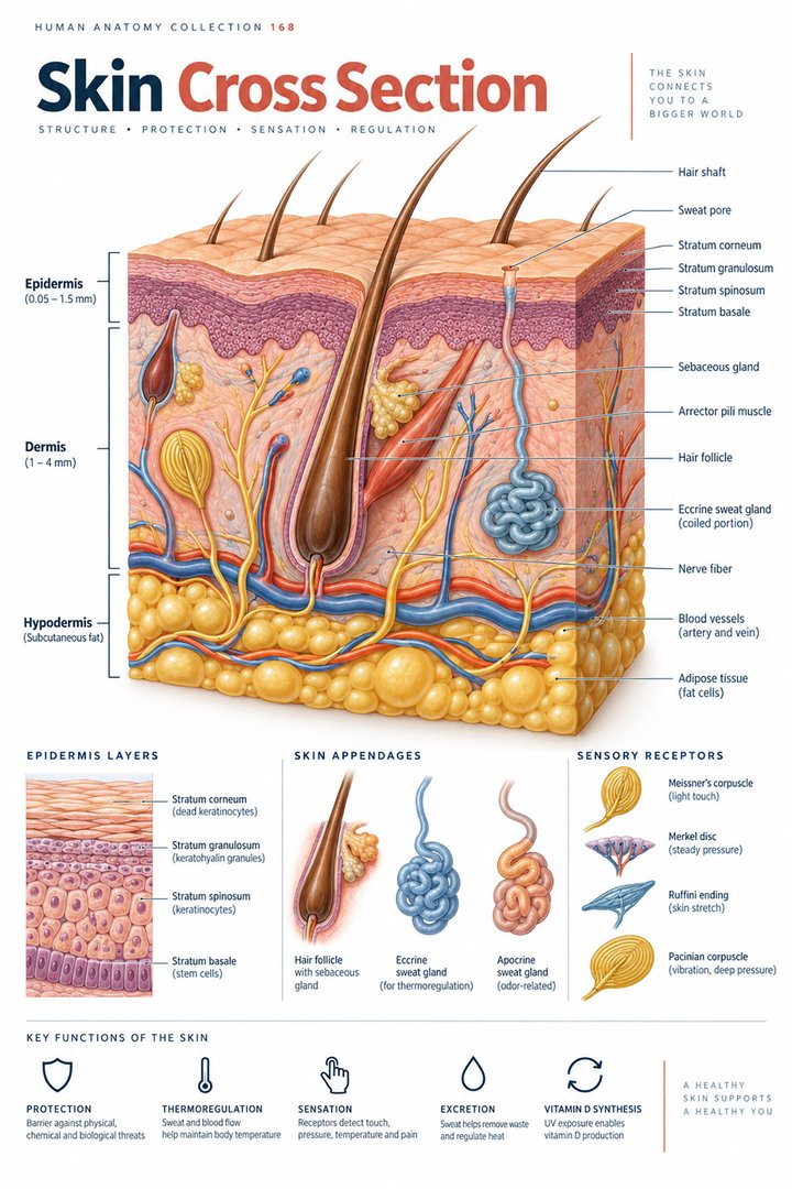 Human Skin Layers ↗
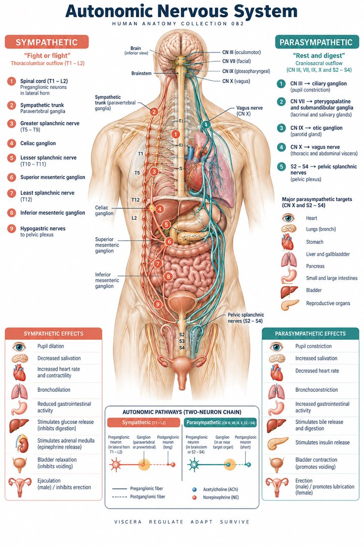 Autonomic Nervous System ↗
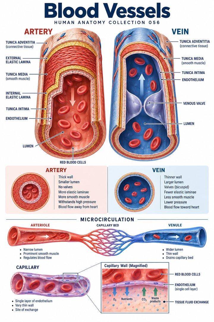 Human Blood Vessels ↗
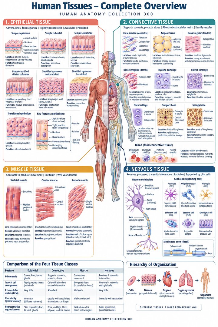 Human Body Tissues ↗
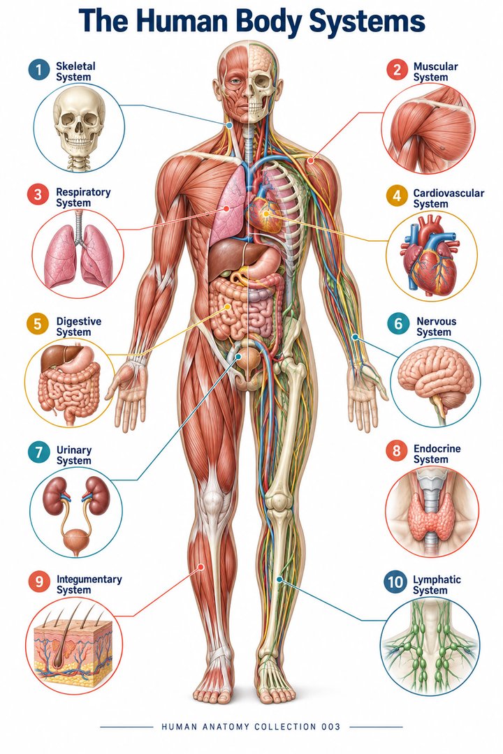 Human Body Systems ↗
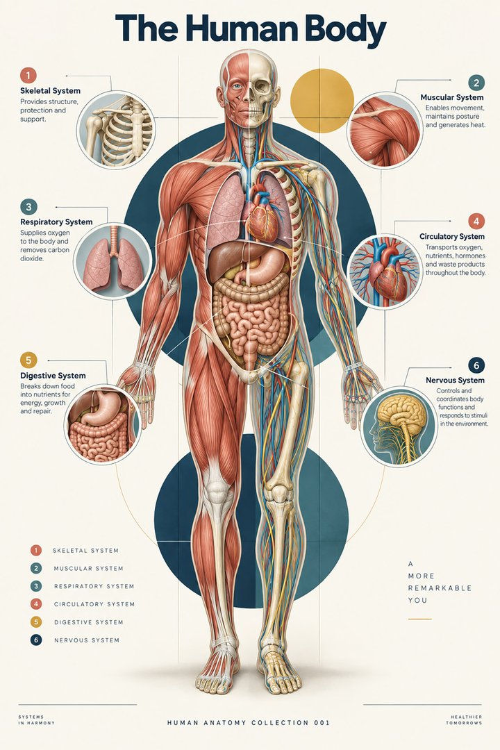 Human Body Facts ↗Human Anatomy and Physiology ↗
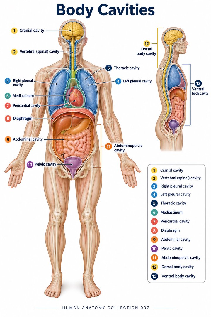 Human Body Cavities ↗Human Torso Anatomy ↗Human Digestive Tract ↗
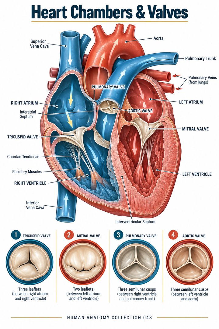 Human Heart Chambers ↗Human Organ Placement ↗
 Human Body Proportions ↗
 Peripheral Nervous System ↗
 Human Endocrine Glands ↗
 Human Bone Density ↗Human Blood Circulation ↗
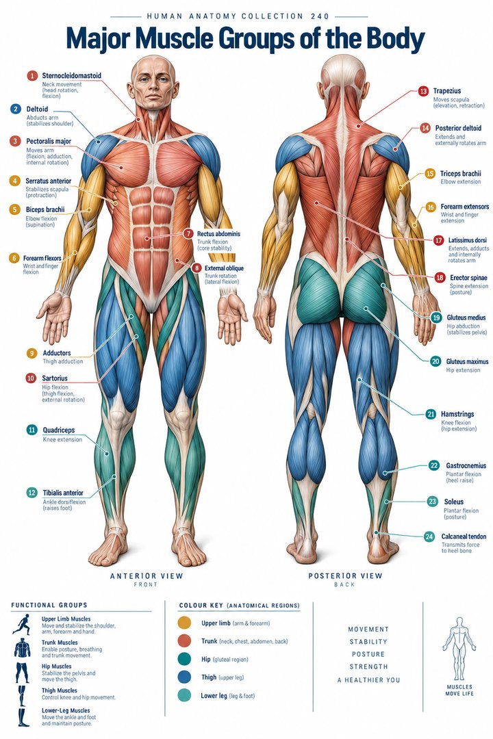 Human Muscle Groups ↗
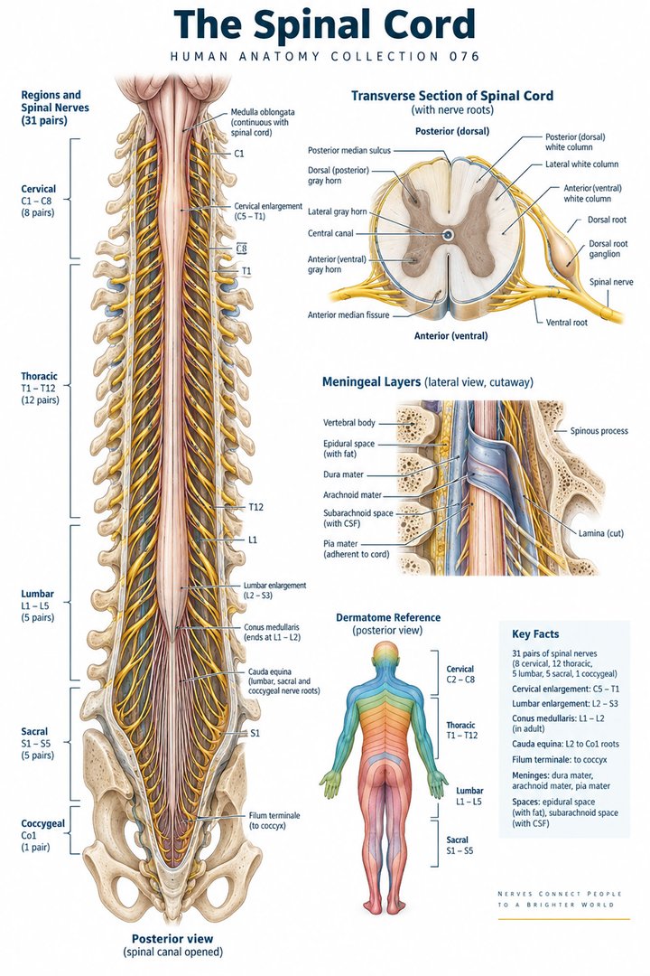 Human Spinal Cord ↗
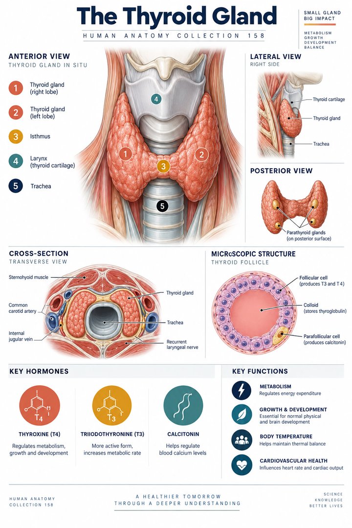 Human Thyroid Gland ↗
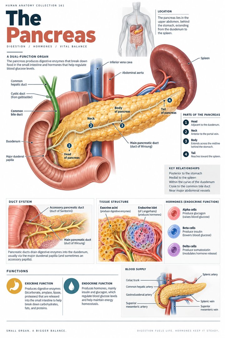 Human Pancreas Function ↗
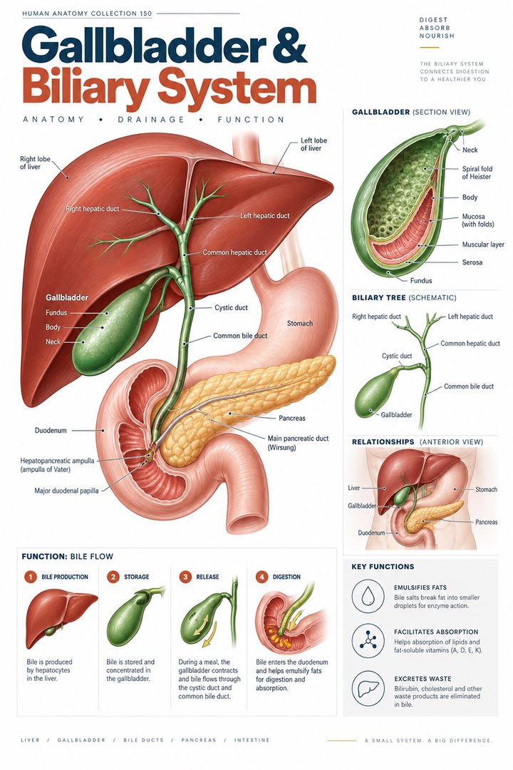 Human Gallbladder Anatomy ↗