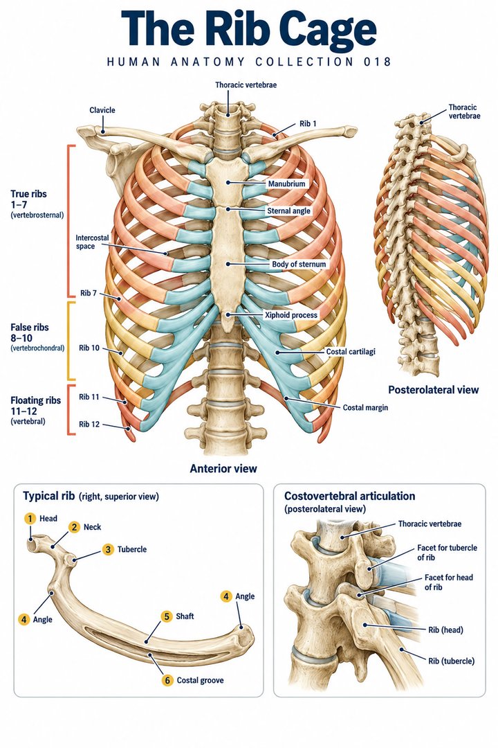
The Rib Cage
Individual poster · 300 DPI print files
Regular $9.90
Explore poster ↗$4.90USD
Offer price · Save $5.00Individual poster · 300 DPI print files
Reduced web preview. Separate high-resolution print files are supplied with the digital download.
ANATOMY 018
A modern anatomy illustration exploring the rib cage, designed for educational and decorative use.
Purchases are not available yet. Checkout and download delivery are being prepared.
Sold by Kunstolymp.de · Germany
Compare prices & the complete collection ↗This is a DIGITAL DOWNLOAD. No physical product will be shipped. After purchase, download your files and print them at home, at a local print shop, or through an online professional printing service.
The separate 300 DPI print files are ideal for 24×36-inch posters (approximately 60×90 cm). ISO and 5×7 formats are included. The white border lets you adjust the layout or trim to fit other frame sizes; check the preview before printing to keep the illustration and labels visible.
The planned bundle delivery includes a PDF information sheet with a link to the relevant Google Drive folder and download instructions. The store is currently in preview and purchases are not yet available.
A science-inspired choice for a student’s study corner, an educator’s classroom, or an anatomy enthusiast’s wall. Final licensing terms will apply.
CLINICAL CONTEXT
The rib cage comprises ribs, costal cartilages, sternum, thoracic vertebrae, intercostal muscles, neurovascular bundles, and the diaphragm’s attachments. It protects lungs, heart, major vessels, and upper abdominal organs while allowing ventilation through coordinated motion. In clinical practice, chest-wall pain may be benign and musculoskeletal, but it must first be distinguished from cardiopulmonary, vascular, pleural, pulmonary, oesophageal, and upper-abdominal emergencies. Anatomical localisation is useful only after considering acuity and physiology.
Rib pain often follows a fall, direct blow, intense cough, or repetitive strain. Patients may report focal tenderness, pain with deep breathing or coughing, bruising, and sleep difficulty. Rib fracture, costochondritis, intercostal strain, and chest-wall contusion are common explanations. Yet chest pain with breathlessness, diaphoresis, syncope, fever, haemoptysis, palpitations, or exertional symptoms needs a broader differential including acute coronary syndrome, pulmonary embolism, pneumothorax, pneumonia, pericarditis, aortic disease, and referred pain. In older adults or people with osteoporosis, low-energy trauma can still cause clinically important fractures; multiple fractures can impair ventilation and cough.
Assessment starts with vital signs, respiratory effort, oxygenation where relevant, mechanism of injury, anticoagulant use, comorbid cardiopulmonary disease, and examination of the chest wall, lungs, heart, abdomen, spine, and skin. Imaging is selected for suspected complications or when it will alter care; absence of a fracture on plain film does not automatically exclude injury, and management is often guided by function and respiratory risk. Conventional treatment for uncomplicated injury focuses on adequate analgesia, deep breathing and cough, mobilisation, smoking cessation, and review for infection or deterioration. More severe trauma, flail segment, pneumothorax, haemothorax, hypoxia, or suspected injury to lung, liver, or spleen requires emergency assessment and may need procedural or inpatient care.
Breathing exercises, physiotherapy, sleep positioning, and carefully graded return to activity can support recovery when serious injury has been excluded. Relaxation or mindfulness can assist pain coping but does not treat intrathoracic injury. Avoid tight chest binding because it can restrict breathing. Manual treatments should not be used over suspected fracture or unstable trauma. If a patient uses herbal analgesics or supplements, review bleeding and drug-interaction risks, particularly with anticoagulants, antiplatelet therapy, renal disease, or surgery.
Worsening shortness of breath, increasing chest pain, coughing blood, fever or purulent sputum, abdominal or shoulder pain after chest trauma, or injury from a high-energy event requires urgent local assessment. The NHS specifically notes that a rib injury can sometimes damage a lung, liver, or spleen.
Further reading: NHS: broken or bruised ribs · WHO: complementary and integrative medicine
Explain the working diagnosis, important alternatives, planned tests, expected course, and specific safety-netting in language the patient can use. Invite questions about treatment goals, capacity, cost, culture, and complementary therapies. Use a professional interpreter when language access could affect consent or understanding, and confirm clear arrangements for timely clinical review in writing. Document the reasoning, reassess response to treatment, and coordinate care across professions when illness affects more than one system. Teaching anatomy is most useful when it supports this partnership: accurate localisation, respectful listening, timely escalation, and a plan that remains responsive to the individual patient.
HUMAN ANATOMY COLLECTION · 001—300
A closer look at what makes us human. Discover anatomy posters for your study space, classroom, and everyday curiosity.
DIGITAL DOWNLOADS · 300 DPI PRINT FILES
EXPLORE BY SYSTEM
LEARN & EXPLORE
Detailed English guides with Latin terminology, diagrams, real images, and clinical learning connections.
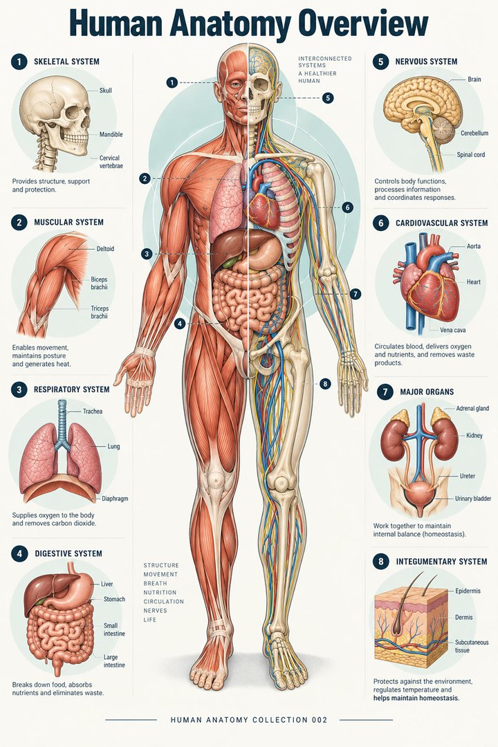 Human Anatomy Diagrams ↗
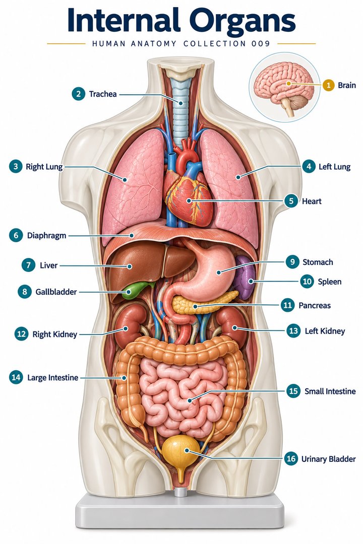 Human Body Organs ↗
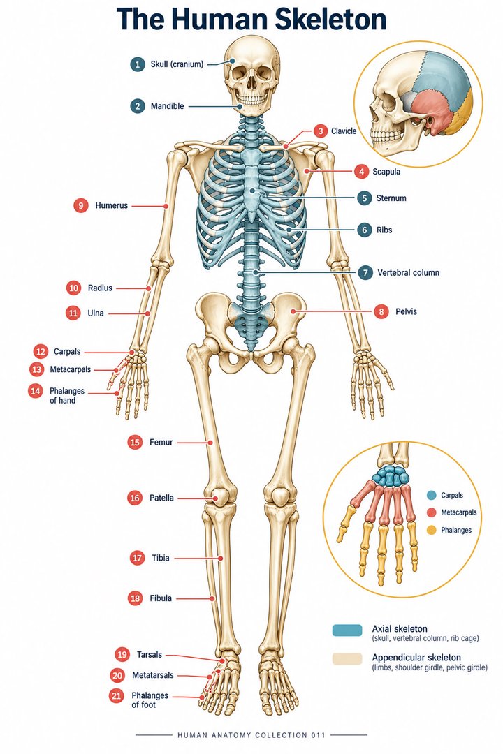 Skeletal System Anatomy ↗
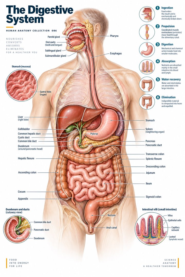 Human Digestive System ↗
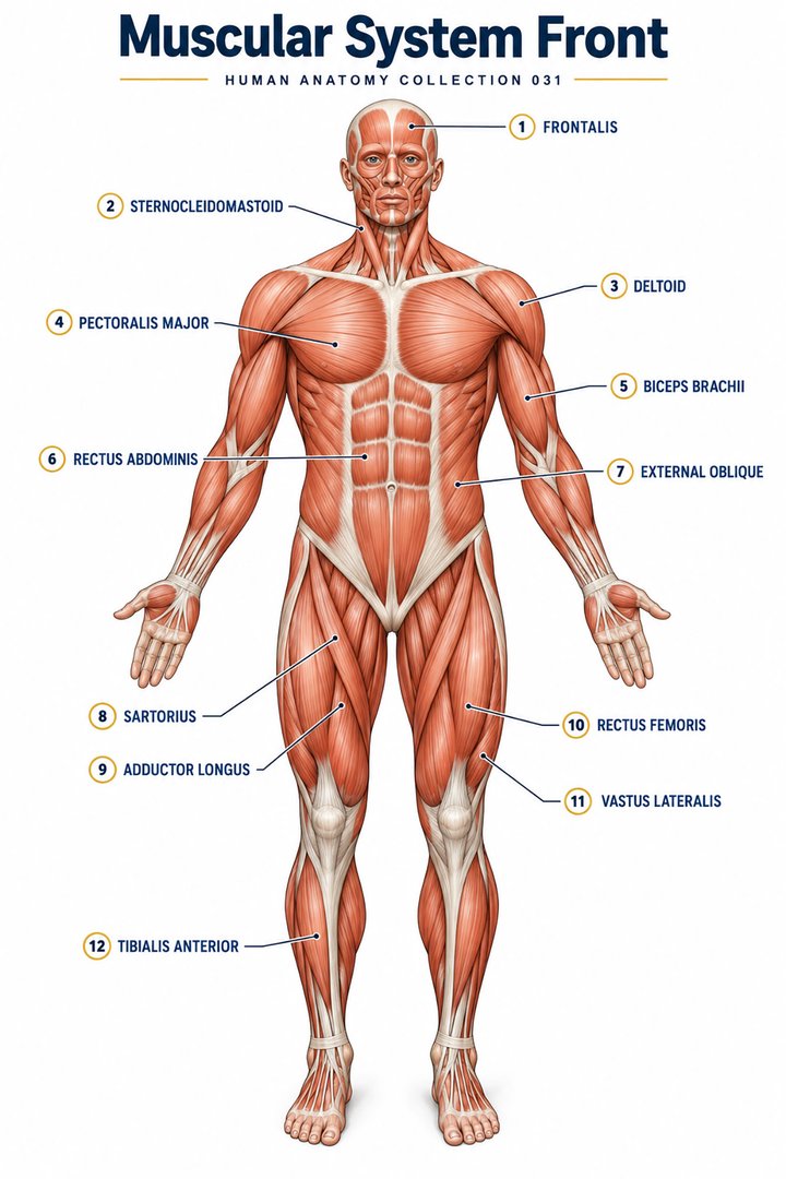 Muscular System Overview ↗
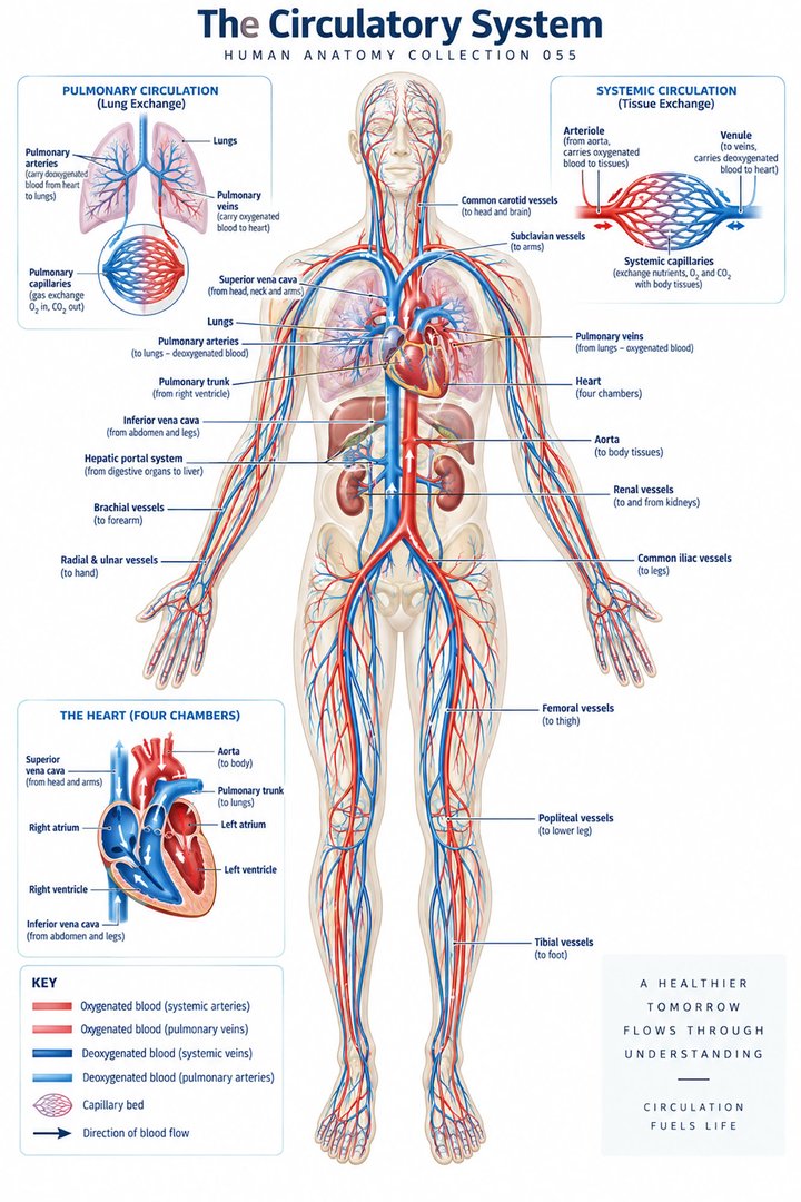 Human Circulatory System ↗
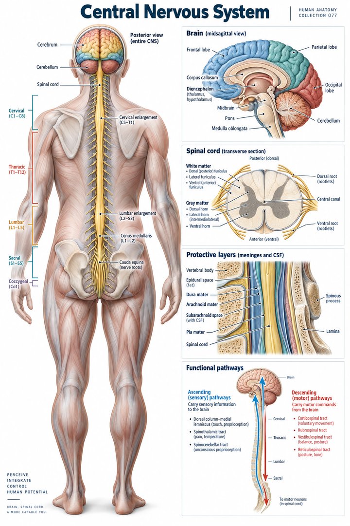 Human Nervous System ↗
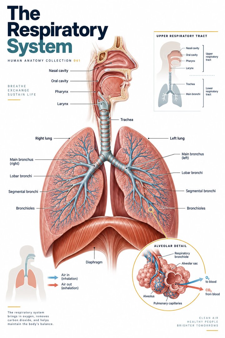 Respiratory System Functions ↗
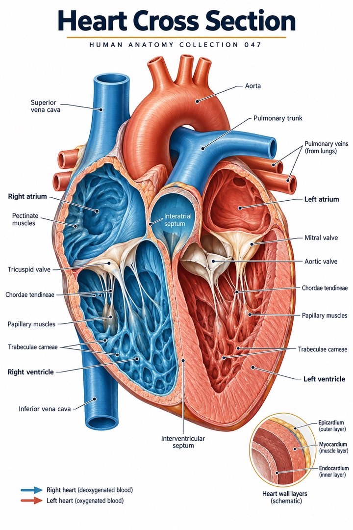 Human Heart Structure ↗
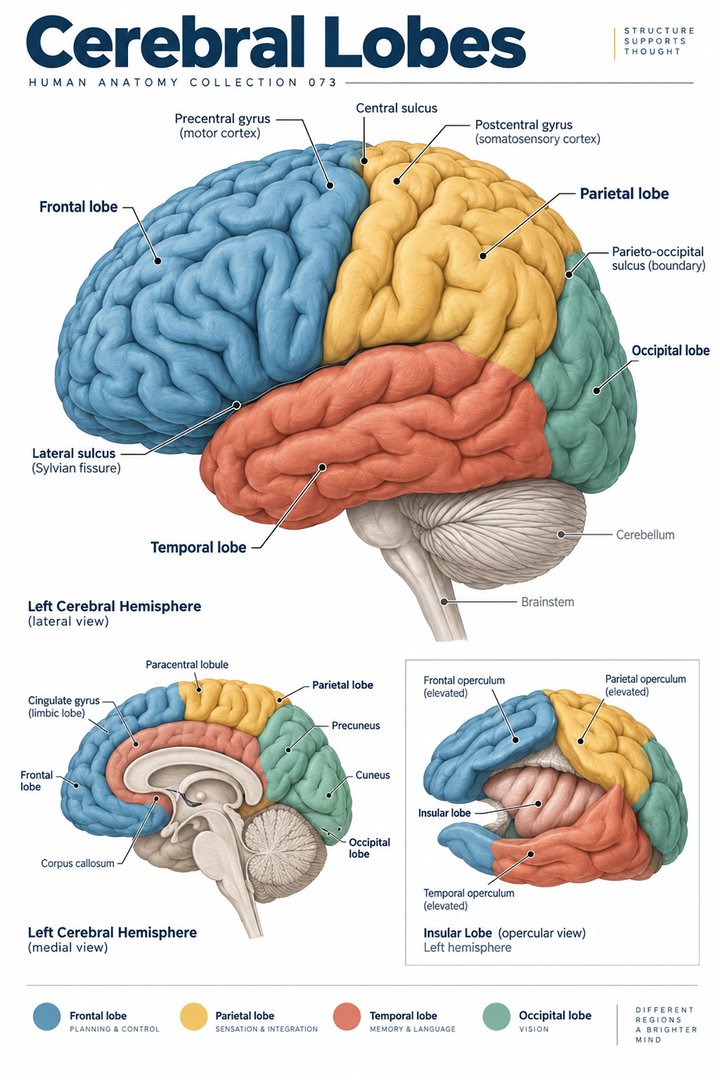 Human Brain Lobes ↗
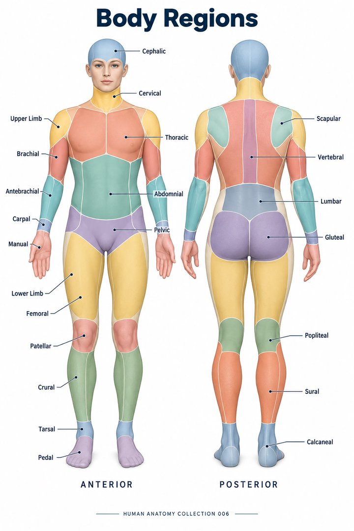 Human Body Parts ↗
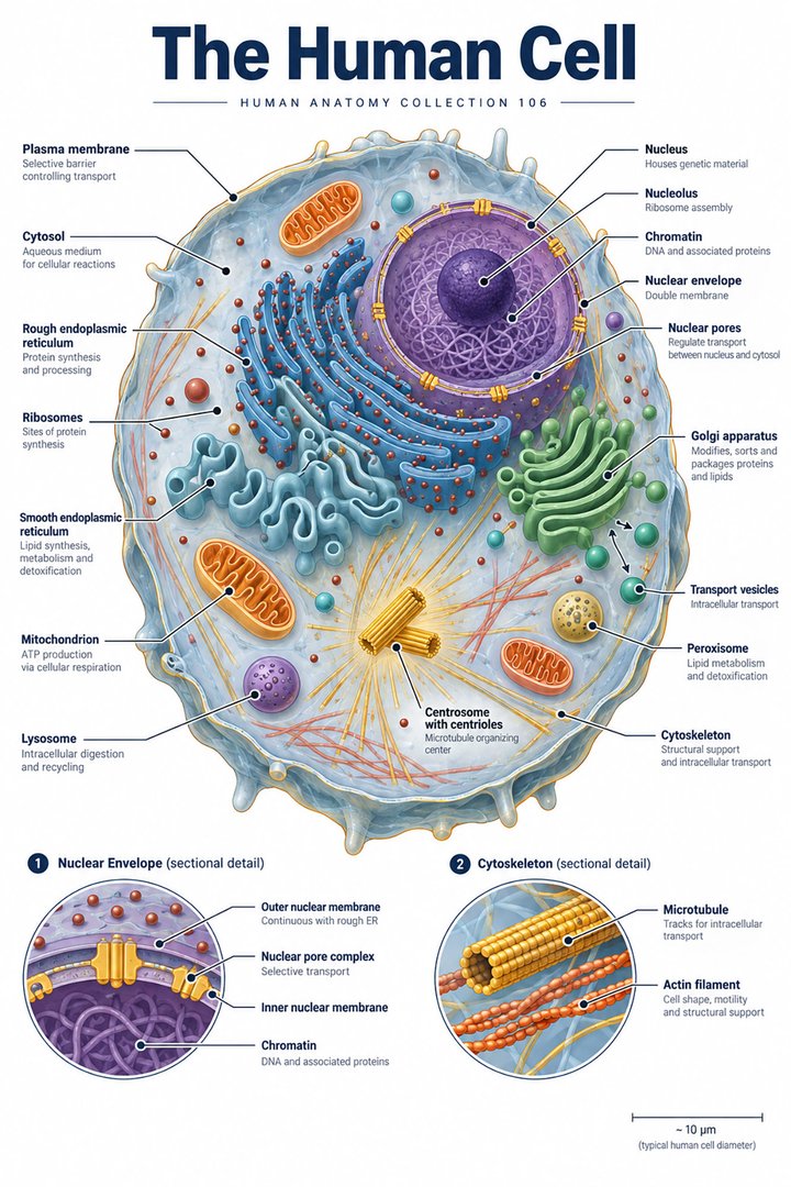 Human Cell Structure ↗
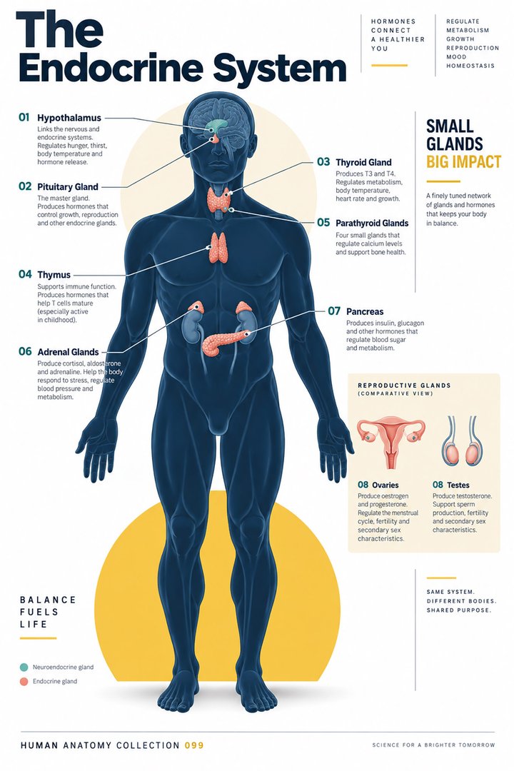 Human Endocrine System ↗
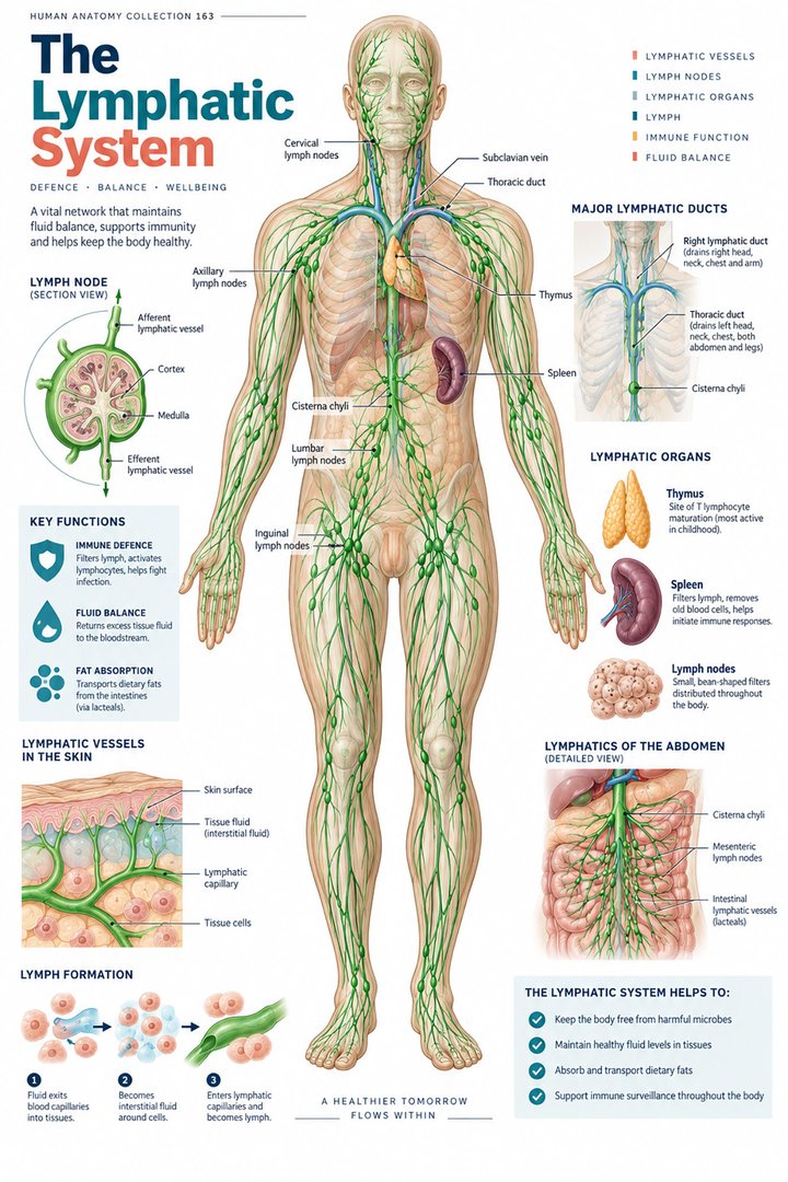 Human Lymphatic System ↗
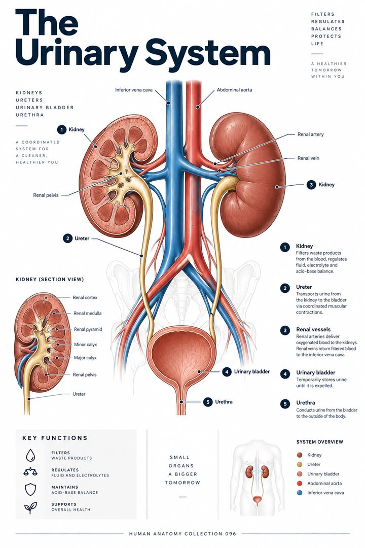 Human Urinary System ↗
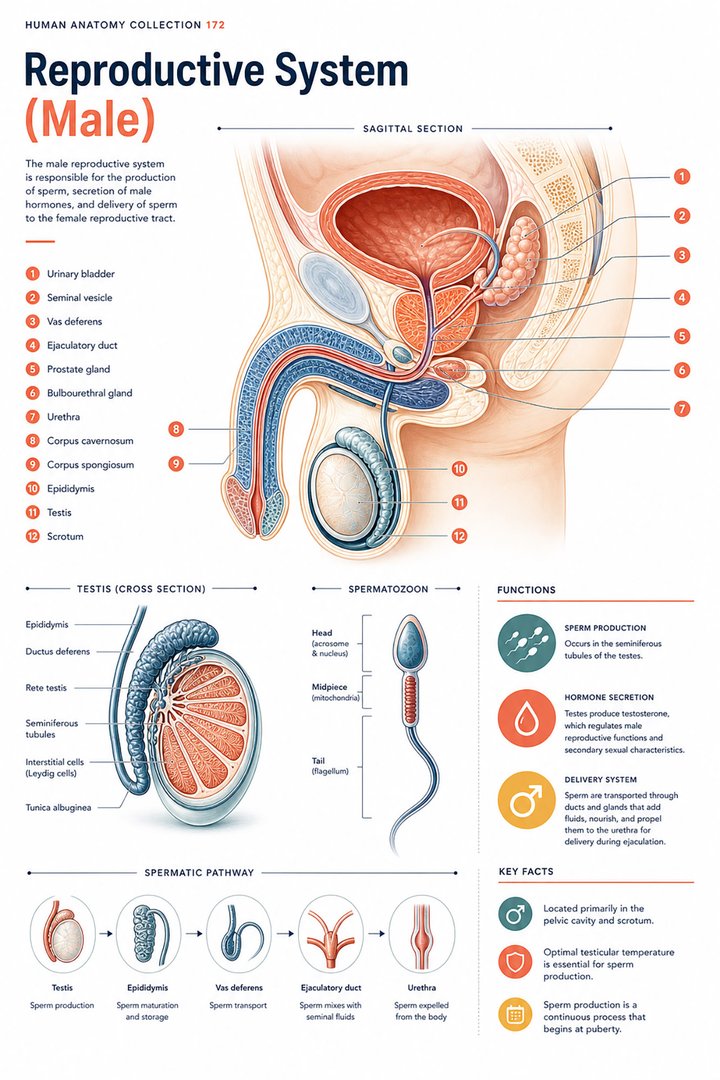 Male Reproductive System ↗
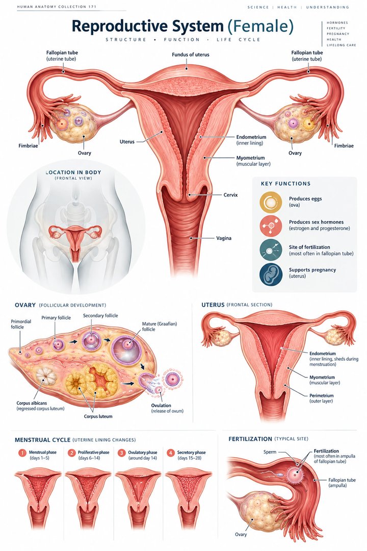 Female Reproductive System ↗
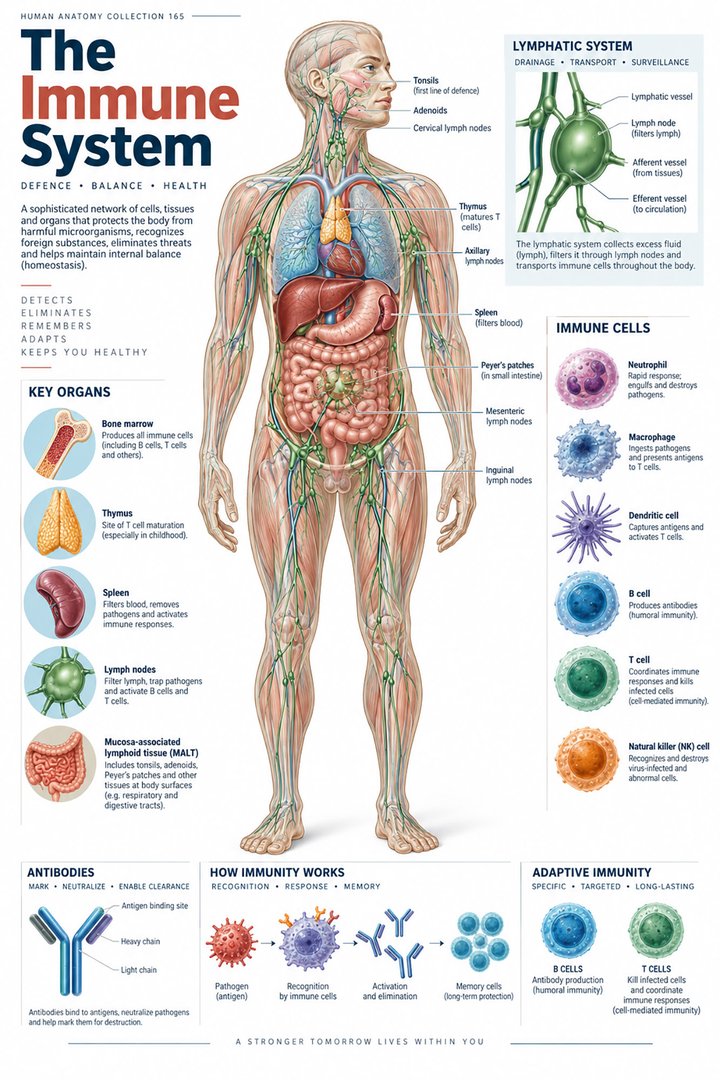 Human Immune System ↗Central Nervous System ↗
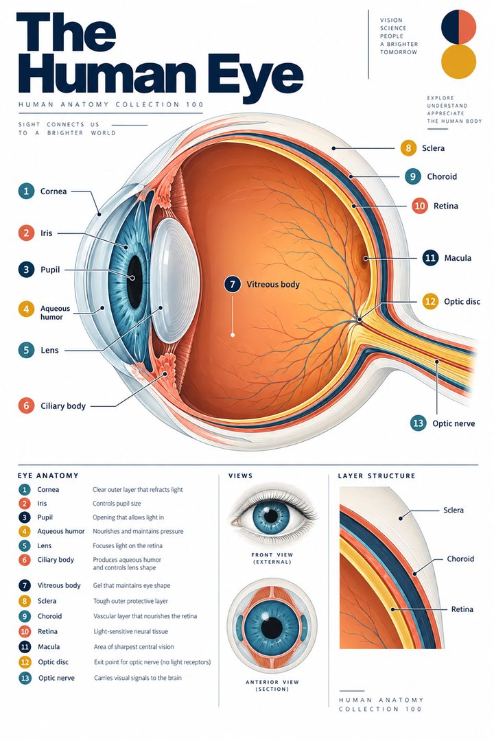 Human Eye Structure ↗
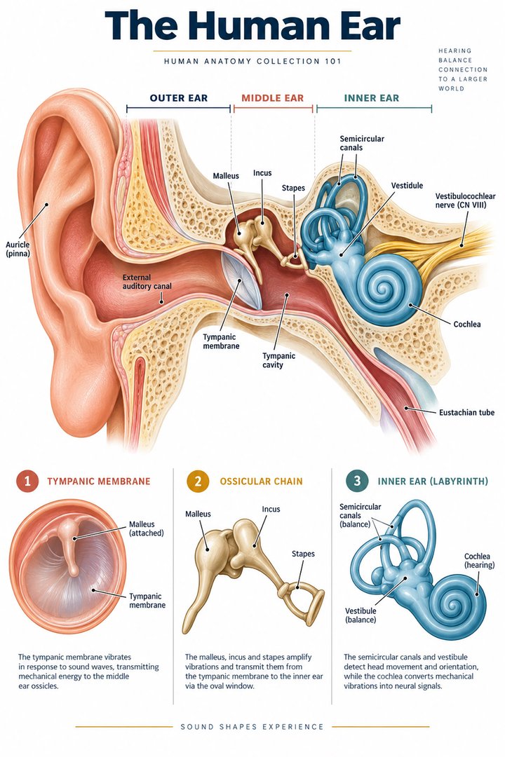 Human Ear Structure ↗
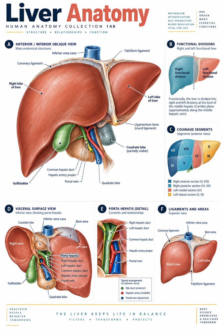 Human Liver Function ↗
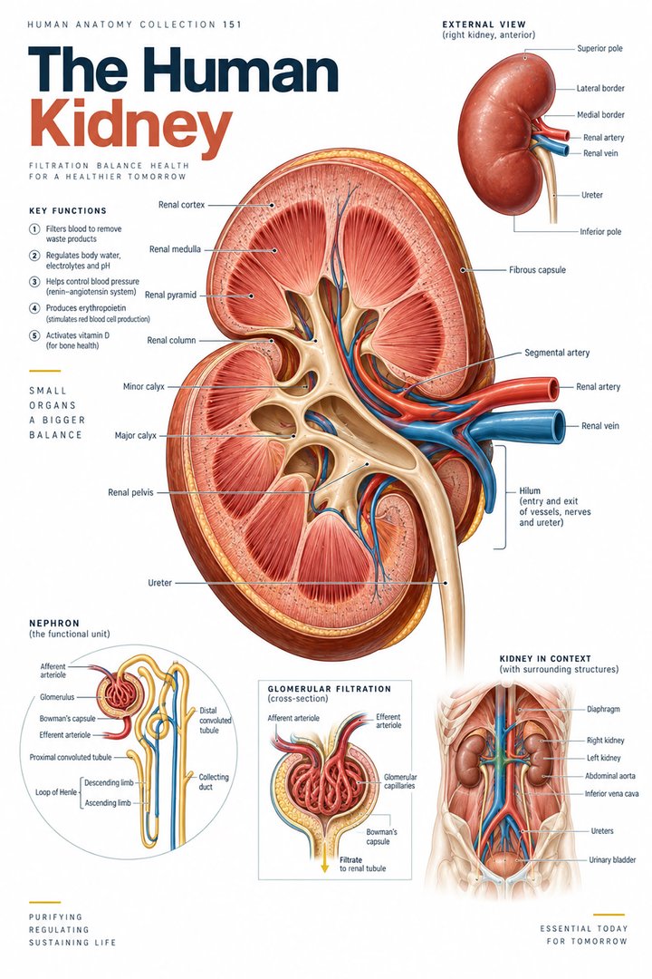 Human Kidney Anatomy ↗
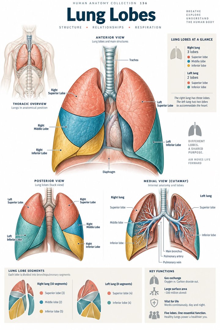 Human Lung Anatomy ↗
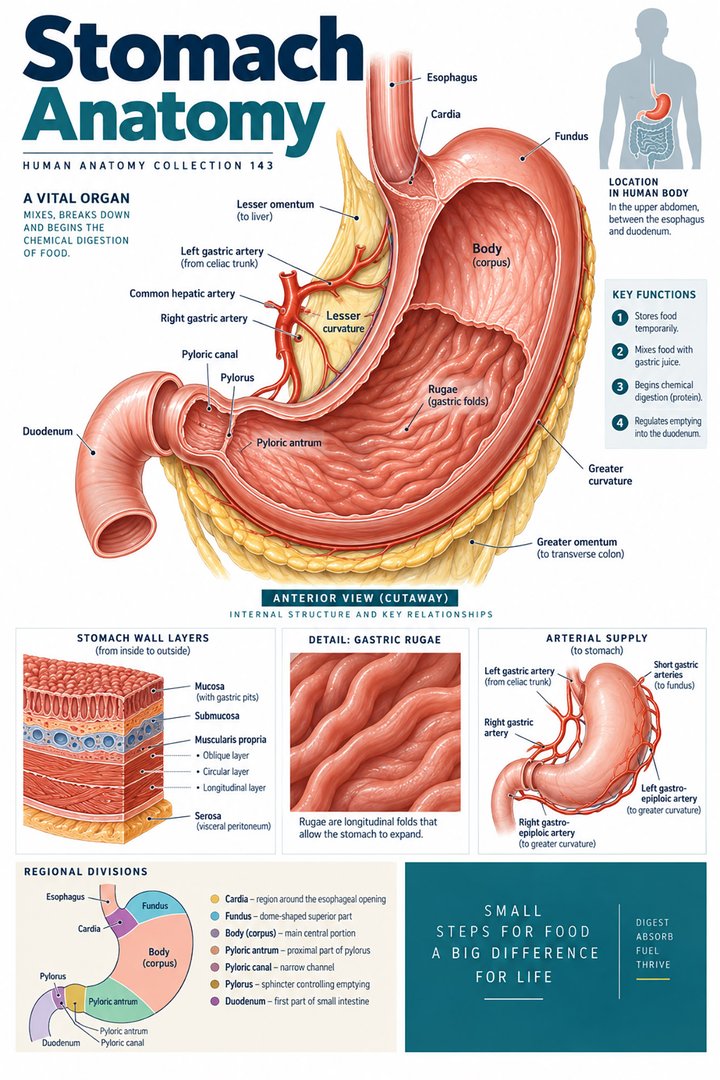 Human Stomach Structure ↗
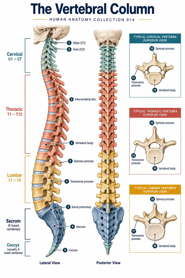 Human Spine Anatomy ↗
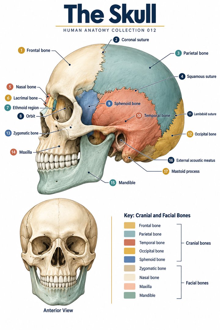 Human Skull Anatomy ↗
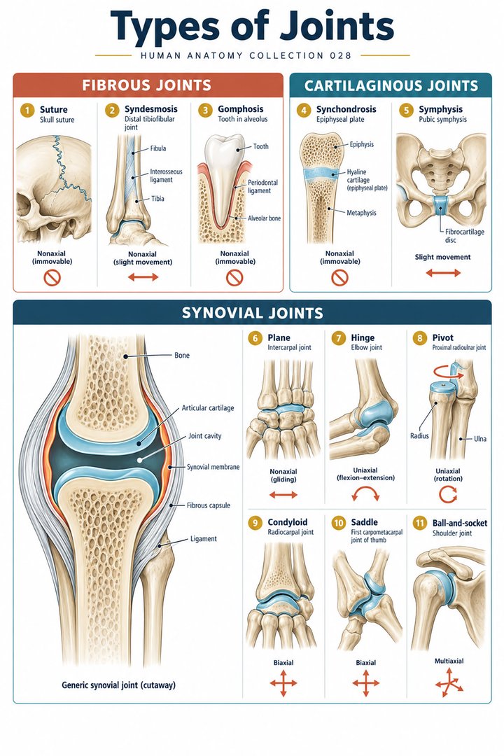 Human Joint Types ↗
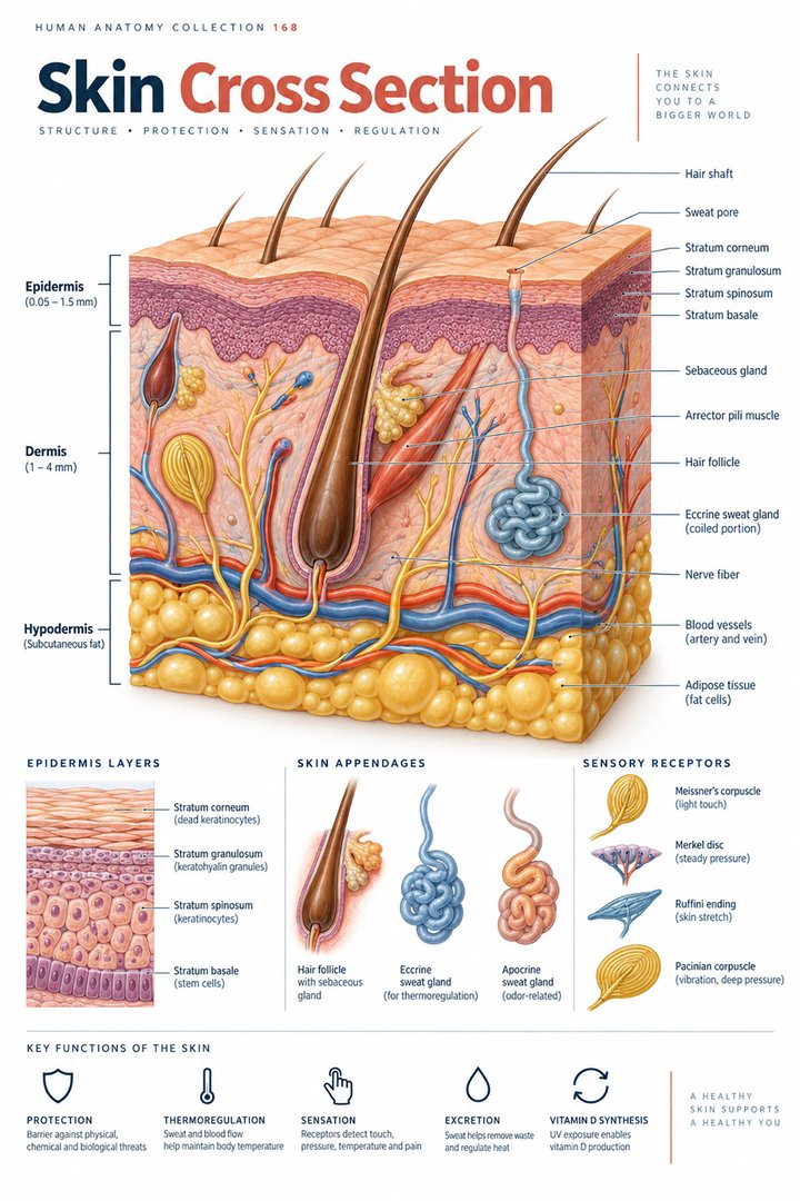 Human Skin Layers ↗
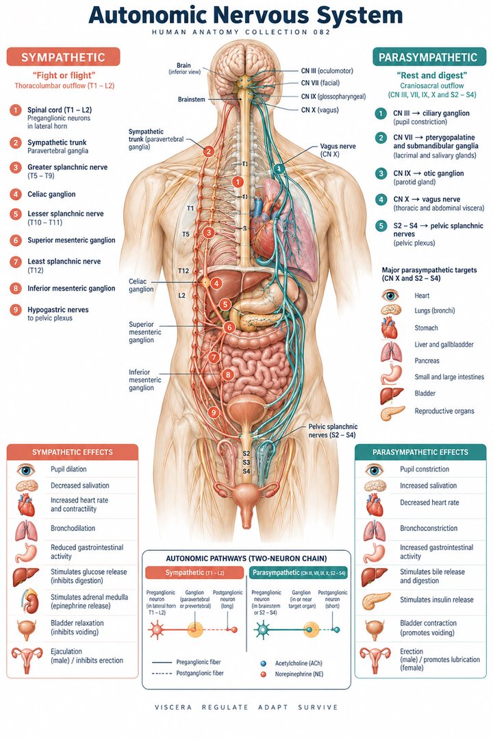 Autonomic Nervous System ↗
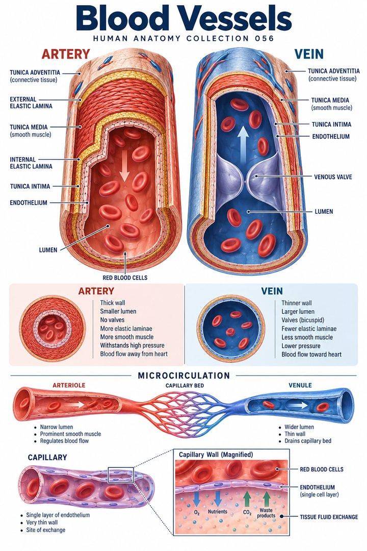 Human Blood Vessels ↗
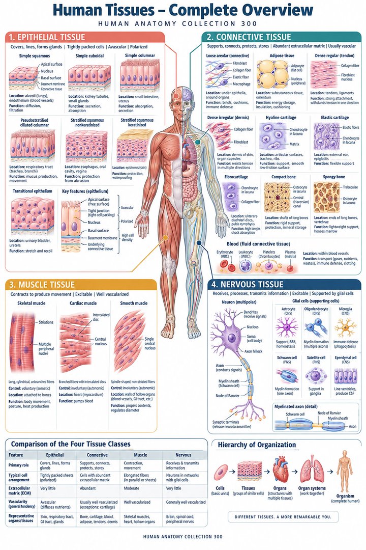 Human Body Tissues ↗
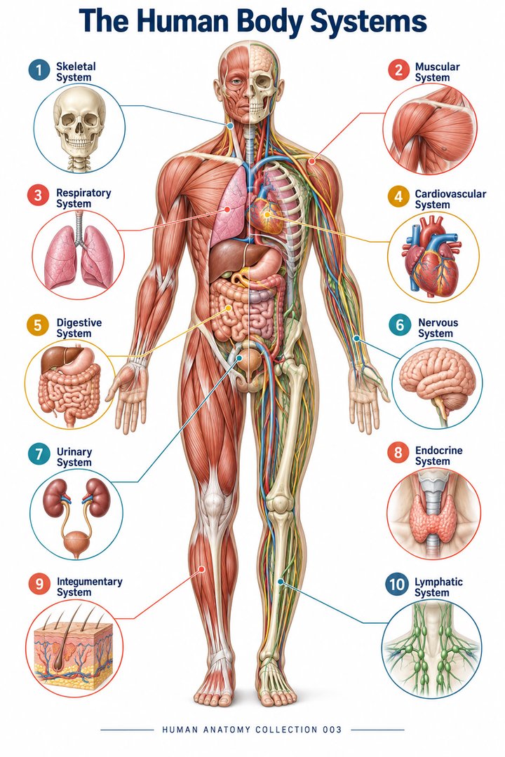 Human Body Systems ↗
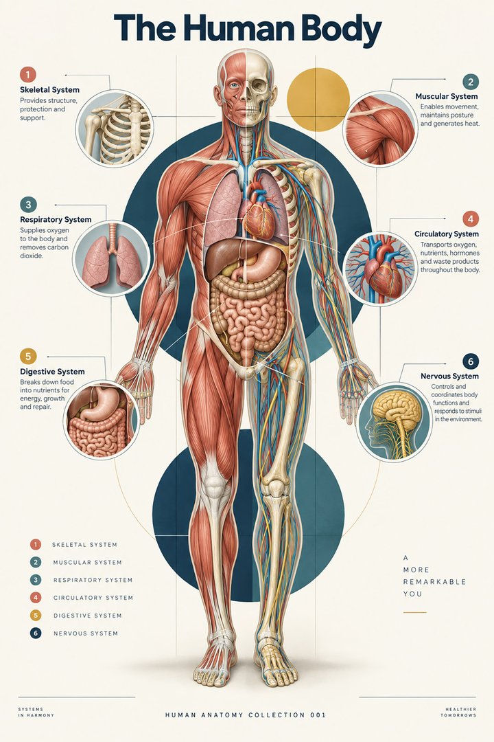 Human Body Facts ↗Human Anatomy and Physiology ↗
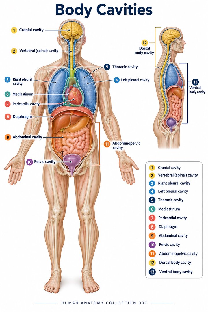 Human Body Cavities ↗Human Torso Anatomy ↗Human Digestive Tract ↗
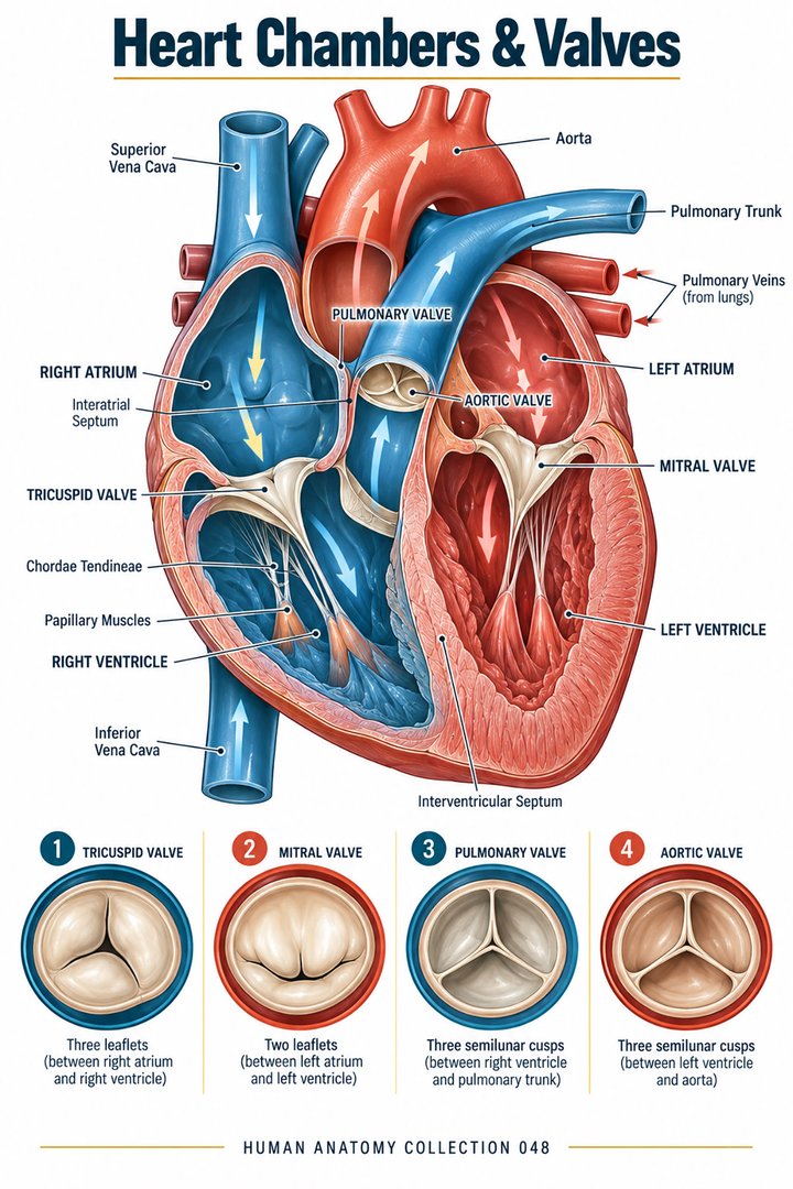 Human Heart Chambers ↗Human Organ Placement ↗
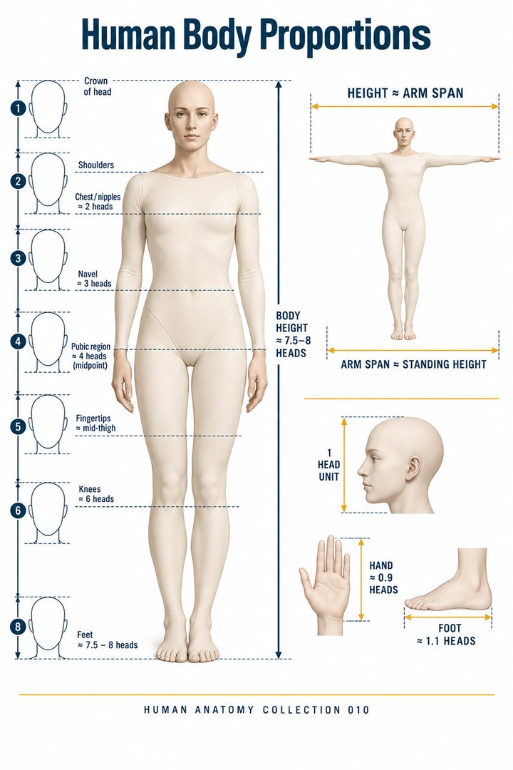 Human Body Proportions ↗
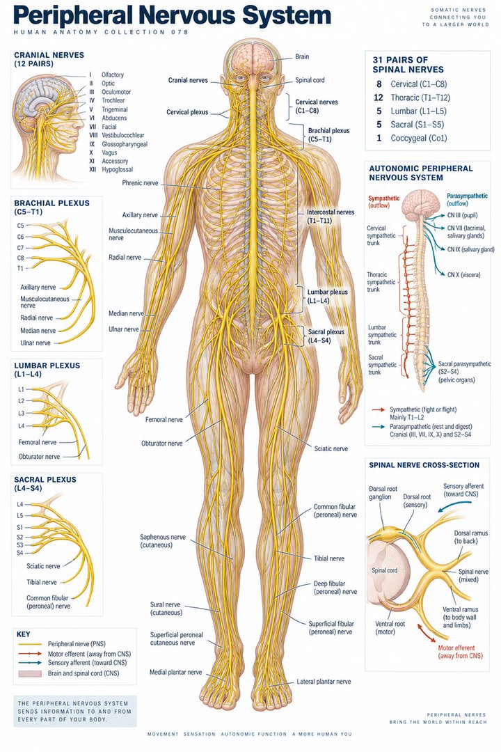 Peripheral Nervous System ↗
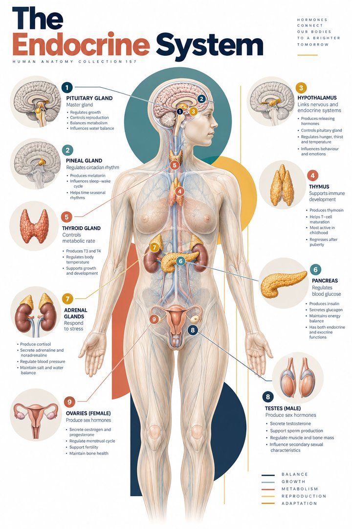 Human Endocrine Glands ↗
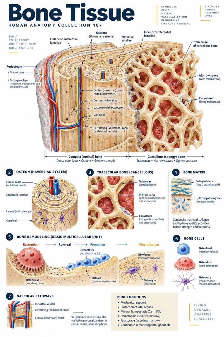 Human Bone Density ↗Human Blood Circulation ↗
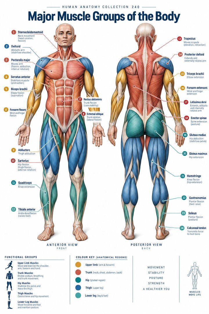 Human Muscle Groups ↗
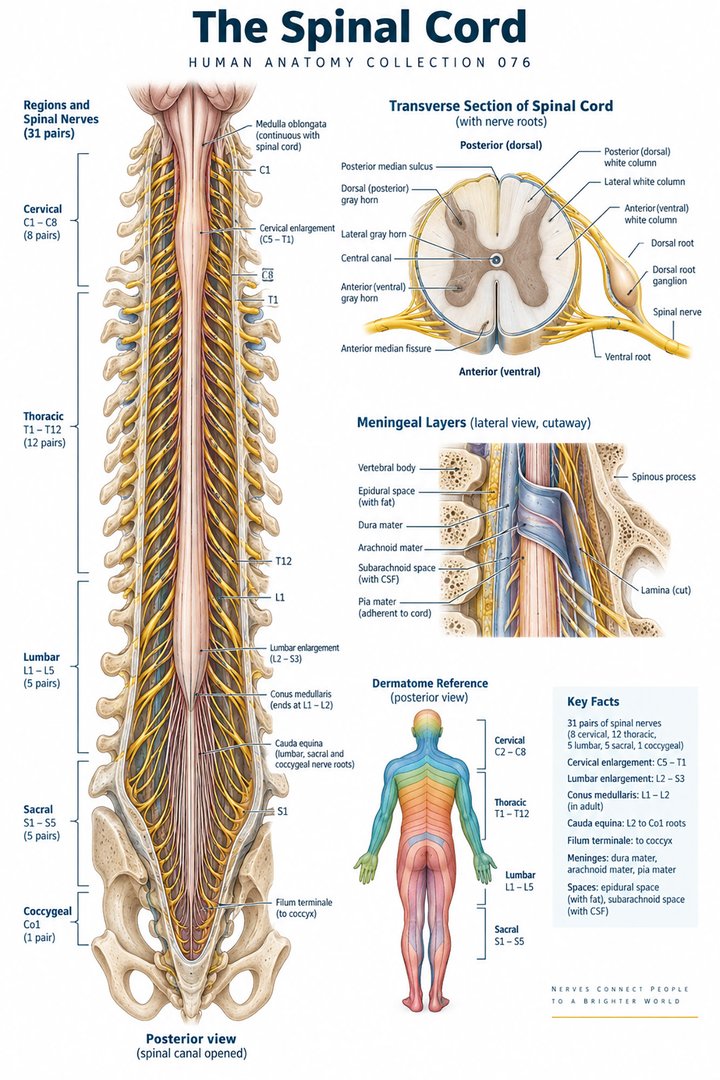 Human Spinal Cord ↗
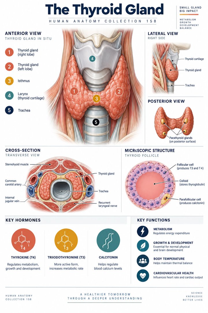 Human Thyroid Gland ↗
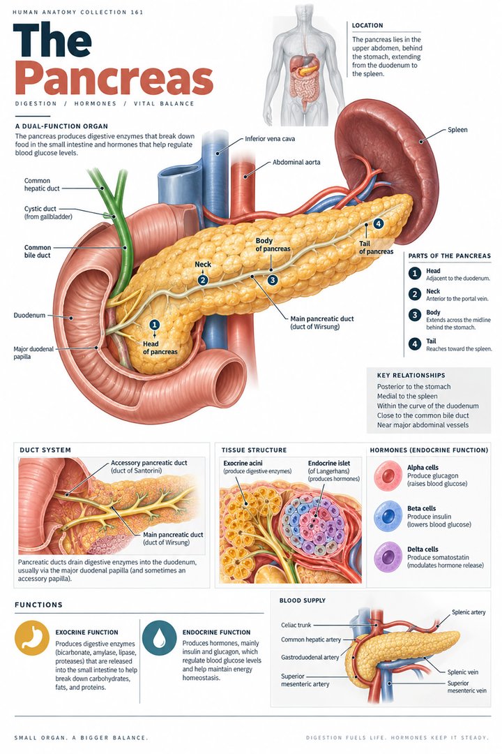 Human Pancreas Function ↗
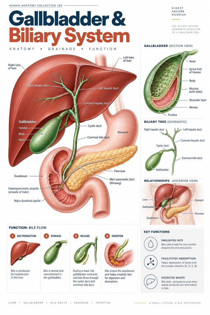 Human Gallbladder Anatomy ↗