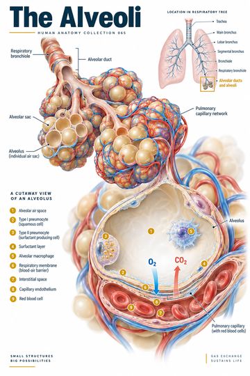
The alveoli are tiny air spaces at the ends of the respiratory passages. Their thin walls and close relationship with pulmonary capillaries make them central to gas exchange. This guide traces the route from the conducting airways to the alveolar surface and explains the structures that help oxygen and carbon dioxide cross between air and blood.
From airways to alveoli
Air travels through the trachea and branching bronchi into progressively smaller bronchioles. The conducting zone moves, warms and humidifies air but does not itself carry out the principal gas exchange with pulmonary capillary blood. Terminal bronchioles lead into respiratory bronchioles, where alveoli begin to appear in the airway walls. These continue into alveolar ducts and alveolar sacs. An alveolus is an individual air space; an alveolar sac is a group of alveoli opening into a shared space.
The branching pattern is useful when explaining why an airway problem and a problem at the gas-exchange surface can affect breathing in different ways. The structures are continuous, however, and real disease may affect more than one level. A simplified poster helps learners locate the parts; it cannot display every airway branch or microscopic relationship.
The respiratory membrane
The alveolar lining lies very close to pulmonary capillaries. At the thinnest regions, the gas-exchange barrier includes the alveolar epithelium, fused or closely associated basement membranes, and capillary endothelium. Oxygen diffuses from alveolar air into blood, while carbon dioxide diffuses in the opposite direction. Diffusion depends on partial-pressure differences and properties of the exchange surface, including its area and thickness.
This process is not the same as ventilation. Ventilation moves air in and out of the lungs; perfusion moves blood through pulmonary capillaries. Effective gas exchange requires both to reach corresponding regions of the lung. An illustration of a single alveolus is a useful starting point, but the relationship between ventilation and perfusion varies throughout the lung.
Cells on the alveolar surface
Type I alveolar cells are thin squamous cells that cover most of the alveolar surface and provide a short diffusion path. Type II alveolar cells secrete surfactant, a mixture that lowers surface tension at the air–liquid interface and helps resist alveolar collapse. Type II cells also participate in repair of the alveolar lining after injury.
Alveolar macrophages are mobile immune cells found on the alveolar surface and in nearby spaces. They ingest particles and microorganisms that reach the distal lung. These cell types have different roles: the macrophage is not a substitute for the epithelial barrier, and surfactant is not itself the respiratory membrane.
Blood flow and the direction of exchange
Blood returning to the right heart travels through the pulmonary arteries to the lungs. In pulmonary capillaries, oxygen enters blood and carbon dioxide leaves it. Blood then returns to the left heart through the pulmonary veins. The pulmonary artery therefore carries relatively deoxygenated blood, while the pulmonary veins carry oxygenated blood under normal conditions. Vessel names describe direction relative to the heart, not oxygen content.
Reading an alveolar diagram
Start with the air space and identify the epithelial lining. Follow the adjacent capillary and locate the blood cells inside it. Arrows can then show oxygen moving toward the capillary and carbon dioxide moving toward the alveolar air. Look separately for a type II cell, surfactant and a macrophage. In many teaching images, cell sizes and spaces are enlarged to make labels legible; they should not be measured as if the drawing were a micrograph.
Clinical and teaching context
Fluid, inflammatory material or other changes affecting alveolar spaces or the interstitium can impair gas exchange. Loss of functioning exchange surface and mismatches between ventilation and perfusion can also affect oxygenation. These are mechanisms, not diagnoses: symptoms and oxygen measurements must be interpreted with the clinical history, examination and appropriate investigations. A diagram cannot determine the cause of a patient's breathlessness.
For patient conversations, a useful explanation is: “Air reaches tiny air sacs surrounded by blood vessels; oxygen moves into the blood and carbon dioxide moves out.” Add detail only as needed for the clinical question. Avoid implying that all respiratory conditions involve damaged alveoli.
Quick review
- Where does most pulmonary gas exchange occur? Across the alveolar–capillary interface.
- What does surfactant do? It lowers surface tension at the alveolar air–liquid interface.
- What is the difference between ventilation and perfusion? Ventilation moves air; perfusion supplies blood.
- Which cells cover most of the alveolar surface? Type I alveolar cells.
From structure to function
Gas crosses the barrier by diffusion rather than by a pump in the alveolar wall. Oxygen that enters pulmonary capillary blood is transported mainly bound to hemoglobin, while carbon dioxide is carried in several forms in blood. The direction of net diffusion depends on the relevant partial-pressure gradients. A drawing that shows only arrows is therefore a model of normal exchange, not a measurement of the gas pressures in an individual patient.